Physical Address
304 North Cardinal St.
Dorchester Center, MA 02124

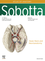
Open full size image Open full size image Overview The neck (collum, cervix) connects the head to the trunk. The respiratory and digestive tracts, the neurovascular pathways and the central nervous system are somatic connections using the neck as a transit route. An osseous base is provided by the cervical spine, on which the head rests and which allows free rotation relative to the trunk of…
Open full size image Open full size image Overview The ear is divided into the outer ear, middle ear and inner ear. Forming part of the outer ear are the auricula (Auricula pinna), the outer ear canal (Meatus acusticus externus) and the eardrum (Membrana tympanica). Behind the eardrum, located in the petrous part of the temporal bone (Pars petrosa ossis temporalis) and belonging to the middle…
Open full size image Open full size image Overview The eye or organ of sight (Organum visus) is often referred to as the ‘gateway to the soul’ and is considered by many people to be the body's most important sensory organ. It includes the eyeball (Bulbus oculi), the optical apparatus and the auxiliary structures (Structurae oculi accessoriae), such as extraocular muscles (Mm. externi bulbi oculi), eyelids…
Open full size image Open full size image Open full size image Overview The head (caput) is flexibly connected to the torso (trunk, Truncus) via the neck area. This allows one to direct the sensory organs of the head towards environmental stimuli without having to move the whole body. The bony skeleton of the head is the skull (cranium). Its posterior section, called the neurocranium, encloses…
Open full size image Open full size image Overview There are good reasons to deal with the retroperitoneal situs of the abdomen (i. e. the organs which are not located in the peritoneal cavity, but at the dorsal wall) in combination with the pelvis. The kidneys, which are the major organs of the retroperitoneal space, initially develop in the pelvis and later ascend to a level…
Open full size image Open full size image Overview Opening the abdominal wall reveals a cavity filled with some smooth and some partially solid organs (viscera). In its entirety it is called the ‘situs’ of the abdominal organs. The inside of the abdominal wall and the surface of the organs are covered with a thin, moist, shiny membrane (peritoneum). The lining on the inside of the…
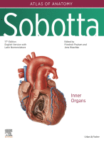
Open full size image Open full size image Overview In a dissection course, the opening of the thoracic cavity is one of the key processes which is met by teachers and students with a mixture of awe, suspense and interest. Exposing the heart and lungs as well as being allowed to grasp (literally and metaphorically) these vital organs of the body with one's own hands is…
Open full size image Overview The lower limb can be divided into the pelvic girdle and the leg. The pelvic girdle consists of the sacrum and the two hip bones. The leg is divided according to its joints into the thigh, the lower leg and the foot. The lower limb functions as a locomotor and a supporting (or weight-bearing) organ. The long bones of the legs…
Open full size image Open full size image Overview The upper limb includes the shoulder girdle and arm. The shoulder girdle consists of the clavicle and the shoulder blade on each side. The arm is subdivided by joints into the upper arm, forearm and hand. The shoulder girdle and thus the whole upper limb are only anchored by the medial clavicular joint directly at the torso.…
Overview The torso supports the head and upper extremities, which are secured in a flexible manner. The spine is the support structure of the torso, with its vertebrae increasing in size from top to bottom to support the weight of the body. To cushion any impact there are intervertebral discs (Disci intervertebrales) between the individual vertebrae which consist of a fibrous ring and a central gelatinous…
Overview The Greek word ‘ανατɛμνɛιν’ (anatemnein) means ‘cut open’ and describes the oldest method in the subject of anatomy, already practised in ancient times. Anatomy is the study of the structure of the healthy body. Without the knowledge of anatomy, one cannot learn about functions, and without the knowledge of structure and function, no pathological changes can be understood. In order to learn a new language,…
You’re Reading a Preview Become a Clinical Tree membership for Full access and enjoy Unlimited articles Become membership If you are a member. Log in here
‘ To practise as a doctor without profound anatomical knowledge is unthinkable. For the diagnosing, treatment and prognosis of illnesses, a detailed knowledge of the structure, positional relationships and the neurovascular pathways which supply the regions and organs of the body is central.’ Anatomical knowledge is gained through cognitive, tactile and especially visual learning processes, and can only be fully acquired when working with the human…
You’re Reading a Preview Become a Clinical Tree membership for Full access and enjoy Unlimited articles Become membership If you are a member. Log in here
You’re Reading a Preview Become a Clinical Tree membership for Full access and enjoy Unlimited articles Become membership If you are a member. Log in here
You’re Reading a Preview Become a Clinical Tree membership for Full access and enjoy Unlimited articles Become membership If you are a member. Log in here

The different diagnoses of diseases and disorders within the nervous system are made using neuropathology in coordination with clinical signs/symptoms and imaging. The imaging techniques discussed in Chapter 9 , coalesced with biopsies, the use of selective antibodies to identify cellular markers, and particular stains to highlight cellular structures, are all used to make a diagnosis. This chapter will present some common neuropathological diseases Stains used…
You’re Reading a Preview Become a Clinical Tree membership for Full access and enjoy Unlimited articles Become membership If you are a member. Log in here
The preceding chapters presented the major structures seen at individual levels of the central nervous system (CNS) or in particular views of the brain. This chapter is complementary, using many of the same sections and views to indicate the structures and connections involved in particular neurological functions. We have taken a “bare-bones” approach to this task and indicated only major pathways and connections. Much of the…
You’re Reading a Preview Become a Clinical Tree membership for Full access and enjoy Unlimited articles Become membership If you are a member. Log in here