Physical Address
304 North Cardinal St.
Dorchester Center, MA 02124

Overview of Headaches Headache is one of the most common reasons for consulting a physician and is one of the top three reasons for lost work days. Rather than a disease, headache is a symptom, frequently providing a valuable warning of hidden pathology. Physicians treating patients for headache must decide whether the headache represents a primary or secondary headache syndrome. Primary headaches are most common and…
Clinical Presentations of Brain Tumors Brain tumors commonly present with symptoms of elevated intracranial pressure or focal neurologic dysfunction. Elevated intracranial pressure can directly result from an enlarging mass or can be secondary to the development of hydrocephalus stemming from obstruction of the ventricular system and cerebrospinal fluid (CSF) flow by the tumor. The specific neurologic dysfunction depends on the local mass effect of the tumor.…
Bacterial Meningitis Pathophysiology. Bacterial meningitis is initially an acute purulent infection of the meninges and subarachnoid space that is followed by an inflammatory reaction in the subarachnoid space, the brain parenchyma, and the cerebral arteries (arteritis) and veins (dural sinus thrombosis and thrombophlebitis). Meningitis is most often the result of bacterial invasion of the subarachnoid space from hematogenous dissemination. Bacterial meningitis may be preceded by colonization…
Multiple Sclerosis: Overview In temperate climates, multiple sclerosis (MS) is the most common episodic neurologic illness of early adult years. The process begins as periodic and focal loss of central nervous system (CNS) myelin and the oligodendrocytes (OGCs) that synthesize myelin. Axons that have lost their myelin function imperfectly or not at all. Accordingly, symptoms ensue, and as episodes recur, disability accumulates. MS is thought to…
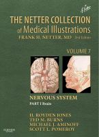
Overview and Approach to Stroke Patient Arterial Supply to the Brain and Meninges Overview and Cervical Segments The brain and meninges are supplied by branches that originate from the aorta. The brachiocephalic trunk (or innominate artery ) divides behind the right sternoclavicular joint into a right common carotid artery (CCA) and a right subclavian artery that supplies the arm. The next aortic branches are the left…
Cerebellum and the Fourth Ventricle The fourth ventricle lies posterior to the pons and upper half of the medulla oblongata and anterior to the cerebellum (see Plate 8-1 ). Its upper and lower ends become continuous, respectively, with the cerebral (sylvian, or mesencephalic) aqueduct and the central canal of the spinal cord in the lower half of the medulla. On each side, a narrow prolongation, the…
Anatomy of the Basal Ganglia and Related Structures Overview of Movement Disorders For the past 30 years, movement disorders have encompassed the study of a group of conditions characterized by poverty of movement, the akinetic-rigid syndromes, and those with excessive movements, the hyperkinetic movement disorders (tremor, dystonia, myoclonus, chorea/ballism, tics, and others). This traditional view, in which disorders of basal ganglia resulted in the aforementioned syndromes,…
Coma The term consciousness refers to a state of awareness of self and one's environment. Assessing consciousness in another person relies on judging that individual's performance or behavior in some mental function and arousal or response of awakening to a stimulus. The word coma originates from the Greek koma (κωμα) and komatos meaning sleep, and deep sleep, respectively. In this section, the use of the word…
Anatomic Relationships of the Hypothalamus The hypothalamus is a small area, weighing about 4 g of the total 1,400 g of adult brain weight, but it is the only 4 g of brain without which life itself is impossible. The hypothalamus is so critical for life because it contains the integrative circuitry that coordinates autonomic, endocrine, and behavioral responses that are necessary for basic life functions, such as thermoregulation,…
Limbic System The limbic system is the only brain area receiving major hypothalamic input and providing interconnection with widespread cortical areas. Major limbic structures include the amygdala, piriform cortex (parahippocampal gyrus, uncus + amygdala) , hippocampus, substantia innominata, and septal area. The limbic system's diverse roles include memory, drive, affect, autonomic tone, endocrine control, and immunoregulation. The amygdala is connected extensively to the hypothalamus and other…
Electroencephalography The electroencephalogram (EEG) is a record of the electrical activity of the nerve cells in the brain. The EEG is based on the measurement of electrical fields generated by volume conduction of ionic currents from nerve cells through the extracellular space. Recorded EEG potentials arise from extracellular current flow from summated excitatory postsynaptic potentials (EPSPs) and inhibitory postsynaptic potentials (IPSPs). The EEG does not record…
Surfaces of Cerebrum The cerebrum is divided into right and left hemispheres by a longitudinal fissure. Each hemisphere has three surfaces—superolateral, medial, and inferior—all of which have irregular fissures, or sulci, demarcating convolutions, or gyri. Although there are variations in arrangement between the two hemispheres in the same brain and in those from different persons, a basic similarity in the pattern allows the parts of the…
Initial Specification of the Nervous System: The Embryo at 18 Days After fertilization and implantation, the embryo consists of a single cell layer called the inner cell mass. The inner cell mass sits at the bottom of a fluid-filled cavity defined by the key extraembryonic membrane, the amnion. Beneath the embryo is another cavity, the yolk sac, lined with a cell layer called the embryonic hypoblast,…
Bones of the Hand Metacarpal Bones Five metacarpals form the skeleton of the hand. They are miniature “long” bones, comprising a shaft, a head, and a base. They are palpable on the dorsum of the hand and terminate distally in the knuckles, which are their heads (see Plates 4-1 and 4-2 ). The shaft is curved longitudinally so as to be convex dorsally and concave on…
Bones and Joints of Forearm and Wrist Distal Parts of Radius and Ulna The distal end of the radius is broadened because its carpal articular surface is the bony contact of the forearm with the wrist and hand. This surface is concave transversely and anteroposteriorly; it is divided by a surface constriction and a slight ridging into a larger triangular portion laterally and a smaller quadrangular…
Bony Anatomy and Landmarks Anteriorly, the contour of the biceps muscle is seen starting in the upper arm and extending distally into the cubital fossa, which is the inverted triangular depression on the anterior aspect of the elbow. The flexion crease along the anterior elbow is in line with the medial and lateral epicondyles and is 1 to 2 cm proximal to the joint line when…
Bones and Joints of Shoulder The function of the upper extremity is highly dependent on correlated motion in the four articulations of the shoulder. These include the glenohumeral joint, the acromioclavicular joint, the sternoclavicular joint, and the scapulothoracic articulation. The glenohumeral joint has minimal bony constraints, thus allowing for an impressive degree of motion. Scapula Ossification centers of the scapula begin to form during the eighth…
Anatomy of the Ankle and Foot Tendon Sheaths at Ankle The extrinsic tendons of the foot originate as muscles in the leg, and as the tendons cross the ankle they must change their orientation. The retinaculum and the corresponding bony anatomy account for the pulley system that allows this to occur and to generate a mechanical advantage for transmission of force. The retinaculum also prevents bowstringing…
Compartments of Leg Fasciae and Compartments The fascia lata of the thigh continues into the leg, where it is designated as the crural fascia (see Plate 4-2 ). At the knee, the fascia has many attachments—the patella, patellar ligament, tibial tuberosity, condyles of the tibia, and head of the fibula—that reinforce the medial and lateral patellar retinacula. The fascia is strengthened by expansions of the tendons…
Anatomy of the Knee Knee Joint The knee is primarily a hinge joint that permits flexion and extension. In flexion, there is sufficient looseness to allow a small amount of voluntary rotation; in full extension, some terminal medial rotation of the femur (conjunct rotation) achieves the close-packed position. The condyles of the femur provide larger surfaces than those of the tibial condyles, and there is a…