Physical Address
304 North Cardinal St.
Dorchester Center, MA 02124

The benefits of youth sports participation go beyond increased health and fitness. Sports participation nurtures the development of skills needed for lifelong physical activity, teamwork, leadership, and peer socialization skills, which leads to increased self-esteem, and higher academic achievements. , Current trends show that youth sports participation and year-round training continues to be on the rise. However, there is evidence that inactivity and lack of physical activity in our youth is also on the rise concomitantly, as school-based physical education programs have decreased, with only 29% of all high school students participating in a daily physical education class. ,
Despite concerns of rising obesity rates and inactivity in present day youth, approximately 27 million youth in the US 6 to 18 years of age participate in team sports. The National Council on Youth Sports 2008 report on trends and participation in organized youth sports reported 60 million youth 6 to 18 years of age participate in some form of organized sports, compared to 52 million in 2000. Participation trends of organized sports in children ages 6 and younger have also increased from 9% in 1997 to 12% in 2008.
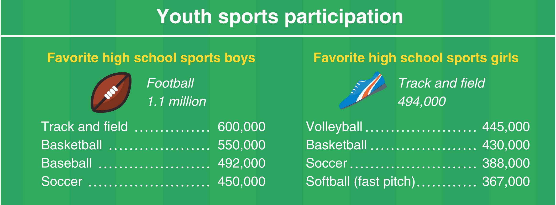
The overall number of young athletes participating in high school sports increased for the 28th consecutive year in 2016–2017, according to the annual High School Athletics Participation Survey conducted by the National Federation of State High School Associations (NFHS). It reached an all-time high of nearly 8 million high school participants (3.4 million girls, 4.6 million boys) along with an all-time high of female participants noted during the 2016–2017 school year. Competitive spirit saw the largest increase in registration among high school girls (see Table 40.1 ). In addition, the survey indicated that greater than 60 different sports were offered from high schools around the country, including rodeo, snowboarding, fencing, judo, rugby, and kayaking. Popular nontraditional high school sports included badminton (17,184), archery (9767), crew (5179), and fencing (4100). Texas and California were the top states with 834,558 and 800,364 participants, followed by New York (367,849), Illinois (341,387), Ohio (340,146), and Pennsylvania (319,153). It is important to note that the above statistics are an underestimate of true athlete participation rates, as they only represent those athletes who participated in the organized sports surveyed by those high schools who are members of the National Federation of High School Sports.
The idyllic days of unstructured child-driven “free play” has shifted to the current sports culture of structured sport specialization patterns, which includes year-round training and participation—often on multiple teams of the same sport, with emphasis on a single sport at an earlier age, at a very high level and intensity. Seventy-eight percent of high school athletic directors surveyed reported an increasing trend in sport specialization, with select/travel leagues starting as young as 7 to 8 years of age on the rise. , The current youth sports culture environment is one of high pressure, often coach and parent driven. Studies have alluded that parents and coaches are the strongest influences in sport selection and specialization of a young athlete. A child’s athletic achievement and success in today’s society is often measured by obtainment of collegiate scholarship and professional contracts rather than emphasis on skill development. This is also contributed to by social media’s widespread attention for successful athletes, with increased recognition and financial rewards that come with elite athlete status.
The concept of specialization was first proposed by Ericsson and his colleagues in 1993. It stated that to reach expert performance, one must practice 10,000 hours over 10 years within that specialized field, adding that one is also more likely to succeed if training is begun at an earlier rather than later age. This similar principle of specialization since then has been adopted in the sports realm by coaches and parents, with a trend of intensive, adult-style training of a single sport at a young age. Cote and his group further emphasized that intense training should be one of “deliberate practice” rather than “deliberate play” or enjoying the activity. In contrast, there is also debate that delayed specialization with early diversification can be more effective in achieving elite athlete status. It is also proposed that those who participate in a variety of sports (sampling, diversification) are more likely to enjoy long-term performance success and have an increased enjoyment for maintaining physical activity long term. The benefits of early diversification include allowing the child to “experience different physical, cognitive, affective and psycho-social environments.” Although there has been no validation that early sport specialization is a requirement for athletic success, and despite the growing evidence that early sport specialization may negatively impact an athlete’s physical and mental health long term, the trend toward early specialization continues to grow.
Sports specialization is defined as “intensive year-round training in a single sport at the exclusion of other sports.” Some advocate that a minimum volume of training is required to meet the definition while others define it as single sport participation year round, regardless of training volume. Sports specialization can be categorized in two groups: (1) early specialization (before puberty) or (2) late specialization with early diversification (sampling). , Sports specialization is often pursued with the goal of obtaining elite athlete status. The reality is a very small percentage of aspiring young athletes reach elite status. Only 3% to 11% of high school athletes go on to compete at the National Collegiate Athletics Association level (NCAA). Nearly 8 million high school students participate in high school athletics, and roughly only 460,000 would go on to play at NCAA schools. Furthermore, only 1% receive an athletic scholarship and a few (0.03% to 0.5% of high school athletes) make it to the professional level ( Fig. 40.1 ). , ,

There is concern that early specialization and intense training may result in negative outcomes such as increased risk of injuries, including overuse injuries and increased psychological stress, potentially leading to burnout and dropout of sports at a young age. , However, there are other contributing intrinsic and extrinsic risk factors that have been identified for overuse injuries ( Box 40.1 ). Overuse injuries occur when the musculoskeletal system is exposed to repetitive loading in the environment of inadequate rest. Skeletally immature athletes are also susceptible to unique overuse injuries involving the physis and apophysis.

Growth-related factors
Susceptibility of growth cartilage to repetitive stress
Adolescent growth spurt
Previous injury
Previous level of conditioning
Anatomic factors
Menstrual dysfunction
Psychological and developmental factors—athlete specific
Training workload (rate, intensity, and progression)
Training and competition schedules
Equipment/footwear
Environment
Sport technique
Psychological factors—adult and peer influences
The risk of injury in a young athlete varies upon several factors including training volume, level of competition, and pubertal maturity. A study looking at high school athletes showed an increased risk of injury when the training volume was greater than 16 hours per week, showing a linear relationship between exposure and risk of injury, which significantly increased when training volume exceeded 16 hours. Another study alluded that sports specialization is an independent risk factor for injury. It showed that athletes who participated in more organized sports compared to free play in a ratio greater than 2:1 had an increased risk for overuse injury. It also suggested that those young athletes who participated in more hours of organized sports per week than their age in years were also at an increased risk for an overuse injury. Other studies have shown a significant increased risk for shoulder and elbow surgery in young baseball athletes pitching greater than 8 months per year. In general, the risks of injury from intense training and specialization seem to be multifactorial and variable, dependent on age, growth rate, pubertal maturation, and level of competition. Furthermore, studies allude that injury rates tend to be higher in athletes older than 13 years of age and those competing at higher levels. Fracture risks seem to be higher during the time of peak height velocity (PHV). Gymnasts were also more likely to be injured during periods of rapid growth (Tanner 2,3).
While most agree that sports specialization leads to a greater chance of achieving athletic success, the optimal timing of when to specialize is debatable. Current evidence suggests that for the majority of sports, delaying specialization until after puberty (15 or 16 years of age) with early diversification and participation in a variety of sports is more favorable for long-term health and future athletic success. , , , The idea is late specialization with early diversification will allow the growing athlete to attain various fundamental skills, requiring less deliberate practice later on should they decide to specialize in a single sport. At the professional level, we find examples of individuals who were multi-sport athletes while in high school and some even in college, and successfully transitioning the acquired skills into their ultimate professional sport, such as Michael Jordan, Tom Brady, Deion Sanders, Jackie Robinson, and Herschel Walker. Studies looking at NCAA athletes have shown that many of them played a variety of sports while in high school and their first organized sport was different than the sport they were playing at the collegiate level.
However, there are certain sports such as gymnastics or rhythmic gymnastics, figure skating, and diving in which early specialization (starting frequently as young as age 5 or 6) may be required as peak performance in these sports occurs before an athlete’s full physical maturation.
As mentioned earlier, early specialization in young athletes may not only put them at risk for physical injury, but also at risk for psychological injury/stress, and can lead to burnout and premature withdrawal from sport. , , Several studies have shown increased anxiety, depression, burnout, and attrition in early specializers. There is data indicating that up to 70% of children drop out of organized sports by the age of 13. , Early specializers tended to withdraw from sport either due to burnout or injury. Burnout is thought to occur as a response to chronic stress that causes an athlete to stop participating in a sport or activity that they previously found to be enjoyable. Burnout was first described in four stages by R.E. Smith in 1986: (1) the young athlete is in a situation that involves varying demands, (2) the demands are perceived by the young athlete as excessive, (3) the young athlete as a result experiences physiological responses, and (4) varying burnout consequences result (i.e., withdrawal). , It is important to keep in mind that excessive athletic stress can manifest as decreased appetite, poor sleep, decreased performance, low self-esteem, and ultimate withdrawal from sport. Restriction in exposure to a variety of sports can prevent an athlete from being exposed to a sport that he or she may excel at, enjoy, or may want to participate in for a lifetime. Specializing too early may also socially isolate the athlete from their family and peers and may interfere with identity development. As a healthcare provider taking care of young athletes, it is important to recognize burnout as a sequela of overtraining and to be aware of the environmental factors and personal characteristics that can contribute to burnout ( Box 40.2 ). The diagnosis of burnout and overtraining is made through the athlete’s history and recognition of various nonspecific symptomatology with which the athlete may present. Further imaging and laboratory studies should only be performed if clinically indicated. While there are no set guidelines for treatment, any diagnosed organic disease should be treated appropriately. Physical and mental rest are paramount components to treatment. A multidisciplinary approach should be taken for treatment, involving the athlete, parents, coaches, treating physician, and sometimes mental health specialists. Compared to adults, children tend to have a more psychological component involved with their burnout. Hence, consultation with a mental health expert such as a sports psychologist should be considered when appropriate. The following recommendations are suggested by the American Academy of Pediatrics and American Medical Society for Sports Medicine to prevent burnout/overtraining and overuse injuries in young athletes: keep training, games, and practices fun; emphasize skill development that is age appropriate; avoid overscheduling and excessive time commitments; have at least 1 to 2 days off per week from their particular sport to allow the body to rest both physically and mentally; and encourage at least 3 months off throughout the year from their particular sport to allow for both physical and psychological recovery and to try other activities. Emphasis should be placed on developing lifelong physical activity skills to enjoy health and fitness for life ( Fig. 40.2 ). ,
Extremely high training volumes
Extremely high time demands
Demanding performance expectations (imposed by self or significant others)
Frequent intense competition
Inconsistent coaching practices
Little personal control in sport decision making
Negative performance evaluations (critical instead of supportive)
Perfectionism
Need to please others
Nonassertiveness
Unidimensional self-conceptualization (focusing only on one’s athletic involvement)
Low self-esteem
High perception of stress (high anxiety)

While there has long been an emphasis on the development of elite athletes, much attention has been focused on developing the youth athlete over the past several decades. Whether the goal is to develop an elite athlete to compete at the collegiate or professional level, or perhaps more importantly to build a foundation for lifelong physical activity and health, developing physical literacy begins at a young age. Physical literacy is defined by the Aspen Institute as “the ability, confidence, and desire to be physically active for life.”
Sedentary lifestyles and physical inactivity have serious negative effects on long-term health. According to the Centers for Disease Control and Prevention (CDC), the prevalence of obesity was 39.8% among adults and 18.5% among children and adolescents in the United States in 2015–2016. Physical activity is a crucial component of obesity prevention and treatment, yet only 1 in 5 adult Americans meets the recommended physical activity guidelines, and only 3 in 10 high school students exercise for the recommended 60 minutes daily.
Athlete development models have traditionally been pyramid shaped with a broad base of participants at the lower levels, and decreasing participation numbers at higher levels of competition, with the ultimate goal of achieving athletic success by those who remain at the top of the pyramid. These models neglect the majority of participants. Balyi and Way developed the Long-Term Athlete Development (LTAD) model in 1995 with the goal of helping all participants have a positive sport experience and reach their potential, not just the best athletes. The LTAD model has evolved to include seven stages ( Table 40.2 ).
| Stage | Ages (Female) | Ages (Male) |
|---|---|---|
|
0–6 | 0–6 |
|
6–8 | 6–9 |
|
8–11 | 9–12 |
|
11–15 | 12–16 |
|
15–21+/− | 16–23+/− |
|
18+/− | 19+/− |
|
Enter at any age | Enter at any age |
The LTAD model forms the foundation of Canadian sport organization and has led to development of similar models in other countries, including the United States. Early models of athlete development utilized chronological age and didn’t take into consideration developmental age. Just as bone age may differ from chronological age, so do physical, emotional, and cognitive development. The LTAD model suggests using the onset of PHV to help determine the athlete’s biological or developmental age and individualize training programs accordingly. They also include the concept of “Windows of Opportunity” which are critical or sensitive periods in development during which a child will better respond to training stimulus.
Lloyd and Oliver suggested that the LTAD model is too simplistic and that most aspects of fitness are trainable throughout childhood and should not be restricted to certain windows, and they proposed the Youth Physical Development (YPD) model. YPD emphasizes that prior to puberty, “strength, fundamental movement skills, speed, and agility should be the main physical qualities targeted and that adaptive responses to the appropriate training methods will be neural in nature. Once the child reaches adolescence, additional components (sport-specific skills, power, and hypertrophy) become more important owing to the increased androgenic internal environment associated with this stage of development.”
The United States Olympic Committee created the American Development Model (ADM) in 2014 based on the LTAD model to create early positive experiences for all athletes with the goals of increasing sport participation among the general population, as well as the pool of elite athletes from which future US Olympians and Paralympians are selected. This model emphasizes fun and enjoyment, particularly in the early and late stages, to encourage lifelong engagement. Stage 4 allows for two different pathways around the time an athlete enters high school, in pursuit of high performance and increased competition, or continued participation for enjoyment and social and health benefits ( Table 40.3 ).
| Stage | Age |
|---|---|
|
0–12 |
|
10–16 |
|
13–19 |
|
15+ |
|
For life |
Although the concept of utilizing developmental age for training has been used for decades, competition has continued to group athletes based on chronological age. There is great variability in the timing of maturation of young athletes which may contribute to unfair competitive advantages for those more mature athletes, and impact athlete safety. Smaller, less mature athletes may have increased injury risks, particularly in collision sports such as American football or hockey. Bio-banding refers to the strategy of grouping athletes based on physical attributes rather than chronological age. Several methods to assess the biological maturity status have been proposed, including percent of predicted adult height (% PAH) and maturity offset (time before or after PHV).
Sports such as soccer have begun initiatives to integrate bio-banding, grouping players based on biological maturity using % PAH, typically within a certain range or “band” (for example, 85%–90% PAH). This reduces the physical advantage of early maturing players and allows both early and late maturing athletes to utilize more technical and tactical skills. Early maturing athletes often have great success and are likely to be viewed by coaches as superior players due to their size and power advantages, thus creating a competitive advantage and selection bias. Goals of the bio-banding initiatives include helping early maturing players further develop their skills, allowing later maturing players to showcase their talents, and ultimately to facilitate development of the greatest potential for each individual athlete.
It is clear that with ever increasing focus on youth sports, the current structure and pressures often lead to overuse injuries, burnout, and dropout. Recent evidence supports changes to the current system with an increased emphasis on enjoyment of sport and development of lifelong physically active individuals, whether that be through early sport sampling, youth development models, or bio-banding.
Unlike other joints in the body such as the knee, the muscles around the shoulder are the main stabilizers for dynamic glenohumeral (GH) joint stability during activity, rather than the ligaments ( Fig. 40.3 ). Hence, when the shoulder muscles become fatigued, the shoulder is at increased risk for injury and may become unstable. The scapula plays a pivotal role in the upper extremity kinetic chain and overall shoulder stability. Static stabilizers of the shoulder include the GH ligaments: superior, middle and inferior, anterior and posterior capsule, labrum, rotator interval, and glenoid. Its function is to limit abnormal humeral head translation. Dynamic stabilizers of the shoulder include the rotator cuff muscles, deltoid, long head of biceps, and scapulothoracic tendons and muscles. Connective tissue and collagen disorders such as Ehlers-Danlos syndrome should be considered in the pediatric patients with shoulder instability and generalized ligamentous laxity. A combination of vulnerable epiphyseal plates and excessive laxity of the surrounding structures that support the shoulder, when placed under strenuous forces, can predispose shoulder injuries in the skeletally immature athlete. With the ever-increasing intense, year-around, and single sports play, especially in baseball, shoulder injuries, including overuse injuries such as Little League shoulder, are being seen more frequently in the modern orthopaedic office. ,

Little League shoulder (LLS), also termed proximal humeral epiphysiolysis, osteochondrosis, apophysitis, epiphysitis, stress fracture, or Salter Harris I injury of proximal humerus, is an overuse injury that affects the proximal humeral physis in skeletally immature athletes, caused by repetitive stress. It is most commonly seen in the throwing arm of young baseball pitchers, but can be seen in other baseball positions, and in other overhead sports, including tennis, gymnastics, swimming, and volleyball. Case series looking at patients diagnosed with LLS showed that the majority of the patients were baseball players (97%): 86% pitchers, 8% catchers, 7% other positions, and found a small group of tennis players (3%).
LLS is thought to be a caused as a result of repetitive micro traction and torque forces placed on the unossified cartilage of the proximal humeral physis in the skeletally immature arm, most commonly caused by the throwing motion. , Studies have suggested that the high torque forces generated during the late cocking phase in the pitching motion are strong enough to weaken the proximal humeral epiphysis, leading to humeral retroversion or proximal humeral epiphysiolysis.
Pediatric overhead athletes typically present with LLS between the ages of 11 and 16 years of age with peak incidence occurring at around 13 to 14 years of age, corresponding to the age in which total shoulder range of motion was found to be significantly decreased in adolescent baseball players. It has been alluded that this decrease in rotational motion may cause increased stress at the proximal humeral physis during the throwing motion.
A high volume of pitches and maximum effort throws, poor throwing mechanics, inadequate rest between pitches, and pitching while fatigued are risk factors which have been associated to contributing stress over the proximal humeral physis. ,
Patients commonly present with a history of progressive worsening upper arm pain with throwing motion. They often report pain exacerbating with harder and more frequent throws. As it progresses, it may even cause diffuse shoulder pain with activities of daily living and at rest. The throwing athlete may also complain of decreased pitching velocity and strength with throwing. It is not uncommon for the patient to present with progressive shoulder pain that has been ongoing for months prior to presenting to the physician’s office for evaluation.
A thorough history including number of weekly games and pitches, types of pitches thrown, and number of rest days between pitches should be obtained. Full-speed pitches thrown during practice, including those thrown in the bullpen and at home, should be included in weekly pitch count. It can also be helpful to ask patient which phase of the throwing motion elicits their symptoms.
On physical exam, the most common finding is bony tenderness to palpation along the proximal humeral physis. Pain may be elicited at terminal ends with shoulder range of motion testing. Patients may also present with decreased range of motion with limited external or internal rotation.
LLS is a clinical diagnosis; however, common radiographic findings may include physeal widening, irregularity, and increased sclerosis compared to contralateral nonaffected shoulder ( Fig. 40.4 ). Lateral fragmentation, calcification, and cystic changes may also be noted. Anteroposterior (AP) Grashey view in external rotation views allows for physis evaluation and contralateral radiographs for comparison can be helpful with diagnosis. Conventional radiographs of the shoulder include AP Grashey, scapular Y, and axillary views. Magnetic resonance imaging (MRI) may be helpful in detecting occult physeal injuries when conventional radiographs are normal and the clinical exam is equivocal.
 Arrow showing widening of the proximal humeral physis, right shoulder. (B) Note comparison of normal view of the left shoulder.")
LLS is almost always treated with conservative management consisting of rest from activity that causes pain and cessation of throwing for approximately 3 months. Wasserlauf and Paletta recommend cessation of throwing for a minimum of 6 weeks after diagnosis, and an additional 6 weeks of no throwing during the strengthening phase of rehabilitation, for a total of minimum 3 months’ rest from throwing. , Physical therapy may begin when pain has subsided and should focus on strengthening of the rotator cuff, periscapular and core musculature, stretching and flexibility of the shoulder capsule, and scapular stabilization. , Proper form and pitching mechanics should be implemented.
Prior to returning to play, the athlete should have pain-free throwing motion and demonstrate full range of motion and strength. If the treating healthcare provider sees fit, they may return the athlete to play earlier in positions in which stresses to the arm are less, such as batting or first or second base. Third base should be avoided due to the long throwing distance to first base. The athlete should not return to full pitching or play that requires hard throwing until they have completed a gradual interval throwing program and are able to throw pain-free and exhibit no pain over the proximal humeral physis on exam. If significant widening of the proximal humeral physis was noted on radiographs, follow-up radiographs are recommended prior to return to full play. Numerous studies have reported an increased risk for shoulder and elbow injuries (upper extremity injuries) in young baseball pitchers based on poor biomechanics, high volume of pitches thrown per game and during season ( Table 40.4 ), and throwing with a fatigued arm more than the type of pitch thrown. , , Diversification in positions (playing other positions besides just pitching) may protect against ulnar collateral ligament reconstructions (UCLRs) and other shoulder elbow injures. A preseason strengthening program focusing on core and rotator cuff strengthening and scapular stabilization is recommended. Patient education on discouraging participation in multiple leagues in the same season, focusing on preseason conditioning, with emphasis on proper form, throwing mechanics, pitch count, and pitch types (avoid throwing curveballs at a young age) are felt to be crucial in the prevention of shoulder injuries in young throwing athletes. Maintaining a healthy shoulder through appropriate stretching and strengthening program regularly both in season and preseason is recommended.
Source: Little League Baseball.
From Feeley BT, Schisel J, Agel J. Pitch counts in youth baseball and softball: a historical review. Clin J Sport Med . 2018;28(4):401–405.
| Pitch Counts | |
|---|---|
| Age | Daily Maximum Pitches Per Day |
| 17–18 | 105 |
| 13–16 | 95 |
| 11–12 | 85 |
| 9–10 | 75 |
| 7–8 | 50 |
| Mandatory Rest | ||
|---|---|---|
| Ages 14 and Under | Ages 15–18 | Mandatory Rest Requirement |
| 66+ | 76+ | 4 days |
| 51–65 | 61–75 | 3 days |
| 36–50 | 46–60 | 2 days |
| 21–35 | 31–45 | 1 day |
| 1–20 | 1–30 | No requirement |
Premature physeal closure and physeal fracture extending into metaphysis have been reported in association with LLS, but is rare. ,
Glenohumeral internal rotation deficit (GIRD) is defined as side-to-side total motion arc difference of more than 25 degrees. GIRD can be a source of shoulder dysfunction and pain in the young throwing athlete, and more recent studies are starting to allude that GIRD may be more common among younger overhead athletes than previously thought. One case series looking at patients ages 8 to 16 years diagnosed with Little League shoulder that showed that one-third of patients with LLS demonstrated GIRD and those with GIRD in their group had approximately three times higher probability of recurrence of LLS compared to those without GIRD. Overhead athletes frequently gain external rotation adaptively due to repeated microtrauma to the anterior capsule during the cocking phase of throwing, thought to be advantageous for achieving maximal external rotation for cocking phase of throwing, allowing the athlete to increase throwing velocity. As external rotation increases, internal rotation tends to decrease. Decrease in internal rotation is caused by tightness and contracture of the posterior inferior capsule, which can lead to altered shoulder mechanics and possible injury, such as superior labrum anterior and posterior (SLAP) tears or posterior shoulder impingement. ,
Patients will complain of shoulder pain with throwing motions and may report decreased performance. They may also report difficulty reaching behind their back or reaching across their body. On exam, external and internal rotation are documented with the patient in supine position to help stabilize the scapula and with the arm in 90 degrees of abduction. GIRD can also be visualized with the patient in seated position ( Fig. 40.5 ).

GIRD is a clinic diagnosis. An MRI can be obtained if an associated shoulder injury or pathology is suspected and can reveal associated pathology such as SLAP lesions or rotator cuff pathology if present.
Physical therapy focusing on stretching of the posterior capsule and rotator cuff is recommended ( Fig. 40.6 ). As a primary component of GIRD may be soft tissue contracture, treatment should involve the soft tissues as well. The sleeper stretch is the most commonly used exercise to increase internal rotation and is preferred in a side-lying position to stabilize the scapula. To place strain on various parts of the posterior capsule and muscles, the arm is placed at 60, 90, and 120 degrees abduction and a rotational stretch is applied. The motion is held at the point of tightness. Various other internal rotation stretching and mobilization of the posterior capsule has been shown to decrease posterior capsule tightness and improve GIRD, with numerous studies showing good results. , Anecdotal and clinical experience suggests that the vast majority (>90%) do improve with rehab programs focusing on various types of stretching and mobilization techniques. Posterior capsule stretching should be part a young throwing athlete’s conditioning program. Several studies have shown that those overhead athletes who maintain a regular posterior capsule stretching regimen were less likely to have shoulder problems and injury. These suggest GIRD prevention may play an important role in preventing shoulder injuries in overhead athletes and GIRD should be screened for during preseason and while in season.
 performing the sleeper stretch to focus on posterior capsule stretching.")
Athletes who do not respond to stretching and do not regain internal rotation with therapy may be candidates for posterior-inferior capsule release. In those athletes who undergo capsular release, the posterior inferior capsule may be found to be thickened and scarred down, off the glenoid rim. Releasing the thickened scar tissue can improve internal rotation. This procedure is not indicated as a solo procedure, but should be done in conjunction as part of repair of the superior labral tear or treatment of internal impingement, as those with persistent pain despite internal rotation improvement may have a SLAP lesion that may need to be addressed operatively.
The shoulder joint is the most unstable joint in the human body. It is important for those who care for pediatric athletes are familiar with the different types of shoulder instability and treatment options available. Shoulder instability in the pediatric skeletally immature athlete can be traumatic or atraumatic in etiology. Patients with traumatic shoulder instability typically have a single traumatic event—either subluxation or dislocation with spontaneous reduction or requiring manual reduction. Traumatic dislocations often are associated with structural damage either at the bony or soft tissue level of the GH joint. They are also related with a high risk of recurrent dislocation in young, active patients. ,
Atraumatic instability is often due to multidirectional instability in the young patient. Atraumatic instability can be congenital (i.e., patients with generalized ligamentous laxity) or acquired (i.e., young overhead throwing athletes whose shoulders are stressed repeatedly in hyperextension, abduction).
The three anatomic GH instability patterns are: anterior, posterior, and multidirectional. Anterior instability is the most common instability pattern in young athletes, comprising 85% to 95% of all instability cases. The main cause of anterior instability is the result of traumatic, acute injury. It is less commonly due to repetitive overuse injury in overhead throwing athletes, such as pitchers.
Posterior instability comprises only 2% to 10% of all patients with shoulder instability. They are frequently associated with seizures, electrocution injuries, and severe trauma.
Multidirectional instability (MDI) is subluxation, dislocation, or instability of the shoulder joint that occurs in more than one direction. It is typically atraumatic in the pediatric population and can present as unilateral or bilateral GH instability. It accounts for less than 5% of all shoulder instability cases. There is varying pathophysiology for MDI, which continues to evolve in literature. It is more common in athletes who perform repetitive overhead activities such as swimmers, throwers, gymnasts, and tennis players.
Pediatric patients can also develop secondary shoulder subacromial impingement syndrome due to MDI. This secondary impingement syndrome is thought to be the result of the humeral head not being centered in the glenoid fossa and its tendency for upward migration into the subacromial space due to ligamentous laxity and weakness of rotator cuff muscles.
The history and physical exam often lead to the diagnosis of shoulder instability. The clinician should take into consideration the mechanism of injury, position of arm at the time of trauma, sports participation, and goals. In traumatic anterior shoulder dislocation, they will report of a shoulder in abduction and external rotation and receiving a hit to the arm, with the arm in full outstretched motion. In a posterior traumatic dislocation, the patient may report a direct blow with the arm in forward elevation, adduction, and internal rotation. A common position is linemen in football.
Frank dislocations often present to the emergency department either before or after reduction of dislocation. Spontaneous reductions can occur, but are more common in chronic, recurrent, anterior instability or with multidirectional instability of the shoulder. The young patient will often report a history of his or her shoulder “coming out” or “slipping out,” then popping back in. This may indicate a possible recurrent shoulder subluxation or instability. Patients may also complain of anterolateral and anterior shoulder pain with overhead activities and motion.
Inspect for any shoulder deformity or muscle atrophy. A thorough neurovascular exam, including assessment of the axillary nerve (the most commonly injured nerve in up to 42% of traumatic anterior shoulder dislocations), should be performed. Physical examination should include a comprehensive shoulder exam.
Certain tests are helpful for evaluating GH shoulder instability. Apprehension and relocation tests assess anterior GH instability. The patient should be lying in supine position on examination table with their arm abducted to 90 degrees and externally rotated. If the patient feels a slipping sensation or fear of a impending dislocation of their shoulder with this motion that is improved with applying a posterior force to the GH joint, it is a positive relocation test. This means that the posterior force applied to GH joint relocated the humeral head in place. The sulcus sign is performed at 0 degrees of abduction ( Fig. 40.7 ). Downward traction is applied on the humerus. Dimpling or a “gap” formed in the GH joint is a positive sign, indicating laxity of the superior GH ligament. , The load and shift test is used to evaluate anterior and posterior GH laxity and is performed while the patient is in a seated or supine position with the humeral head centered in the glenoid fossa and performed. Grade 0 means normal translation. Grade 1: translation to rim and back, less than 1 cm. Grade 2: translation over the rim followed by spontaneous reduction, 1 to 2 cm. Grade 3: translation over the rim without spontaneous reduction, greater than 2 cm. Generalized joint laxity should also be assessed using the Beighton score (0–9 point scale). ,

Subacromial impingement secondary to MDI may pre-sent as tenderness to palpation over the supraspinatus tendon insertion site on the greater tubercle. Patients may have scapular winging and pain with impingement maneuvers such as the Hawkins and Neer tests. A positive Neer test is when pain over the subacromial joint space is elicited with forcing the arm into position of maximal forward elevation. A positive Hawkins test is when pain is elicited with the arm in internal rotation with the arm forward elevated to 90 degrees and producing pain as the supraspinatus tendon impinges on the coracoacromial ligament or anterior acromion. Strength testing typically also reveals weak external rotators and supraspinatus.
Radiographic evaluation should include: AP Grashey, Y-scapular lateral views, and axillary lateral view to evaluate the relationship of the humeral head to glenoid and possible posterior humeral head compression fracture (Hill-Sachs lesion) or fracture of the glenoid rim (a bony Bankart lesion). High rates of glenoid bone loss are noted in adolescents with a traumatic shoulder dislocation and are often not seen on conventional x-rays. If further shoulder capsule or labral tissue injury is suspected, an MRI can be helpful. An MR arthrogram is the gold standard for imaging traumatic capsular or labral pathology. SLAP (superior labral anterior to posterior) tears have been found to be commonly associated with shoulder instability patterns in adolescents. If a bony Bankart or large Hill Sachs lesion is noted on radiograph, a CT can be helpful in evaluating and quantifying bone loss. Large bony defects may change surgical management and may require additional or alternative procedures rather than just a capsulorrhaphy, labral repair, or capsulolabral reconstruction.
The first line of treatment for anterior GH instability in the young athlete with a traumatic anterior GH dislocation is controversial. High rates of recurrent instability have been reported after first-time dislocation in patients less than 20 years of age. Patients with traumatic anterior shoulder subluxation events may have associated labral injuries as well. The mainstay treatment for young patients with multidirectional instability is typically conservative. Nonsurgical management has shown overall good results. Physical therapy should focus on periscapular and rotator cuff strengthening to optimize dynamic stabilizers, as static ligamentous stabilizers are deficient in these patients. Good to excellent results in 80% of the patients with atraumatic shoulder subluxations treated conservatively with an exercise program focusing on strengthening dynamic stabilizers of the shoulder—including scapular stabilizers, trapezius, rhomboids, serratus anterior—have been shown.
Initial GH dislocation management is discussed in Chapter 29 . In the pediatric population, traumatic anterior shoulder dislocation accounts for greater than 90% of shoulder dislocation. , The first line of treatment for traumatic anterior GH shoulder dislocations is controversial. The rate of recurrent dislocation/instability following a traumatic shoulder dislocation in patients less than 20 years of age is found to be very high (75%–90%) in those treated nonoperatively. , , Traumatic anterior shoulder dislocations in skeletally immature patients have also been shown to be associated with a humeral avulsion of the glenohumeral ligament (HAGL) injury. The causes of re-dislocation are likely multifactorial, including ligamentous laxity and participation in collision sports/high-demand activities.
A prospective series looking at 252 patients (15–35 years old) showed 56% of the patients had recurrent instability episodes at a mean follow-up of 13 months after being treated nonsurgically.
Hence, the idea of early surgical intervention with a stabilization procedure in these patients following a traumatic dislocation is often considered. Recurrent dislocations are not only painful, but may lead to cartilage damage, and limit activities of daily living and sports participation in these young patients. Several factors should be taken into consideration following traumatic shoulder dislocation and should include: the sport the patient is involved in (contact, throwing, upper extremity weight bearing sport), patient’s goals of continuing athletic participation at a high level, and possible career aspirations long term.
Surgical treatment for primary traumatic GH anterior dislocation can be arthroscopic or open. Instability recurrence rates after arthroscopic repair with suture anchors are now equal to classic open Bankart procedure, due to improved technology and advances in equipment, implants, and methods. Currently, there are few studies of arthroscopic repair of adolescent shoulder instability in literature. A retrospective study of 32 patients (11–18 years of age) showed 5 re-dislocations (16%) at 2 years after arthroscopic repair. Eleven patients returned to sports. Another retrospective study reported that although not statistically significant, post-surgical patients who underwent an open Latarjet technique showed better signs of shoulder stability and higher return to same level of sport (92%) compared to the nonsurgically treated group (52%).
Presently, there is no widely accepted guideline for surgical management for patients with MDI laxity of shoulder joint. However operative treatment can be considered for those patients with debilitating symptoms or limit activity after at least a six-month trial of dedicated rehabilitation. Excessive external rotation (>130 degrees) on clinical exam may be an indication for anterior capsular plication. Open and arthroscopic procedures are both options for those patients who fail nonoperative treatment. Both open and arthroscopic procedures have a similar reported success rate of 88% to 100%, with good to excellent results, and a recurrence rate up to 10%. , , Open capsulorrhaphy or arthroscopic procedure should focus on the direction of instability. Thermal-assisted capsular shrinkage has been used to treat shoulder instability, but its utility has been questioned. There have been reported complications of capsular ablation, chondrolysis, and axillary nerve palsies. ,
Injuries to the skeletally immature patients are unique and oftentimes may be sport specific. Many respond well to conservative and nonsurgical management, but surgical intervention may be necessary for certain circumstances. There is still a paucity of studies focusing on surgical intervention and outcomes in the young pediatric patient. In the meantime, injury prevention is key to helping keep our young patients active yet healthy.
The term Little League elbow has been used to describe a multitude of lesions about the elbow, usually medial epicondylar apophysitis/avulsion, osteochondritis dissecans (OCD) of the capitellum, but also Panner disease and stress lesions of the olecranon apophysis and radial head epiphysis. The casual application of this term is unfortunate in that it accurately describes neither the pathology nor the mechanism. These overuse syndromes can be seen in any overhead athlete. Patients usually have localized pain that is activity related. Radiographs may be normal or reveal characteristic changes consistent with osteonecrosis or epiphysiolysis. Treatment consists of nonsteroidal antiinflammatory drugs (NSAIDs) and activity modification. Young athletes may require immobilization to ensure compliance with rigid activity restrictions. Once symptoms have abated, a carefully designed, well-controlled return to athletics should be implemented. Persistent symptoms or instability may require surgical intervention.
The ossification center of the medial epicondyle of the humerus appears between 5 and 7 years of age and unites with the humeral diaphysis between 18 and 20 years of age. The common tendon of the flexor muscles of the forearm and ulnar collateral ligament of the elbow insert on the medial epicondyle. The ulnar collateral ligament is the primary soft tissue stabilizer to valgus stress and originates from the distal aspect of the medial epicondylar apophysis. The ulnar nerve runs in a groove in the posterior aspect of this epicondyle. The medial epicondyle is an apophysis and does not contribute to longitudinal growth of the humerus.
The mechanism of repetitive stress or acute injury is a valgus stress producing traction on the medial epicondylar apophysis through the flexor muscles. In sports, this may occur traumatically with a fall in an upper extremity weight-bearing athlete such as gymnast, or in repetitive or an acute or chronic avulsive fashion as in a baseball player engaged in valgus mechanics during throwing. ,
The hallmark activity for most medial elbow apophysitis in adolescents is youth baseball. A history of repetitive throwing, often year-round or on more than one team, as well as over-representation of symptoms in the pitching and catching positions is common. Poor form and lower-body mechanics during transitional growth years may contribute to an increased valgus position during throwing that increases symptoms. Pain during and after throwing at the medial elbow, tenderness to medial flexor muscle palpation, and direct tenderness over the epicondyle is seen. Pain with valgus testing is usually less than with direct palpation and the patient may occasionally present with loss of full elbow extension. ,
The epicondyle may enlarge, exhibit distal traction related avulsive changes, or may have increased apophyseal cartilage width. While an MRI is often not indicated in the absence of an acute event, edema at the medial epicondyle or sublime tubercle, and occasionally periosteal thickening or layering, may be seen. ,
Treatment for medial apophysitis is focused upon forced rest from the repetitive event and physical therapy. While avoidance of repetitive throwing and dynamic valgus is of primary importance, shoulder and arm physical therapy is also prescribed. Relative internal rotation deficit of the shoulder may be present, and shoulder stretching to balance range of motion is followed. Periscapular strengthening followed by rotator cuff strengthening is prescribed. Forearm flexor repetitive strengthening with avoiding of the eccentric load is also employed. Following a minimum of 6 weeks and often as much as 12 weeks of forced rest, return to throwing or upper extremity weight bearing is allowed via a graduated protocol. A maintenance program of this therapy may be required over several years for some patients.
Information specific to upper extremity athletes will be covered here. For more detailed information, please refer to the upper extremity trauma section.
There may or may not be a history of antecedent pain with throwing or upper extremity weight bearing. This may be seen as a sequela of ongoing throwing despite symptoms discussed above. The patient often reports a pop and sudden medial pain. The physical findings depend on the degree of displacement of the medial epicondyle. Generally, the elbow is held in flexion and any motion is painful. There is tenderness over the medial epicondyle that is exacerbated with valgus stress. Ulnar nerve dysesthesias may be present. Medial contusion is common at 24 to 48 hours.
Unfortunately, no widely accepted classification of medial epicondyle fractures has been presented, and most investigators have described unique systems based on what they consider critical information. All the established classification systems consider whether the fracture is displaced or nondisplaced but there is no agreement on what constitutes a displaced fracture and accuracy and reliability on plain radiographs also makes agreement difficult. Although less commonly employed, oblique and axial views have been proposed to improve measurement accuracy of displacement. , , , ,
Plain radiographs are still most often employed; however MRI of the elbow and the upper extremity athlete may be employed or used to identify partial or periosteal sleeve avulsions of the medial epicondyle.
In the upper extremity athlete, the anatomic position of the medial epicondyle as the origin of the medial elbow flexor musculature and ulnar collateral ligament is felt to be important for future throwing or upper extremity weight bearing. As such, little to no displacement of the fracture fragment is desired if non-operative treatment is being considered. Athletes may be returned to upper extremity sport following nonoperative treatment; however minimal displacement is most often required for an excellent result. , For a nondisplaced or minimally displaced fracture (<2 mm), immobilization in a posterior splint, long-arm cast, or sling for 1 to 2 weeks, followed by early active range-of-motion exercises, is recommended. At 3 to 4 weeks, a physical therapy program should focus on strengthening of shoulder, elbow, and wrist muscular associated with throwing and avoid wrist flexor strengthening for 6 to 8 weeks and any motion causing a valgus moment. At 8 to 12 weeks and based on radiographic and clinical healing, a throwing program can be initiated. There should be absolutely no throwing until pain free at the fracture site.
While acceptable results for function in the nonoperative extremity athlete may be commonly reported following nonoperative treatment of displaced fractures, we advocate an operative treatment for any displacement beyond 2 mm in the upper extremity athlete. , ,
For operative management, the patient may be positioned either supine or prone. We have found the prone approach to be most useful for ease of reduction and intraoperative management. In this position the avulsion bed is more easily identified and the tension of the medial flexor musculature is decreased. Open reduction is performed through a medial longitudinal skin incision. The ulnar nerve is identified and retracted posteriorly. The fractured medial epicondyle is identified and is anatomically repositioned with a towel clip. We favor fixation with a partially threaded screw (most often 4.0 mm with or without a washer), often using a cannulated system to achieve temporary fixation ( Fig. 40.8 ). Care must be taken in young patients to prevent comminution of the predominantly cartilaginous distal fragment during fixation. After open reduction we immobilize the elbow in 90° flexion for 1 to 2 weeks, after which active range-of-motion exercises are initiated. We occasionally splint the wrist for an additional 3 to 4 weeks after cast removal to prevent active contraction of the flexor muscle mass, which might displace the distal fragment.
 Displaced medial epicondyle fracture in a 14-year-old Little League pitcher. The injury was sustained during pitching. (B) Anteroposterior radiograph obtained after open reduction and fixation of the medial epicondyle fragment.")
With advanced imaging following an elbow dislocation, or a known significant medial elbow valgus overload in an upper extremity athlete, a partial apophyseal avulsion or periosteal sleeve avulsion of the distal aspect of the medial epicondyle may be identified. This fragment may often have the majority of the origin of the ulnar collateral ligament attached. When displacement of this osteocartilaginous avulsion is felt to be significant and at risk for nonunion or malunion of the origin of the ulnar collateral ligament, open reduction and fixation may be employed. In these select cases, small 2 to 3 mm suture anchors may be placed in the distal aspect of the remaining intact epicondyle and suture fixation of the small partial or periosteal sleeve avulsion may be employed. In such cases, a similar postoperative and return to play protocol is followed as above.
We favor a brief period of immobilization (no more than 1 week), followed by early active range-of-motion exercises. At 3 to 4 weeks, physical therapy can be initiated and follow a similar program as stated for non-operative treatment.
Complications from medial epicondyle fractures include stiffness, ulnar neuritis, missed incarceration, and symptomatic nonunion. Stiffness is the most common complication and is best prevented by avoiding prolonged immobilization. It is important to remember that the soft tissue injury is usually much more significant than the radiographic abnormality. Early motion is employed as above.
Symptomatic nonunion in a high-performance athlete is difficult to treat. We have had some success in establishing union in symptomatic patients. Our approach is to stabilize the fragment with in situ fixation and a local bone graft. We do not attempt to mobilize the fragment and reduce it anatomically. Others have advocated simple excision of the symptomatic nonunion with reattachment of the ulnar collateral ligament. We prefer an initial attempt at establishing union.
The anterior bundle of the medial ulnar collateral ligament (UCL) is the primary static stabilizer of valgus stress at the elbow. It originates on the distal osteocartilaginous portion of the medial epicondyle apophysis with an origin center point approximately 3 mm from the lateral edge of the bony apophysis. The ligament inserts on the medial aspect of the proximal ulna at and just distal to the sublime tubercle.
A history of repetitive overuse with throwing as described above is most often present in baseball players with a finding of ulnar collateral ligament injury of the elbow. High repetition of in-season throwing, play on multiple teams, positions of pitcher and catcher, and continued play through medial elbow pain may be significant correlations. , , The ulnar collateral ligament may also be injured acutely with a valgus weight-bearing event during upper extremity gymnastic weight bearing or with an elbow dislocation.
Similar to medial elbow flexor tendinitis and apophysitis, the patient with an ulnar collateral ligament injury may present with local tenderness to palpation and loss of elbow extension. Additionally, pain with valgus stress tests and local pain focally and distal to the ulnar collateral ligament origin and extending to the sublime tubercle region is common.
Plain radiographs may often show chronic changes of valgus stress in the ulnar collateral ligament injured elbow. In the case of a partial or osteocartilaginous sleeve avulsion, this radiodense fragment or fleck may be identified on radiographs.
Ultrasound or MRI may be employed to investigate the soft tissue portion of the ulnar collateral ligament. , , MRI with joint arthrogram is most commonly used. A low-grade (I or II) partial injury may be difficult to definitively assess. Complete mid-substance or origin, insertional injuries (III) may be more clear. a
a References , , , , , .
Definitive complete injury of the ulnar collateral ligament requires correlation of history, physical exam, imaging, and often failure of return to sport. Particularly in the adolescent population, nonoperative modalities should often be employed prior to consideration of operative treatment. Prolonged rest from throwing, stretches to address internal rotation deficit of the shoulder, periscapular and glenohumeral strengthening, arm strengthening, and a graduated return to throwing program is required. This may require several months of rest from desired throwing. Adolescent patients may also be counseled regarding transition to a less demanding position that will allow them to play and avoid surgery in some cases. ,
A detailed description of perioperative care and surgical technique for reconstruction of the ulnar collateral ligament of the elbow is beyond the scope of this age-specific text. Operative treatment should be reserved for select athletes who have failed all nonoperative measures and following shared decision making regarding the significant time in rehabilitation commitment required following this procedure to return to high-level upper extremity sport. Excellent outcomes can be achieved in the adolescent population with ligament reconstruction, with or without augmented internal bracing. In addition to meticulous surgical technique, supervised physical therapy and graduated return to sport over 9 to 18 months is required for successful outcomes. b
b References , , , , , .
The exact processes leading to capitellar and repetitive radial head lesions are unclear. Osteochondrosis may occur without any distinguishing history. Capitellar OCD and radial head stress fractures occur most often in valgus-mechanics throwing or upper extremity weight bearing. It is postulated that repetitive compression and sheer stress on the incompletely ossified capitellum and proximal radial epiphysis may play a role in the development of these lesions.
Panner first described a lesion in the epiphysis of the capitellum similar to Legg-Calvé-Perthes disease in 1927. In 1964, Smith reviewed the literature and proposed that Panner disease was a self-limiting condition that was nontraumatic in origin. Histologic studies of Panner disease have documented focal areas of avascular necrosis, with repair and revascularization.
The entity described by Panner and subsequently referred to as Panner disease affects children younger than 10 years and is associated with pain and stiffness in the elbow. These patients do not have constitutional symptoms and rarely have a history of trauma. There may be a flexion contracture and diffuse synovitis.
Radiographically, the capitellum will show irregular areas of radiolucency with areas of sclerosis. An effusion may be noted. An MRI will demonstrate areas of edema surrounding poorly vascularized epiphyseal bone. These lesions may also be evaluated by ultrasound. The articular cartilage has been noted to be normal.
Treatment usually consists of reassurance, activity restrictions, and NSAIDs. Occasionally extremely symptomatic patients benefit from a period of immobilization in a splint or cast if the symptoms fail to resolve with conservative measures. Panner syndrome that does not follow this course should be re-evaluated critically for signs of capitellar OCD.
Schenck and colleagues have proposed a repetitive microtrauma mechanism for the development of OCD of the capitellum. In a cadaver study, they noted that the central section of the radial head was significantly stiffer than the lateral capitellum. Presumably, the disparity in the mechanical properties of the central radial head and lateral capitellum would increase strain in the lateral capitellum. During activities associated with high valgus stress, such as throwing, this increased strain may be a factor in the development of OCD of the elbow. , ,
OCD of the capitellum is distinguished from Panner disease in that it occurs in older children, is generally associated with overhead athletes (usually baseball players and gymnasts), and is more likely to require surgical treatment. As with Panner disease, the initial symptoms are typically pain, loss of extension, and occasionally mechanical symptoms.
Radiographically, OCD of the capitellum may be similar to Panner disease ( Fig. 40.9 ). MRI, CT, and ultrasound may be used to further delineate the stability of the lesion. Lesion stability, and subsequently consideration of nonoperative and operative treatment, is primarily dictated by the status of the articular surface. If there is a breach in the cartilage and subchondral bone indicating in situ instability, the lesion is unlikely to heal with nonoperative treatment. These advanced imaging modalities have all been shown to be useful but not entirely specific in detection of instability, and CT and MRI may be employed without intra-articular contrast to aid in detection of surface anatomy. c
c References , , , , , , .
 of the capitellum in a 12-year-old.")
Initial treatment of OCD is dictated by the stability of the lesion and integrity of the cartilage surface. Small lesions (less than 8 mm in diameter), with no signs of instability, in young patients with short symptom duration may have the best opportunity to heal with complete forced rest from upper extremity sport. , , , Hinged range of motion bracing may be employed to augment activity restrictions. We employ shoulder and arm strengthening and ensure full wrist and elbow range of motion prior to graduated return to activity.
Patients in whom conservative treatment fails can be treated with arthroscopy for the assessment of articular cartilage, followed by treatment dictated by the stability of the lesion. For stable lesions, transarticular fine wire (.045 or.062 K-Wire) drilling is employed. Drilling at 2- to 3-mm intervals throughout the lesion using arthroscopic visualization and lavage is advocated. Treatment by injection of the subchondral bone with blood or marrow elements, or introduction of bone graft, has little current support in the literature. For unstable lesions, fragment excision and marrow stimulation using multiple drilling techniques has been advocated with reasonable results. Attention to débridement of all necrotic bone, restriction to use in smaller more central lesions, and use in nonupper extremity athletes may yield improved results (  ). , Osteochondral grafting has been extensively studied and advocated for larger lesions in higher demand patients. Both auto and allograft osteochondral techniques have been described. , , , , Single plug fixation of 8- and 10-mm diameters may often be utilized. Mosaic-plasty of the larger lesions with 6-, 8-, and 10-mm grafts may be employed to reconstitute the curvature of the articular surface.
Early range of motion is employed following treatment. As in marrow stimulation techniques and other parts of the body, continuous passive motion may be employed for defects treated with marrow stimulation in the elbow; however the utility of this has not been well studied.
A 6- to 9-month healing and rehabilitation is recommended prior to attempt to return to sport. , Periscapular, glenohumeral, arm, and wrist strengthening are employed. A graduated return to activity is recommended.
Results after surgical treatment may be favorable following either excision and marrow stimulation, or grafting, although some have reported an inability to return to high-level competition. , , Larger and more lateral (particularly those uncontained by the capitellar lateral margin) lesions may have residual symptoms and decreased return to play. , , , , Return to activity levels and outcomes are available in the early and intermediate terms only. Further study will be required for longer-term outcomes of capitellar OCD treatment.
Despite good short-term results, permanent deformity and disability can develop. A number of studies have documented permanent changes in the radial head, presumably the result of growth stimulation because of hypervascularity. The radial head has also been noted to dislocate over time. Two long-term studies (17- and 23-year follow-up) reported pain and decreased motion in 50% of patients with OCD as adolescents. ,
Table 40.5 demonstrates common hand and wrist conditions and injuries seen in youth athletes.
| Mechanism of Injury | History/Exam | Imaging | Treatment | |
|---|---|---|---|---|
| De Quervain tenosynovitis | Inflammation of the tendons from the forearm, wrist, and thumb caused by repetitive ulnar while grasping. Abductor pollicis longus and extensor pollicis brevis tendons repetitively glide over radial styloid leading to shear microtrauma. | Radial styloid swelling and tenderness. Positive Eichhoff’s, Finkelstein’s tests, and Wrist Hyperflexion and Abduction of the Thumb (WHAT) test. | Ultrasound or MRI of the first dorsal compartment retinaculum will reveal tenosynovial effusion, soft tissue edema, and hypertrophy. | Antiinflammatories, immobilization with thumb spica splinting in neutral position, and cortisone injection (recommended first-line treatment). In severe cases, releasing the sheath. |
| Distal radial epiphysitis “gymnast wrist” | Repetitive force across the wrist, which creates hyperextension and distraction of the radial physis at the volar aspect. This can lead to positive ulnar variance. | Bilateral or unilateral wrist pain for several weeks, especially with load bearing over the distal radial physis. | Radiographs of the wrist show irregular widening of the distal radial physis. MRI of the wrist significant for failure of ossification of the physis. | Rest from upper extremity impact. Temporary relief can be achieved with range of motion limiting wrist braces. |
| Triangular fibrocartilage complex injuries (TFCC) | Chronic, repetitive loading, forced dorsiflexion, ulnar-sided distraction force and trauma. | Clicking or locking with pronation and supination, decreased range of motion, decreased strength, pain with gripping, ulnar-sided tenderness. Positive press test or fovea sign. | Bilateral radiographs for comparison. MR arthrogram and CT allow for improved diagnosis of foveal tears and small bony avulsions. Wrist arthroscopy is considered gold standard. | Immobilization with soft bracing, antiinflammatories, and physical therapy. Long arm cast for four to six weeks for more significant tears. Surgical intervention may be necessary for instability and high-level athletes. |
| Basketball finger | Hyperextension or axial loading leading to a sprain or dislocation of the proximal interphalangeal joint. | Contact with a ball. | Radiographs are able to detect a bony avulsion; however, MRI or US can better assess ligament tear. | Reduction, immobilization. Swelling can last 6–12 months. |
| Game keeper’s thumb (skier’s thumb) | High-energy injury that results in forceful radial deviation of an abducted thumb resulting in a tear of the ulnar collateral ligament at the metacarpophalangeal joint. | Oftentimes a skier gripping the pole and falling with the thumb in abduction. Decrease motion and strength as well as pain with pinch. Stener lesion with palpation. | Radiographs to identify MCP joint subluxation and bony avulsion. Stress radiograph can identify complete rupture. MRI can be used to identify partial versus complete ligament tear. | Sprains and incomplete tears can be treated in thumb spica. Complete tears require ligament reattachment. |
| Jersey finger | Forced hyperextension of the distal interphalangeal joint causing the flexor digitorum profundus tendon to be avulsed from its insertion at the distal phalanx. Most commonly the ring finger. | Attempted to tackle another player or grabbed the jersey. Lack of active DIP motion. Tenderness and lump over the FDP tendon. | Radiographs determine the amount of osseous injury. MRI is useful in determining the extent of the tendon damage and retraction. | May require open reduction and internal fixation. |
| Mallet finger | Direct blow to tip of finger or getting caught in helmet causing forced flexion and a terminal extensor tendon disruption at the insertion on the distal phalanx. | Unable to actively extend the distal interphalangeal joint. | Radiographs should be used to determine amount of articular surface involved. | Stack splint for six to eight weeks. Starts over if the finger flexes. Fixation may be required if the articular surface involved is greater than one-third. |
Unique challenges to an athlete with hip pain are primarily due to the significant developmental changes that occur in the hip during peak growth velocity which frequently occurs during vigorous athletic involvement. Hip conditions that may occur in a developing hip in an athlete are listed on ( Table 40.6 ). Oftentimes, conditions such as a slipped capital femoral epiphysis, hip dysplasia/instability, osteonecrosis of the hip, and sacroiliitis will present in an adolescent athlete with hip pain and should be critically investigated if suspected (see Chapters 13 , 14 , and 15 ).
| Condition | Mechanism of Injury | History/Exam | Imaging | Treatment |
|---|---|---|---|---|
| Coxa sultans externus or external snapping hip | OVERUSE: Iliotibial band (ITB) snaps over greater trochanter |
Lateral hip popping, +/- pain, visible or palpable while walking | Not indicated for diagnosis Anteroposterior pelvis and frog pelvis radiograph Rarely indicated: MRI of the pelvis |
|
| Coxa sultans internus or internal snapping hip | OVERUSE: Iliopsoas snapping across anterior femoral head |
Deep audible popping with rotation of the hip | Not indicated for diagnosis Anteroposterior pelvis and frog pelvis radiograph Rarely indicated: MRI of the pelvis |
|
| Femoral stress fracture | OVERUSE: Repetitive loading |
Runner, dancer, female triad | Anteroposterior pelvis and frog pelvis radiograph OR anteroposterior and lateral femur radiograph MRI of the pelvis if suspected and radiograph is negative Follow-up of MRI may be needed to confirm resolution |
For femoral shaft or compression-side femoral neck:
For tension-side femoral neck:
Consider metabolic evaluation |
| Femoroacetabular impingement (FAI) | Acquired in athletes involved in repetitive jumping or hip flexion | Deep hip pain with squats or sitting for prolonged periods Impingement sign | Anteroposterior pelvis and 45-degree Dunn radiograph MRI +/- arthrogram to evaluate for labral tear and intra-articular pathology |
|
| Hip dysplasia/instability | Congenital but may be acquired | Apprehension with extension, external rotation, and abduction | Not indicated for diagnosis Anteroposterior pelvis and frog pelvis radiograph Rarely indicated: MRI of the pelvis |
See Chapter 13 |
| Hip flexor or soft tissue strain | Eccentric contraction of hip flexors, adductors, or hamstrings | Tenderness and weakness over suspected muscle group | Not indicated for diagnosis Anteroposterior pelvis and frog pelvis radiograph Rarely indicated: MRI of the pelvis |
|
| Iliotibial band (ITB) syndrome | OVERUSE: Inflammation of the ITB | Lateral hip pain; may mimic greater trochanter apophysitis or bursitis | Not indicated for diagnosis Anteroposterior pelvis and frog pelvis radiograph Rarely indicated: MRI of the pelvis |
|
| Labral tear | Acute injury from a land or acquired from FAI | Difficult walking, + impingement sign | Anteroposterior pelvis and 45-degree Dunn radiograph MRI +/- arthrogram to evaluate for labral tear and intra-articular pathology |
|
| Sports hernia/athletic pubalgia | Acute or acquired | Dull pain centered over the pubis | Anteroposterior pelvis and 45-degree Dunn radiograph MRI of the pelvis |
|
Both acute and nontraumatic, or overuse, conditions occur in young athletes and should be considered when evaluating hip pain in a growing athlete. Several conditions that were previously thought to occur in skeletally mature athletes are now commonly identified and treated in the growing athlete ( Box 40.3 ). From soft tissue strains to snapping hip to femoroacetabular impingement, a complete hip evaluation for common hip problems is necessary.
Sprains/strains
Apophysitis
Snapping hip/coxa sultan (internal and external)
Hip impingement
Labral tear
Hip dysplasia/instability
Apophyseal avulsion
Stress fracture
Athletic pubalgia
Back pain/strain/spondylolysis
Arthritis/synovitis or other inflammatory conditions
Intra-abdominal problems (appendicitis/ovarian cyst)
Osteonecrosis (Legg-Calves-Perthes disease)
Slipped capital femoral epiphysis (SCFE)
Epiphyseal dysplasia
Infection
Fracture/dislocation
Neoplasm
Apophysitis is inflammation on the apophysis as a result of a traction injury of the cartilage in the secondary ossification center where a tendon attached. The most common form of apophysitis is located on the tibial tubercle in the knee, called Osgood Schlatter or tibial tubercle apophysitis. The hip and pelvis have several apophysis that are at risk of developing apophysitis in the growing athlete.
The bony prominences (ASIS, iliac crest, greater trochanter, ischial tuberosity) allow for a straightforward diagnosis with tenderness upon direct palpation along with resisted activation of associated muscle group. Apophysitis presents with an insidious onset but can become very painful within a few weeks. If a sudden pop or severe pain occur, consideration for a nondisplaced apophyseal avulsion should be considered. Radiographic changes can be visible with irregularity of an apophysis ( Fig. 40.10 ).
. Red circle outlines the irregularity of the secondary ossification center at the origin of the sartorius consistent with radiographic apophysitis of the ASIS.")
The most common location of apophysitis in the hip and pelvis is located on the anterior superior iliac spine (ASIS) and may extend posterior along the iliac crest. ASIS apophysitis is due to repetitive hip flexion, oftentimes as a result of tight hip flexors. This is commonly seen in long distance runners and responds well to rest, antiinflammatories, and a stretching and strengthening program. Other forms of apophysitis include ischial tuberosity from hamstring insertion often found in gymnast, and greater trochanter that will mimic trochanteric bursitis.
If apophysitis is suspect, rest from the associated activity is the mainstay of treatment. Stretching and antiinflammatories can also be useful and prevent recurrence. Symptoms, depending on severity, can remain for up to 18 months. There is no associated risk of fracture or long-term sequelae noted with apophysitis of the pelvis.
As described in adults, femoroacetabular impingement (FAI) occurs when there is irregular contact between the femoral neck and the rim of the acetabulum. Typically radiographs demonstrate either too much bone of the femoral neck (CAM impingement) ( Fig. 40.11 ), the acetabulum (PINCER impingement), and mixed (CAM and PINCER impingement).
 and a hip with an area of bone protuberance that is consistent with CAM impingement. Note the area in red where there is abnormal bone growth that can cause further intra-articular insult including a labral tear of cartilage injury.")
Special consideration in the growing athletes with FAI should be considered. The incidence of radiographic findings consistent with either cam or pincer impingement are similar for adolescents and adults. , , Thus, the radiographic appearance of hip impingement without symptoms does not warrant treatment as FAI is a clinical and not a radiographic diagnosis. If symptomatic, physical therapy may be useful and provide symptomatic relief. Arthroscopy and open treatment in adolescents is effective, , , including an osteochondroplasty to the level of the proximal femoral physis, with little added risk of physeal arrest, physeal instability, or osteonecrosis. , , , However, recurrence of the bony protuberance will occur in 30% of patients undergoing an osteochondroplasty while skeletally immature.
The formation of osseous impinging lesions in FAI is developmental and thus pertinent in the growing athlete. In 2004, Siebenrock originally described an etiology of the CAM lesion to be an extension of the proximal femoral physis that occurs during peak adolescent growth. Since then, multiple studies have identified that the formation of the CAM lesion on the femoral head neck junctions occurs primarily in athletes, compared to nonathletic controls, and occurs during peak growth velocity in adolescents. , Vigdorchik and colleagues in a systematic review reported that active males were two to eight times more likely to develop a CAM impingement lesion compared to nonactive males. A role to avoid vigorous at-risk activity during an adolescent growth spurt or peak growth velocity may be indicated.
Become a Clinical Tree membership for Full access and enjoy Unlimited articles
If you are a member. Log in here