Physical Address
304 North Cardinal St.
Dorchester Center, MA 02124
In 1910, Hans Christian Jocobaeus, a Swedish physician, started exploring the abdomen and thoracic cavity with the use of a laporothoracoscope. The Danish surgeon Severin Nordenthoff was the first to use endoscopic instrumentation in a knee joint and presented a paper at the German Society of Surgeons in Berlin in 1912, using the term “arthroscopy.” Arthroscopy was first performed in 1918 by Takagi in Tokyo when he inspected a cadaver knee with a cystoscope. This eventually led to the development of a 7.3-mm arthroscope in 1920 and a 3.5-mm arthroscope in 1931. Kreuscher became the first American to report the use of an arthroscope in the knee joint in 1925. In 1931, Burman reported his experience of arthroscopic examination of multiple joints in a cadaver. At that time, the ankle was not believed suitable for arthroscopic examination because the joint space was too narrow.
In 1939, Takagi developed a standard method of arthroscopic examination of the ankle that was published in the Journal of the Japanese Orthopedic Association . In 1972, Watanabe reported on 28 ankle arthroscopies performed with the newly developed fiberoptic arthroscope. In this report, the anteromedial, anterolateral, and posterior approaches to the ankle during arthroscopic examination were described in detail. In 1976, Chen reported his experience with ankle arthroscopy in 67 clinical and 17 cadaver cases. In this report, Chen offered an extensive description and discussion of the compartments within the ankle and their surgical anatomy. Subsequently, Ikeuchi reported on his results of ankle arthroscopy at the International Arthroscopy Association in 1977. Over the years, Andrews, Boynton, Drez, Guhl, Ferkel, Jerosch, Ogilvie-Harris, van Dijk, Parisien, and others have described their techniques and approaches for arthroscopy of the ankle joint.
Since 1980, clinical experience with ankle and foot arthroscopy has significantly increased. Arthroscopy has become an important diagnostic and therapeutic technique for documenting and treating disorders of the ankle and foot. Arthroscopic techniques have continued to advance with the development of small-joint arthroscopes and instrumentation that have allowed improved visualization and surgical access to the foot and ankle. Guhl, van Dijk, and Ferkel have published textbooks on foot and ankle arthroscopy to help orthopedic surgeons learn the techniques.
Arthroscopy of the ankle and foot allows direct visualization of all intraarticular structures without an extensive surgical approach, arthrotomy, or malleolar osteotomy. Direct inspection of the ankle provides the best assessment of articular surface changes and damage. Ligamentous structures may be observed directly by using the arthroscopic approach. Intraoperative stress-testing maneuvers can be performed to determine the specific sites of laxity or incompetence of the ligamentous structures.
A plethora of surgical procedures can be performed by using arthroscopic techniques, including biopsy, debridement, synovectomy, loose body removal, ligament repair, and cartilage procedures. The potential postoperative advantages of arthroscopy include decreased patient discomfort and morbidity as well as faster rehabilitation, leading to an earlier return to daily and athletic activities.
Cartilage and soft tissue injuries of the ankle may be associated with recurrent effusion, nonspecific tenderness, restricted range of motion, popping, or a feeling of instability. These findings can pose a diagnostic challenge for the orthopedic surgeon. Arthroscopic examination of the ankle and foot provides the opportunity to directly visualize and evaluate articular cartilage and soft tissue pathology.
Disadvantages of ankle and foot arthroscopy are similar to those for arthroscopy of other joints. These include the steep learning curve, the potential for numerous complications, the need for special equipment and operating team, a propensity for equipment failure, and expense.
Ankle arthroscopy has numerous indications ( Box 39-1 ). The relative contraindications for ankle and foot arthroscopy include moderate degenerative joint disease with restricted range of motion, a significantly reduced joint space, severe edema, tenuous vascular status, and complex regional pain syndrome.
Soft tissue injury and impingement
Bony impingement
Arthrofibrosis
Instability
Fracture
Synovitis
Biopsy of intraarticular soft tissues
Loose bodies
Osteophytes
Tendinitis
End-stage arthritis requiring fusion
Unexplained pain/swelling
Mechanical symptoms (locking, catching, painful popping)
Osteochondral injuries
The absolute contraindications include localized soft tissue infection, severe degenerative joint disease not amenable to arthroscopic arthrodesis, and other generalized medical conditions precluding surgical intervention. In severe degenerative joint disease, it can be difficult to achieve successful joint distraction and adequate range of motion for arthroscopic visualization of the joint. In the case of localized soft tissue infection, there is a potential for intraarticular dissemination and thus septic arthritis. Arthroscopy may be an excellent diagnostic and treatment option in the setting of septic arthritis without extension into the soft tissue envelope.
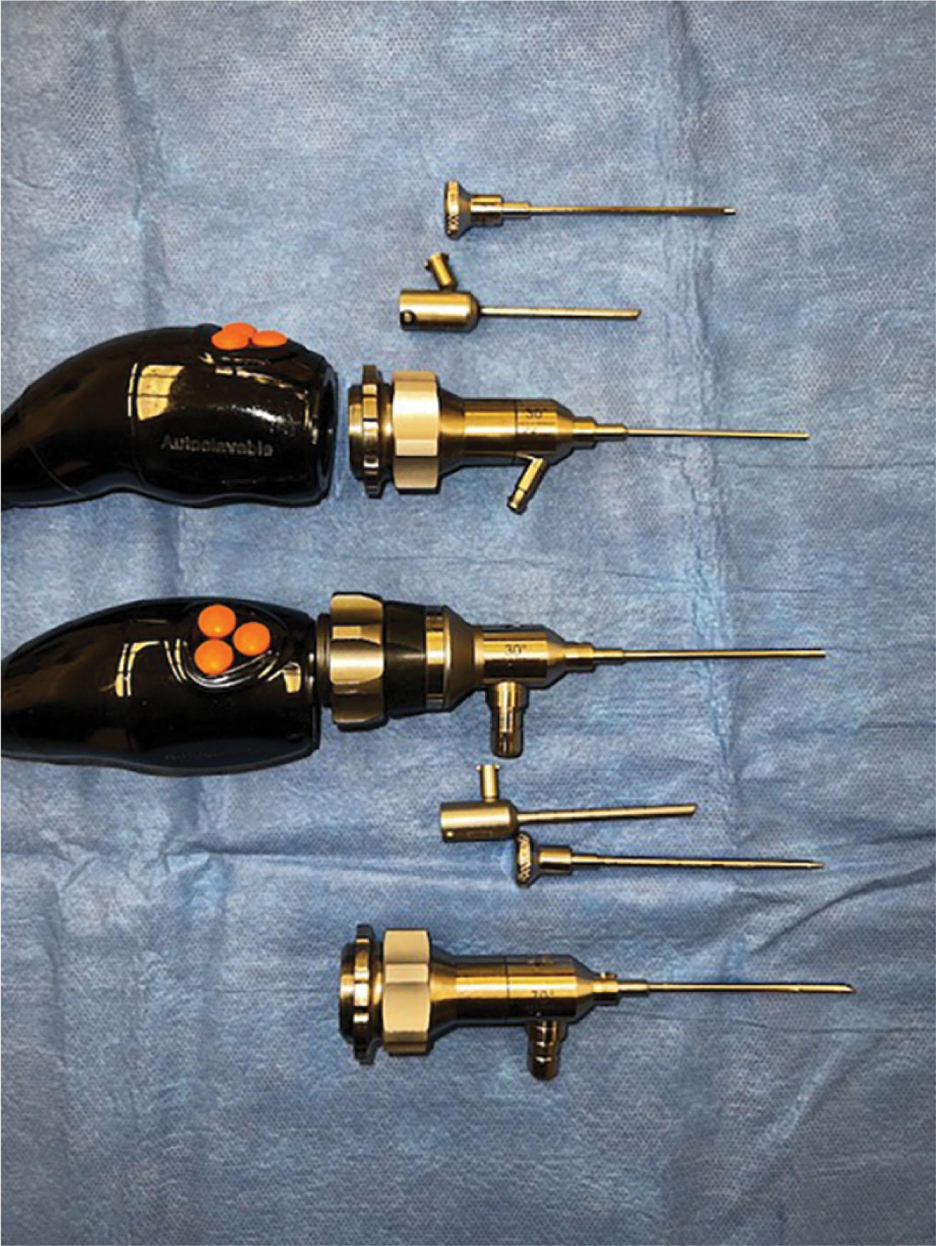
The 1.9- and 2.7-mm video arthroscopes are used for ankle arthroscopy ( Fig. 39-1 ). Rarely, the 4.0-mm arthroscope is used; however, the authors do not routinely recommend its use because its larger size prevents safe maneuverability within the joint. In general, the 4.0-mm arthroscope provides a large, clear picture and is more resistant to bending or breakage. Although the 2.7- and 1.9-mm short video arthroscopes are more delicate, they provide an excellent picture and a wide-angle field of vision. They are also preferred because the shorter lever arm reduces iatrogenic cartilage injury or instrument breakage.
Currently, the quality of the arthroscopic image is quite good using a 4 K arthroscope and television monitor. The images are saved digitally and can be inserted into the operative report and shared with the patient.
A “videoscope” has a telescopic extension that is directly connected to a camera without an intermediary eyepiece. This accommodates a camera that is lightweight, does not fog, is easy to maneuver, and generates less torque because of its short lever arm.
Arthroscopes provide excellent visualization throughout the ankle, particularly in the tighter spots, such as the medial and lateral gutters and posterior aspect of the ankle, as well as smaller joints. The 1.9- and 2.7-mm arthroscopes have a 30-degree obliquity for improved field of vision. The 2.7- and 4.0-mm 70-degree arthroscopes are particularly helpful in seeing over the medial or lateral domes of the talus and looking into the gutters as well as evaluating certain osteochondral lesions of the talus, tibia, and calcaneus. Surgeons must be cautious with use of 70-degree arthroscopes because they are not commonly used. The 70-degree arthroscopes have a central blind spot, making it more challenging to safely navigate the joint without causing iatrogenic cartilage injury.
For the extremely tight ankle, a 1.9-mm 30-degree arthroscope allows increased maneuverability. The small size of the 1.9-mm arthroscope decreases the amount of instrument crowding and inadvertent chondral damage. However, the 1.9- and 2.7-mm arthroscopes carry a higher risk of equipment failure because of their propensity to bend and break.
Regardless of the arthroscope used, it is important to have an interchangeable cannula system that allows the arthroscope and equipment to be switched from portal to portal without repeated reinstrumentation of the portals (see Fig. 39-1 ). This minimizes the potential for nerve and wound damage. The cannula size for the 2.7-mm arthroscope is 2.9 mm, and for the 1.9-mm arthroscope, it is 2.1 mm.
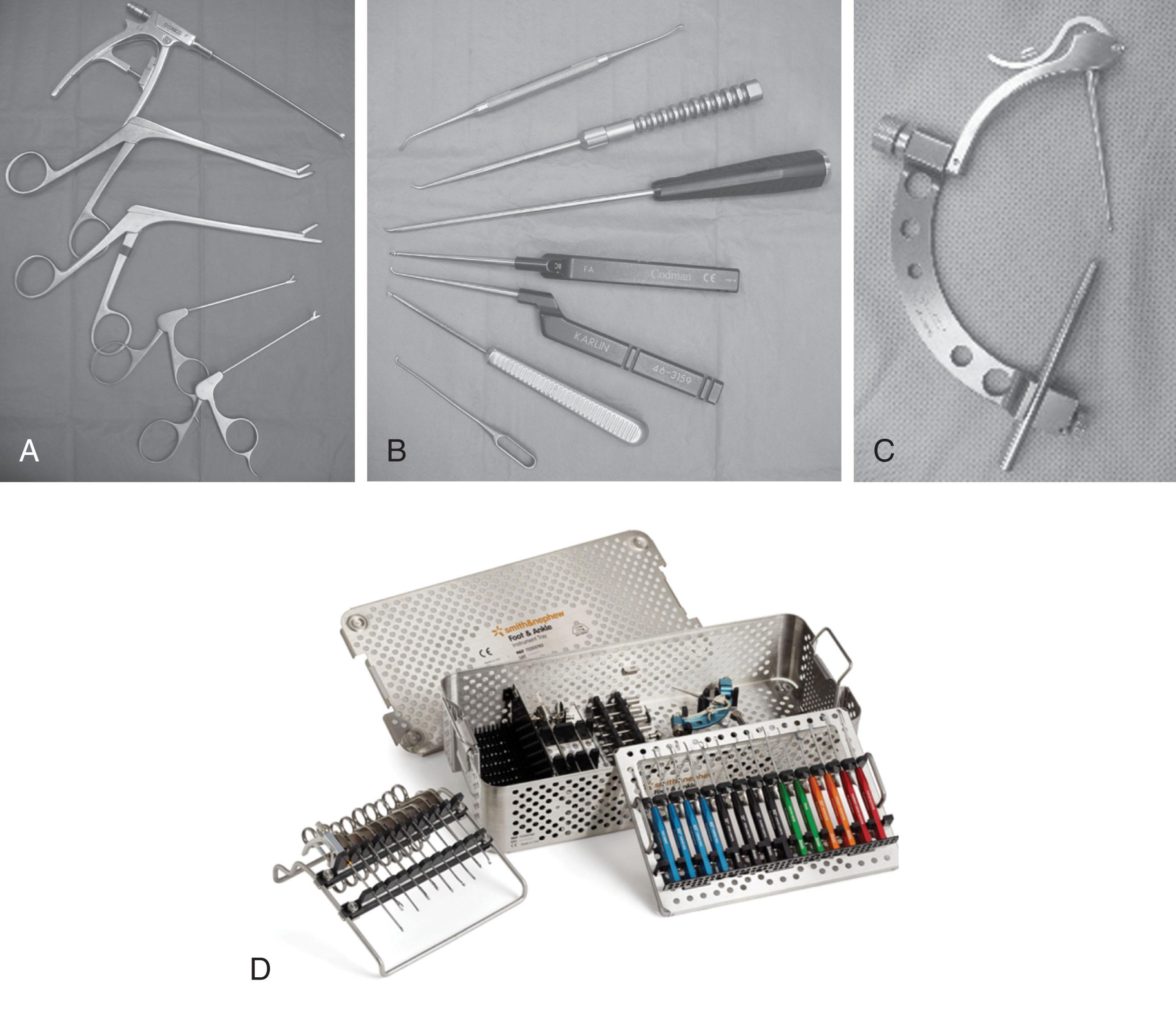
Although standard-size instruments can be used for ankle arthroscopy, small-joint instruments are preferred. These instruments are easier to maneuver within the tight joint spaces and are much more efficient to use than larger ones. Instruments typically used include the 2.0-, 2.9-, and 3.5-mm shavers and burrs; 3.5- and 4.5-mm ring and cup curettes; 1.5-mm probes; 2.9- and 3.5-mm graspers and baskets; 2.9-mm osteotomes; and pituitary rongeurs, microfracture picks, and banana blades ( Fig. 39-2 and Box 39-2 ).
30- and 70-degree 2.7-mm arthroscopes and a 30-degree 1.9-mm arthroscope
3 three-chip cameras with cords
Compatible light source
High-definition monitor with digital image-capture system
Thigh holder
Noninvasive ankle distractor
Disposable soft tissue distractor strap
Adjustable intravenous (IV) poles for gravity fluid management
Small-joint shaver system
2.9-mm full-radius disposable blade, 2.9-mm barrel abrader, 3.5-mm incisor blade, and 3.5-mm abrader
Skin marking pen and ruler
No. 11 scalpel and mosquito clamp
22-gauge 1.5-inch needle
18-gauge spinal needle
Two 19-gauge 1.5-inch needles
10-mL syringe with IV extension tubing
Two 50-mL syringes
Small-joint instrumentation set with tray
Power reamer with Jacobs chuck and key
Wire driver (power)
0.045- and 0.062-inch Kirschner wires (smooth with trochar tips)
2.7- and 1.9-mm cannulas and trochars
Small-joint banana blade
Micro Vector drill guide system
Mallet
IV extension tubing
An ankle distractor can significantly improve ankle visualization by increasing the space between the tibia and the talus. Without the distractor, certain areas of the ankle, such as the central tibial plafond and talar dome, the posterior tibiofibular ligament (PTiFL), transverse ligament, and flexor hallucis longus (FHL), are poorly seen.
Distraction methods applied to the ankle may be either noninvasive or invasive ( Fig. 39-3 ). Noninvasive techniques include manual distraction and gravity distraction, which are uncontrolled methods. Other types of noninvasive methods, such as the modified clove-hitch knot around the ankle, are semicontrolled. Several controlled devices allow the ankle to be distracted noninvasively while also permitting the amount of pressure and force to be monitored and maintained mechanically ( Fig. 39-4A ). To minimize the potential for nerve injury, Dowdy et al have recommended limiting the distraction force to less than 30 lb for less than 1 hour when using noninvasive distraction. Recently, we showed using somatosensory evoked potential (SSEP) that noninvasive distraction with 30 lb of force did not result in nerve injury at 1 hour. Other techniques for noninvasive distraction involve using a strap around the surgeon and around the ankle.

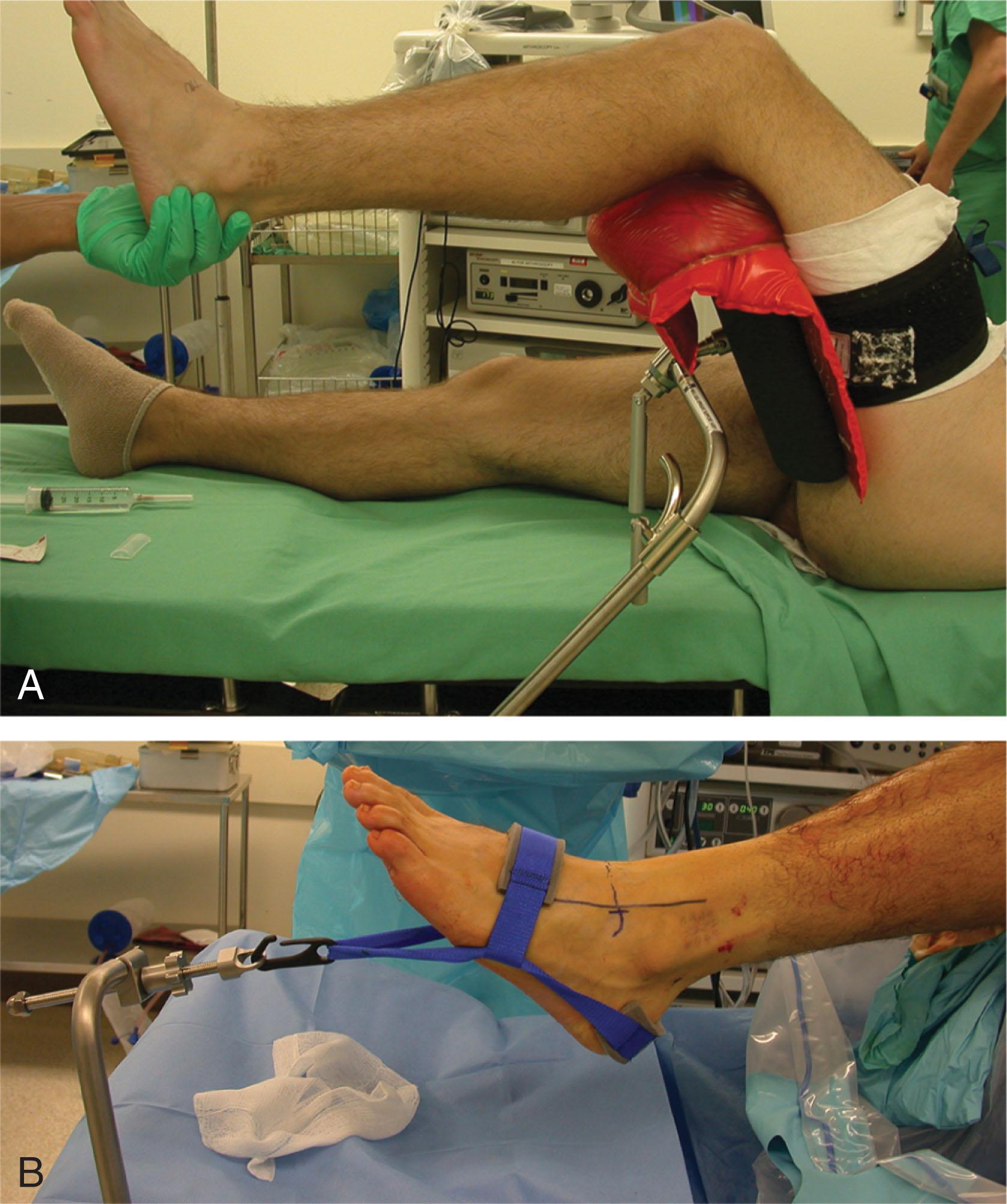
Invasive distractors are also available that use pins in the tibia and talus or calcaneus to provide mechanical distraction ( Fig. 39-4B ). Some of these devices have a strain gauge to measure the amount of force and permit some degree of freedom within the ankle joint. These have fallen out of favor in recent years because of risks of neurovascular injury, infection, fractures, scarring, and wound issues. In addition, the amount of distraction obtained is very similar with the noninvasive and invasive distractors.
Contraindications for noninvasive and invasive distraction include complex regional pain syndrome and open tibial epiphysis. Invasive distraction should also be avoided in patients with pyarthrosis or chronic infection and in high-performance athletes who need to return to their sport quickly. Pins should be inserted unicortically to reduce the risk of fracture.
Over the last 25 years, the authors have used noninvasive distraction (see Fig. 39-4A ) without significant complications and have not had to use invasive pins for any cases in the supine position.
Several positions are described in the literature. The three most common include supine, lateral decubitus on a bean bag, and prone. Some surgeons prefer the supine position, with the thigh secured by a nonsterile thigh holder as is commonly used with knee arthroscopy, to flex the knee 90 degrees over the end of the table. An alternative method is to use a urology leg holder positioned in the popliteal fossa; however, the surgeon must be very careful to keep the pressure in this area to a minimum. The authors prefer the supine position with a special nonsterile thigh support ( Fig. 39-5 ). The supine position is preferred for several reasons, including:
Almost all procedures can be performed in a supine position
Ease of access to anterior, lateral, medial, and posterior portals of the ankle and foot
Adequate posterior access may still be achieved
Difficulty in arthroscoping both the tibiotalar and subtalar joints with patient prone without repositioning
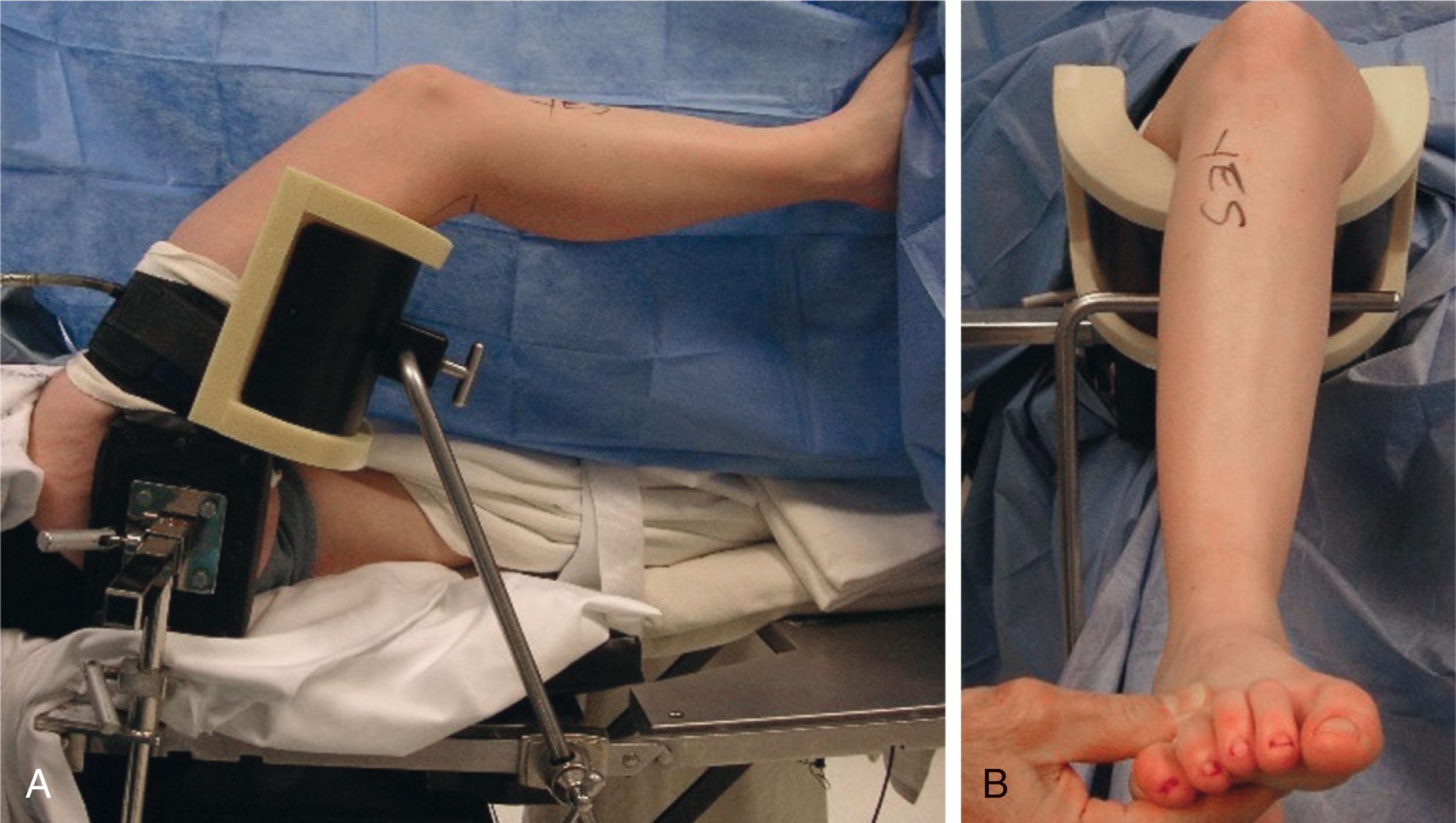
A tourniquet is applied on the proximal thigh, and the thigh holder is placed superior to the popliteal fossa to keep the popliteal fossa free from pressure. The tourniquet is used at the surgeon’s discretion. The thigh holder must be well padded to prevent injury to the sciatic nerve and is set to flex the hip at approximately 45 to 50 degrees. The operating table foot pad is removed to provide more room for posterior hindfoot access .
Because the thigh support is nonsterile, it should be applied before preparing the leg. A thigh post is positioned over the greater trochanteric region to help facilitate and maintain proper foot and ankle position and prevent hip external rotation. The patient is rotated internally to position the knee and ankle in a neutral orientation so that they both face straight up. With this setup, the surgeon can sit or stand during surgery, and both the anterior and posterior portions of the hindfoot are easily accessible without further manipulation of the patient’s extremity. The nonoperative lower extremity is placed on a well-padded, fully extended table and secured. Special care should be taken to pad all areas of bony prominence to prevent potential neurovascular compromise secondary to pressure. The patient is then prepared and draped in the standard fashion.
In ankle arthroscopy, it is critical to have a high inflow and outflow system. In general, this can be accomplished with gravity drainage into the posterolateral portal of the ankle. However, in situations where a posterior portal is difficult to obtain or maintain or higher fluid pressure is desired, an arthroscopy pump can be used. This provides a high flow volume and maintenance of pressure around the ankle. The pump is also helpful in obtaining hemostasis, permits improved visualization, and can be adjusted throughout the operative procedure.
Caution: Arthroscopy pumps can be very dangerous in the foot and ankle if they are not monitored carefully and a good outflow system is not maintained. The fluid can extravasate into the foot or up into the anterolateral compartment, and the potential for increased compartmental pressure can be significant. Surgeons must be especially aware of this potential complication if a peripheral block has been performed because the patient may not be able to sense neurovascular compromise.
General anesthesia with complete paralysis in addition to a regional block provides excellent anesthesia, facilitates distraction, and is the authors’ preference. A popliteal regional block offers excellent postoperative pain relief and is performed preoperatively with the aid of ultrasonography guidance. The block may be placed at the conclusion of the arthroscopy as well. The authors prefer postoperative local anesthesia with 0.50% bupivacaine without epinephrine in the distribution of the saphenous nerve if a popliteal block has been used, because the popliteal block does not anesthetize this area.
A thorough understanding of the extraarticular anatomy of the ankle is essential for placing the arthroscopic portals. The greatest concern is injury to neurovascular structures; however, the surgeon must also be aware of potential injury to the tendons traversing the ankle joint ( Table 39-1 ). It is imperative to mark out all structures at risk before portal placement. It is useful to mark out the superficial peroneal nerve branches PRIOR to prepping the ankle, since it is best seen at this time.
| Portal Site | Landmarks | Structures at Risk |
|---|---|---|
| Anteromedial | Medial to anterior tibialis tendon |
|
| Anterolateral | Lateral to peroneus tertius tendon |
|
| Anterocentral | Between tendons of EDC |
|
| Medial midline | Between EHL and anterior tibialis tendon |
|
| Posterolateral | Lateral to Achilles tendon |
|
| Posterocentral (trans-Achilles) | Through center of Achilles tendon | Achilles tendon |
| Posteromedial | Medial to Achilles tendon |
|
| Transmalleolar |
|
|
| Transtalar |
|
The three most commonly used anterior portals are the anteromedial, anterolateral, and anterocentral ( Fig. 39-6 ). The anteromedial portal is placed just medial to the anterior tibial tendon at the joint line. Care must be taken to not injure the saphenous vein and nerve traversing the ankle joint along the anterior edge of the medial malleolus.
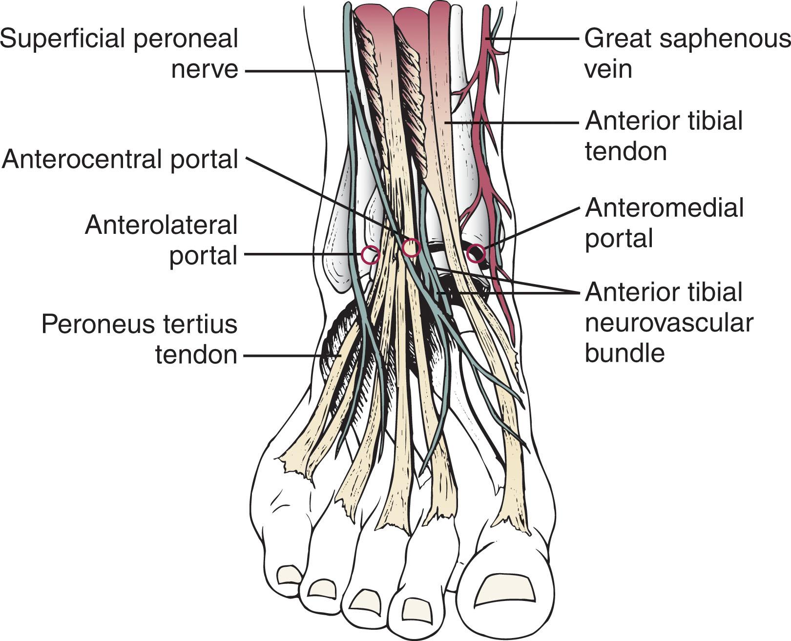
The anterolateral portal is placed just lateral to the peroneus tertius tendon. This is at a level at or slightly proximal to the joint line. Care must be taken to avoid injury to the superficial peroneal nerve, which is the most commonly injured nerve during ankle arthroscopy. The superficial peroneal nerve has four main variations. The superficial peroneal nerve most commonly divides approximately 6.5 cm proximal to the tip of the fibula into the intermediate and medial dorsal cutaneous branches. The intermediate dorsal cutaneous nerve passes over the inferior extensor retinaculum, crosses the common extensor tendons of the fourth and fifth digits, and then runs in the direction of the third metatarsal space before dividing into dorsal digital branches. The medial terminal branch of the superficial peroneal nerve—the medial dorsal cutaneous nerve—passes over the anterior aspect of the ankle overlying the common extensor tendons. It runs parallel to the extensor hallucis longus (EHL) tendon and divides distal to the inferior extensor retinaculum into the three dorsal digital branches.
Between these portals, an anterocentral portal may be established between the tendons of the extensor digitorum communis (EDC). This portal is placed between tendons of the EDC to avoid possible injury to the nearby neurovascular structures, including the dorsalis pedis artery and the deep branch of the peroneal nerve. The dorsalis pedis artery and the deep branch of the peroneal nerve lie deep in the interval between the EHL and the medial border of the EDC tendons. Medial branches of the superficial peroneal nerve must also be avoided when using this portal. Use of this portal is uncommon and rarely necessary, and the authors strongly discourage its routine use because of the increased potential for complications, including trauma to the dorsalis pedis artery.
A medial midline portal has been described between the tendons of the EHL and anterior tibialis tendon. A study found this portal to be farther from the superficial peroneal nerve branches and the dorsalis pedis artery than the anterocentral portal. The authors advocate the use of the anteromedial and anterolateral portals because they afford the most visualization and utility in ankle arthroscopy while minimizing potential complications.
Accessory anteromedial and anterolateral portals can be made over the medial and lateral talar dome to facilitate the treatment of specific lesions. These are most commonly used when working in the tight confines of the medial or lateral gutters. The accessory anteromedial portal is established 0.5 to 1.0 cm inferior and 1 cm anterior to the anterior border of the medial malleolus. It is especially useful in facilitating the evaluation of the deltoid ligament, particularly for removal of ossicles adherent to the deep portion of the ligament. The accessory anterolateral portal is established 1 cm anterior to and at, or just below, the tip of the anterior border of the lateral malleolus, near the anterior talofibular ligament.
Posterior portals are also useful during arthroscopy of the ankle ( Fig. 39-7 ). These portals are commonly placed directly medial to, lateral to, or traversing the Achilles tendon, just distal to or at the joint line. The posterolateral portal is established in the soft spot just lateral to the Achilles tendon, 1.2 cm (0.5 inch) above the tip of the fibula. Branches of the sural nerve and the small saphenous vein must be avoided with the posterolateral approach. It is the most commonly used and safest of the posterior portals.
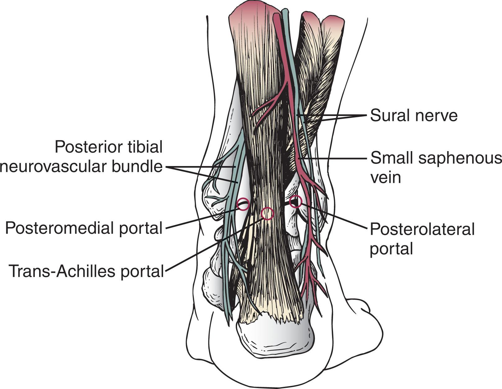
The trans-Achilles portal is established at the same level as the posterolateral portal but through the center of the Achilles tendon. In the authors’ experience, this portal does not allow easy mobility of the instruments and may lead to increased iatrogenic damage to the Achilles tendon. For these reasons, the authors recommend against its routine use.
Several authors have discussed the merits of the posteromedial portal, which is made just medial to the Achilles tendon at the joint line. With the posteromedial portal, the tendons of the FHL and flexor digitorum longus (FDL) must also be protected. In addition, the posterior tibial artery and the tibial nerve with its branches must be avoided. The calcaneal nerve branches from the tibial nerve proximal to the ankle joint and traverses the interval between the tibial nerve and the medial border of the Achilles tendon. Because of the potential for serious complications and difficulty in maneuvering the arthroscope, the trans-Achilles and posteromedial portals are rarely used during supine arthroscopy but are routinely used during prone arthroscopy, which will be discussed in detail later in this chapter. On occasion, accessory posteromedial or posterolateral portals are used to facilitate treatment, especially for posterior osteochondral lesions of the talus (OLT). The accessory posteromedial portal is made in the soft spot between the posterior tibial tendon and flexor digitorum longus tendon. Allegra and Maffulli have described double posteromedial portals to visualize and work posterior with the ankle in the supine position.
During surgery, the anterolateral, anteromedial, and posterolateral portals are routinely used. On occasion, the accessory posteromedial portal is used between the posterior tibial tendon and flexor digitorum longus to treat very posteromedial osteochondral lesions. The transmalleolar portals are also occasionally used for operative techniques, particularly for drilling of Kirschner wires under fluoroscopic and arthroscopic control through either the tibia or fibula into the talar dome, for the purpose of establishing new vascularity into an osteochondral lesion. Transtalar portals can also be used for drilling and possibly bone grafting osteochondral lesions of the talus. Prone portals will be discussed later in the Hindfoot Arthroscopy and Endoscopy in the Prone Position section.
There are several reports of neurovascular injury secondary to portal placement. Ferkel et al reported that neurologic injuries made up nearly half (49%) of all ankle arthroscopy complications, with the superficial peroneal most commonly injured, followed by the sural, saphenous, and deep peroneal nerves. All injuries were attributed to portal or distractor pin placement.
The anterocentral and posteromedial portals place the superficial and deep peroneal nerves and dorsalis pedis artery at increased risk. Because of the challenges in achieving adequate direct visualization of the posterior anatomy in the supine position, the posterior tibial nerve, posterior tibial artery, and calcaneal nerve may be at risk when a posteromedial portal is placed. Amendola et al proposed placing the patient prone when using posteromedial and posterolateral portals to reduce the risk of neurovascular injury. Acevedo et al have recommended coaxial posterior portals. The posterolateral portal is placed first, posterior to the peroneal tendons, followed by the posteromedial portal. This latter portal is placed using an inside-out technique behind the medial malleolus and adjacent to the posterior tibial tendon. Injury to the ligaments and tendons can be prevented by using meticulous placement of the portals and avoiding over-distraction.
Wound complications can be minimized through careful surgical technique. The use of interchangeable cannulas or a dedicated disposable cannula can minimize the trauma associated with the passage of instruments. Portal closure with a nonabsorbable vertical mattress suture, application of a compressive dressings, and postoperative immobilization in a well-padded splint can further decrease wound complications.
The intraarticular ankle anatomy during the arthroscopic examination has been described extensively by Chen. Drez et al have divided the ankle joint into an anterior and a posterior cavity, each of which is subdivided further into three compartments for a methodical inspection of the joint. The chapter authors have devised a 21-point systematic examination ( Table 39-2 ) of the anterior and posterior ankle joint that increases accuracy and reproducibility of the arthroscopic examination.
| Location | Point of Examination |
|---|---|
| Anterior ankle |
|
| Central ankle |
|
| Posterior ankle |
|
![]() As with other joints, it is imperative to use a methodical, systematic approach during arthroscopic examination of the ankle. This allows the surgeon to document the arthroscopic findings in a reproducible fashion, to accurately diagnose any potential intraarticular pathology, and to improve the quality of future clinical studies of the ankle arthroscopy patient population. The 21-point examination consists of three phases: the eight-point anterior examination, the six-point central examination, and the seven-point posterior examination.
As with other joints, it is imperative to use a methodical, systematic approach during arthroscopic examination of the ankle. This allows the surgeon to document the arthroscopic findings in a reproducible fashion, to accurately diagnose any potential intraarticular pathology, and to improve the quality of future clinical studies of the ankle arthroscopy patient population. The 21-point examination consists of three phases: the eight-point anterior examination, the six-point central examination, and the seven-point posterior examination.
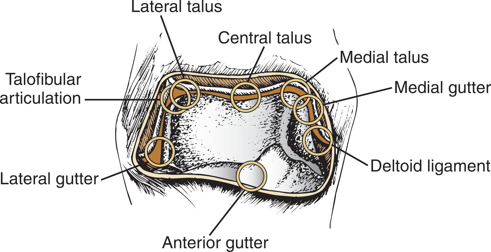
The eight-point anterior examination ( Fig. 39-8 ) includes the deltoid ligament, medial gutter, medial talus, central talus, lateral talus, talofibular articulation (trifurcation of the talus, tibia, and fibula), lateral gutter, and anterior gutter.
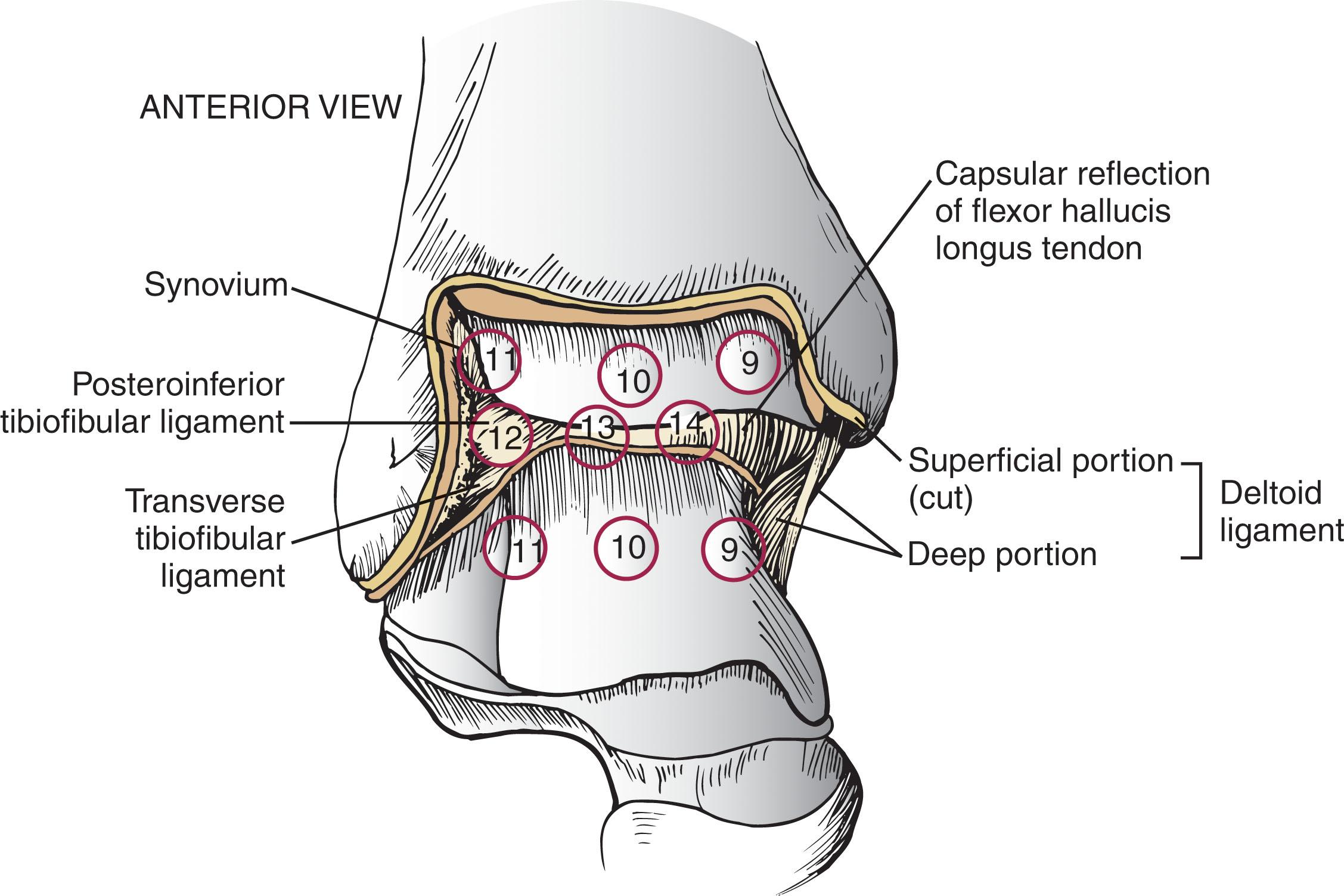
The six-point central examination ( Fig. 39-9 ) is performed by maneuvering the arthroscope through the notch of Harty. The notch of Harty is an anatomic elevation of the anteromedial distal tibia. The central examination includes the medial central tibiotalus, middle tibiotalus, lateral tibiotalus, capsular reflection of the FHL tendon, transverse tibiofibular ligament, and posterior tibiofibular ligament (PTFL). Pathology of the posterior ligaments can be seen here as well.
The seven-point posterior examination is shown diagrammatically in Fig. 39-10 and includes the medial gutter, medial talus, central talus, lateral talus, talofibular articulation, lateral gutter, and posterior gutter.
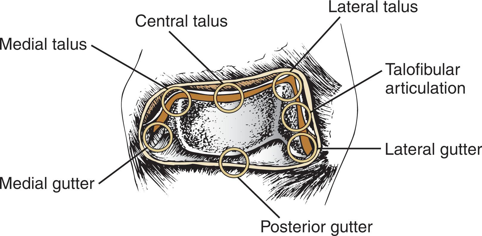
In general, the combination of the anteromedial, anterolateral, and posterolateral portals allows excellent visualization of the entire joint. However, if an area is not well seen, the other described portals may be used to improve visualization and access. Recent studies have described the joint access using the various portals for ankle arthroscopy. Hirtler et al have written extensively about arthroscopic access to the talar dome. They indicate maximum ankle plantar flexion and dorsi flexion allowed for the most optimal arthroscopic access.
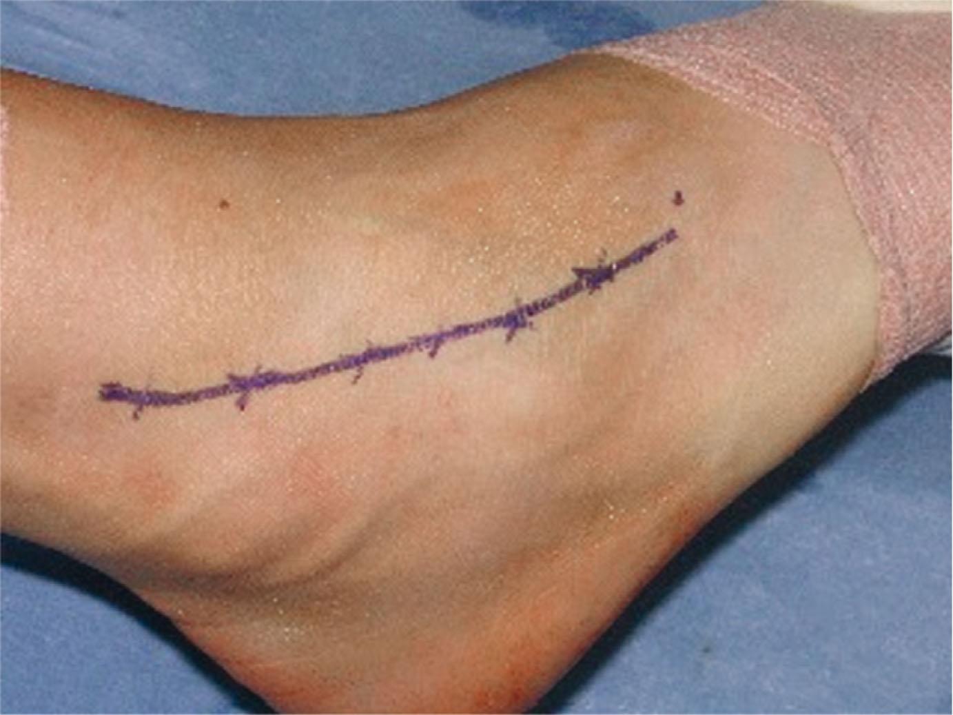
Before preparing and draping the area, the physician should localize the course of the superficial peroneal nerve because it is easier to see at this time. The nerve can often be palpated and visualized along its course anterior and inferior to the lateral malleolus by placing the foot into inversion and plantar flexing it (especially the fourth toe). Its course should be marked with a surgical pen before preparing and draping and re-marked before applying joint distraction ( Fig. 39-11 ). As stated earlier, the superficial peroneal nerve is the most commonly injured nerve during ankle arthroscopy, and every effort should be made to protect it during the surgery.
The technique for noninvasive distraction, as described by Ferkel, is performed as follows:
The patient is secured on the nonsterile thigh holder, and the leg is prepared and draped in the usual manner.
The sterile noninvasive clamp is attached to the operative table over the sterile drape.
The tourniquet is inflated at the discretion of the surgeon.
The noninvasive strap is then placed around the ankle and foot, and soft tissue distraction is applied (see Fig. 39-4A ).
It is important not to overdistract the ankle, and the authors recommend decreasing the distraction one click if the case requires more than 90 minutes of operative time. In addition, during a long case, the fine distraction screw on the distraction device can be used to ease up the distraction without taking it off completely because the viscoelastic stretch of the ligaments will help to maintain a good joint space opening. It is important during surgery to make sure that the noninvasive strap does not exert excessive pressure over the anterior tibial neurovascular bundle and that the padding on the device is not bent or crimped. If concurrent subtalar arthroscopy is performed, it is imperative that the strap is placed below the tip of the fibula to prevent obstruction of the posterior subtalar portals.
After applying distraction, the dorsalis pedis should be carefully palpated, and its position should be marked.
Likewise, the saphenous vein and the anterior tibial and peroneus tertius tendons are outlined over the surface of the ankle. (Marking the superficial peroneal nerve branches is particularly important because it is the structure most at risk for injury when the anterolateral portal is created. It should be identified before joint distraction.)
The joint line is then identified anteriorly by palpation with dorsiflexion and plantar flexion of the ankle.
The anteromedial portal is established first. To establish the position of this portal, an index finger is placed on the anterior tibialis tendon. Using the contralateral hand, a 22-gauge needle is introduced medial to the anterior tibialis tendon at the level of the tip of the medial malleolus. It is important to stay as close as possible to the anterior tibialis tendon and to systematically progress proximally until the distal tibia is palpated. Ideally, the needle should aim slightly up and over the dome of the talus to allow adequate access to the posterior joint.
Sterile lactated Ringer solution is infused into the ankle joint through the 22-gauge needle. Backflow suggests entry into the joint.
A No. 11 scalpel is used to make a vertical skin incision through the skin only while palpating the anterior tibial tendon.
Blunt dissection using a mosquito clamp is performed, first gently spreading through the subcutaneous tissue and then into the capsule ( Fig. 39-12 ). The authors refer to this as the “nick and spread” technique.
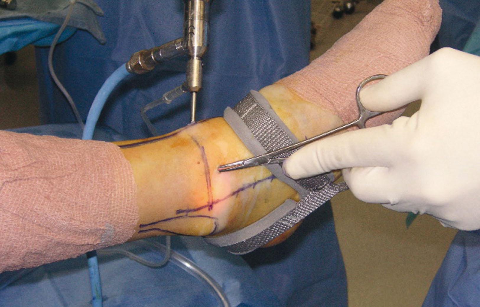
A blunt trochar with attached arthroscopic cannula is carefully placed into the ankle joint.
Additional lactated Ringer solution is infused into the joint through the arthroscopic portal cannula side post using a 50-mL syringe and extension tubing. The arthroscope is inserted, and the joint is visualized from the anteromedial portal.
A 22-gauge spinal needle is inserted into the ankle joint to locate, under direct vision, the position for the anterolateral portal. This portal is placed based on the location of the ankle disease. Typically, the needle should be parallel to the distal tibial surface and just medial to the intraarticular border of the fibula to allow access to the posterolateral joint space.
Using a similar technique, the skin is incised and bluntly dissected with a mosquito clamp, followed by insertion of an inflow cannula into the ankle joint.
Sequential examination of the anterior ankle joint and its structures is performed using the eight-point anterior examination system.
The six-point central examination is performed by carefully maneuvering the arthroscope through the notch of Harty to sequentially examine the structures, as previously outlined.
Commonly, the posterolateral portal is used for inflow or visualization. The key to establishing the posterior portal is good joint distention with constant distraction. The posterolateral portal is established under direct visualization, if possible, by maneuvering the arthroscope through the notch of Harty and using an 18-gauge spinal needle. This portal should be placed in the “soft spot” just lateral to the Achilles tendon, approximately 1.2 cm above a line drawn parallel to the distal tip of the fibula. The needle should be aimed roughly 45 degrees toward the medial malleolus and slightly inferior. Backflow through the needle indicates joint penetration ( Fig. 39-13A ).
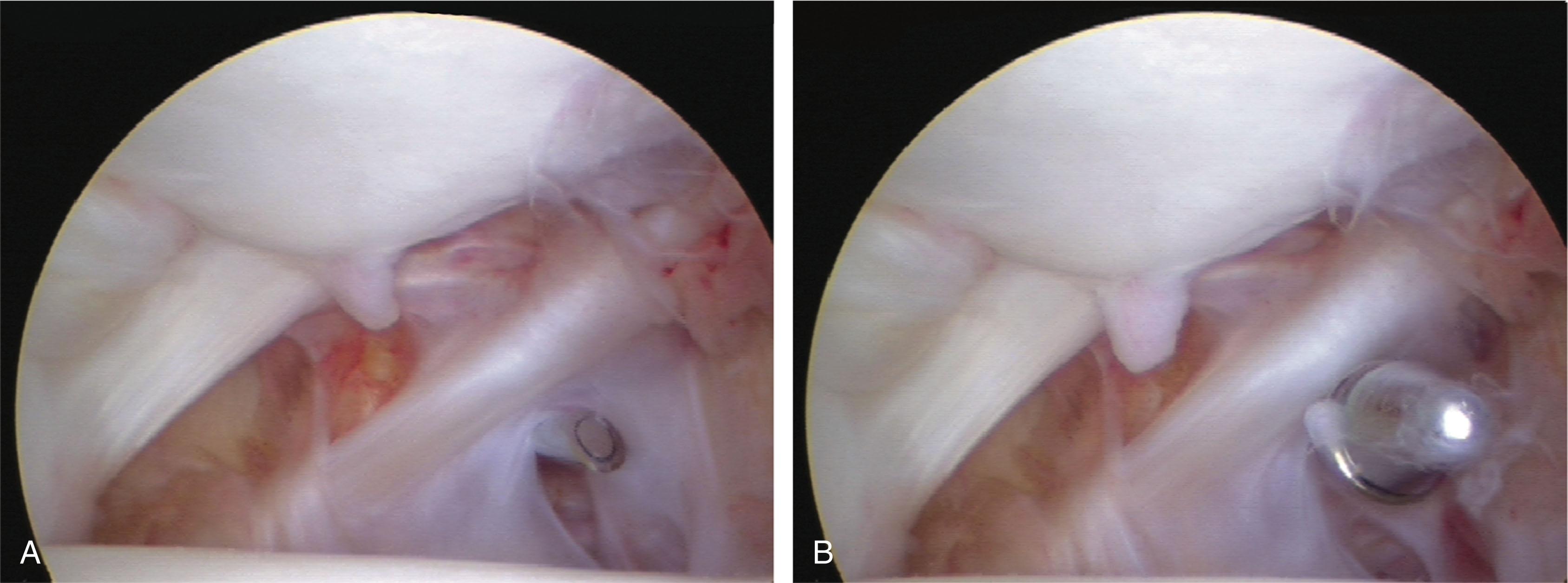
Normally, the cannula should penetrate the posterior capsule medial to the posterior tibiofibular ligament and under the transverse tibiofibular ligament ( Fig. 39-13B ).
The cannula should be inserted with care to avoid injury to the branches of the sural nerve and the short saphenous vein.
The posterolateral portal is initially used as the primary inflow portal and may be subsequently used for visualization or instrumentation by means of an interchangeable system of cannulas using a switching stick ( Fig. 39-14 ). Using the posterior portal for visualization allows the surgeon to perform the seven-point posterior examination. Figure 39-15 depicts the three-portal setup with interchangeable cannulas.
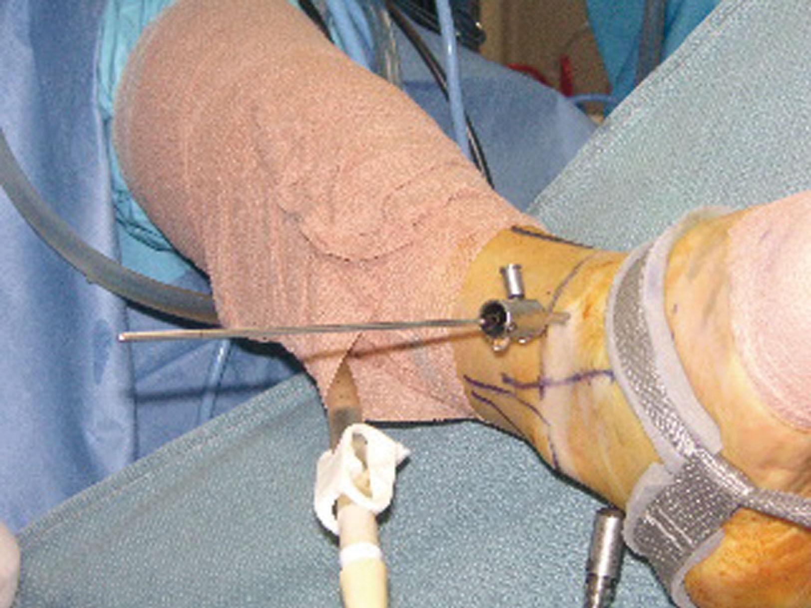
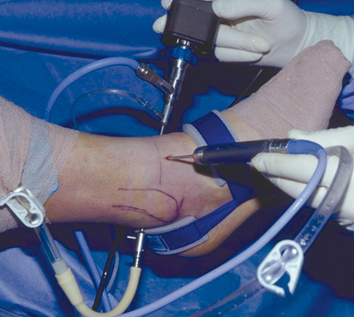
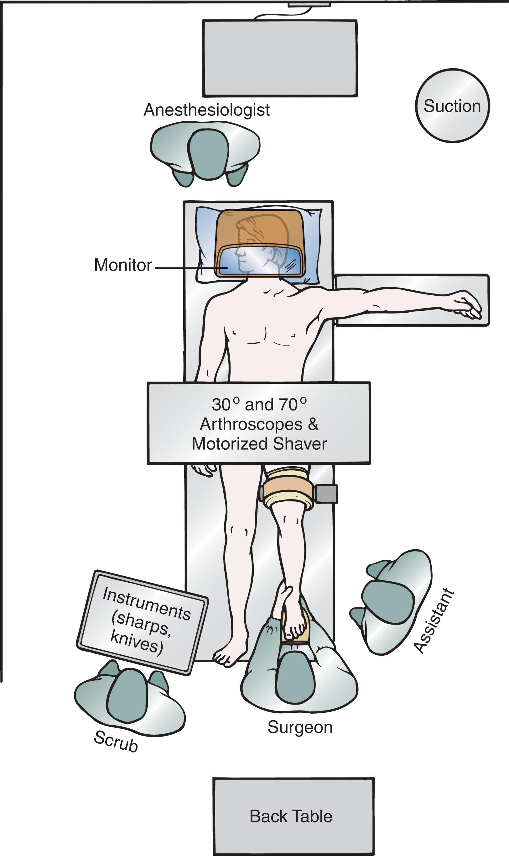
Positioning of the patient, equipment, scrub nurse, and surgeons is critical to a successful surgery. Figure 39-16 demonstrates the authors’ preferred operating room setup.
The arthroscopic portals are closed using 4-0 nylon in a vertical mattress suture pattern. Betadine or Neosporin ointment is placed over the wounds. A nonadhering petrolatum gauze dressing and 4 × 4 gauze pads are placed, and the ankle is immobilized in a well-padded splint in neutral dorsiflexion. Immobilization allows healing of the arthroscopic portals and discourages the formation of a synovial sinus. The sutures and splint are removed in 7 days. Subsequently, postoperative bracing or casting and rehabilitation depend on the pathology and treatment.
Ankle arthroscopy can be used to treat a wide variety of pathologic conditions ( Box 39-3 ). These are individually discussed in the chapter.
Soft tissue lesions account for approximately 30% to 50% of disease in the ankle joint. These have traditionally been a diagnostic challenge to the orthopedic surgeon because the definitive disease is often not apparent by history, physical examination, or radiologic examination. Since the advent of ankle arthroscopy, the ability to diagnose and treat soft tissue lesions around the ankle has increased significantly. In general, soft tissue lesions of the ankle involve the synovium. However, the capsule and the ligamentous tissues of the ankle may also be affected. The sources of synovial irritation are summarized in Table 39-3 and can include congenital, traumatic, rheumatic, infectious, degenerative, neuropathic, and miscellaneous causes.
| Cause of Irritation | Location or Type of Irritation |
|---|---|
| Congenital | Plica |
| Congenital bands within the ankle | |
| Traumatic | Sprains |
| Fractures | |
| Previous surgeries | |
| Rheumatic | Rheumatoid arthritis |
| Pigmented villonodular synovitis | |
| Crystal synovitis | |
| Hemophilia | |
| Synovial chondromatosis | |
| Infectious | Bacterial |
| Fungal | |
| Degenerative | Primary |
| Secondary | |
| Neuropathic | Charcot joints |
| Miscellaneous | Ganglions |
| Arthrofibrosis | |
| Gout | |
| Chondrocalcinosis | |
| Seronegative spondyloarthropathies |
Congenital plicae have been well described in the knee. Little is known about the plicae in the ankle, but they appear to be more congenital than traumatic in origin. The authors have had success with debridement of ankle plicae in patients who have had a careful workup to rule out other pathologies and have failed all conservative treatments.
Rheumatoid arthritis is a systemic inflammatory disease affecting 1% to 2% of the general population. Rheumatoid arthritis involves antibody-mediated inflammation of the synovial tissues, with eventual pannus formation and gradual destruction of the articular cartilage. Foot and ankle surgeons must understand the diagnosis and treatment strategies of this complex disease because nearly 90% of all rheumatoid patients will have some involvement of the foot and ankle during the course of disease. Ankle and subtalar involvement is thought to be more common than forefoot disease. Because of the recent advancements in medical management of this challenging disease, fewer patients are presenting with advanced deformities; therefore, more patients may be candidates for arthroscopic procedures now than in the past.
Surgery in the rheumatoid patient is based on careful timing and evaluation of the condition of the joint as well as the patient’s overall medical condition. If the patient fails conservative therapy, ankle arthroscopy may be considered in close consultation with a rheumatologist. The indications for surgery include pain, swelling, and mechanical symptoms of locking and catching. Contraindications include local infection, vascular insufficiency, and other medical comorbidities that preclude operative intervention. If preoperative radiographs demonstrate significant joint space narrowing, synovectomy should be avoided because the results are poor with only short-term benefit.
A careful physical examination of the ankle joint, tendons, ligaments, and soft tissue envelope is essential. A computed tomography (CT) scan or magnetic resonance imaging (MRI) can help assess articular damage, effusion, synovitis, tendinopathies, and other unsuspected abnormalities. A preoperative ankle and subtalar aspiration may be diagnostic and can rule out other inflammatory arthritides and infection. Close consultation with the patient’s rheumatologist is essential. The surgeon should carefully assess other joints and medical comorbidities.
The technique is as follows.
The patient is positioned supine.
An ankle distractor may be essential to perform the 21-point examination and a thorough debridement. Caution: The force of distraction must be carefully monitored to avoid possible complications.
A synovectomy is performed using a 2.9-, 3.5-, or 4.5-mm full-radius shaver. Use a “trap” in the suction to capture the synovium and send for pathologic analysis.
The surgeon should be especially careful when shaving near the thinned, friable joint capsule in these rheumatoid patients to avoid injuring the nearby neurovascular structures. The surgeon should never direct the shaver blade upward toward the capsule. Articular cartilage necrosis might exist on the tibia or talus joint surfaces and may be the primary disease. The subtalar joint may also be affected, and arthroscopic subtalar synovectomy can provide some relief.
Early synovectomy has yielded better surgical results than delayed synovectomy because better results correlate with less articular cartilage involvement. Early synovectomy can slow the progression of joint degeneration; however, even in the best of circumstances, debridement and synovectomy do not prevent articular destruction. Some investigators have questioned the benefit of synovectomy. Complete synovectomy of the rheumatoid granulations is impossible. Even with a thorough debridement, 5% of the original granulations can remain.
Tenosynovial giant cell tumor (pigmented villonodular synovitis) is postulated to be a benign neoplasm of the synovium. It occurs most commonly in the knee but can occur in the ankle and hindfoot. The neoplasm can occur in a generalized or a localized form, with the localized form being more common in the ankle.
Arthroscopy may be indicated to confirm the diagnosis and establish treatment. Several features must be seen to establish the diagnosis, including synovitis, papillary formation, and hemosiderin deposits. The contraindications are similar to those for general ankle arthroscopy.
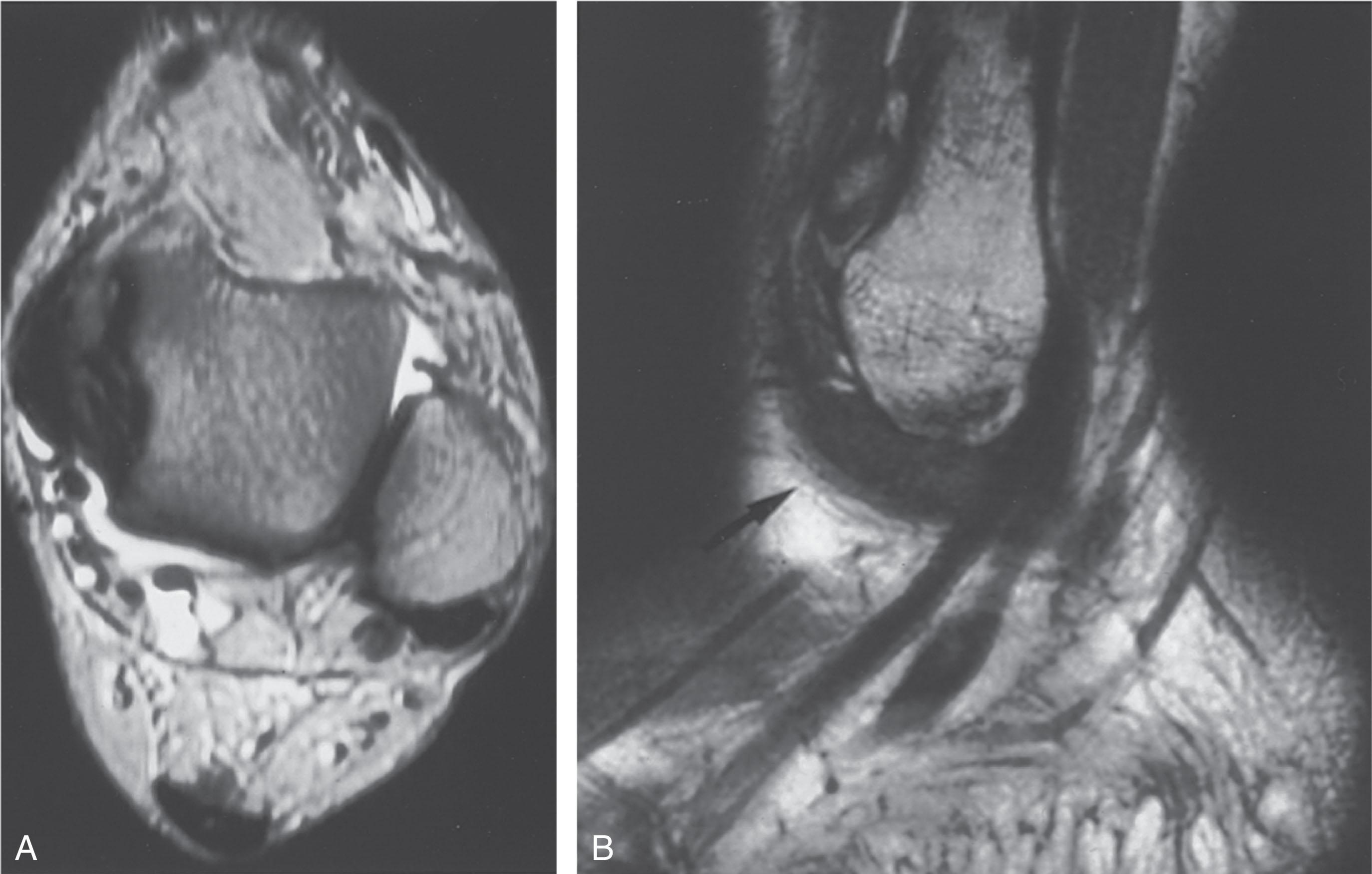
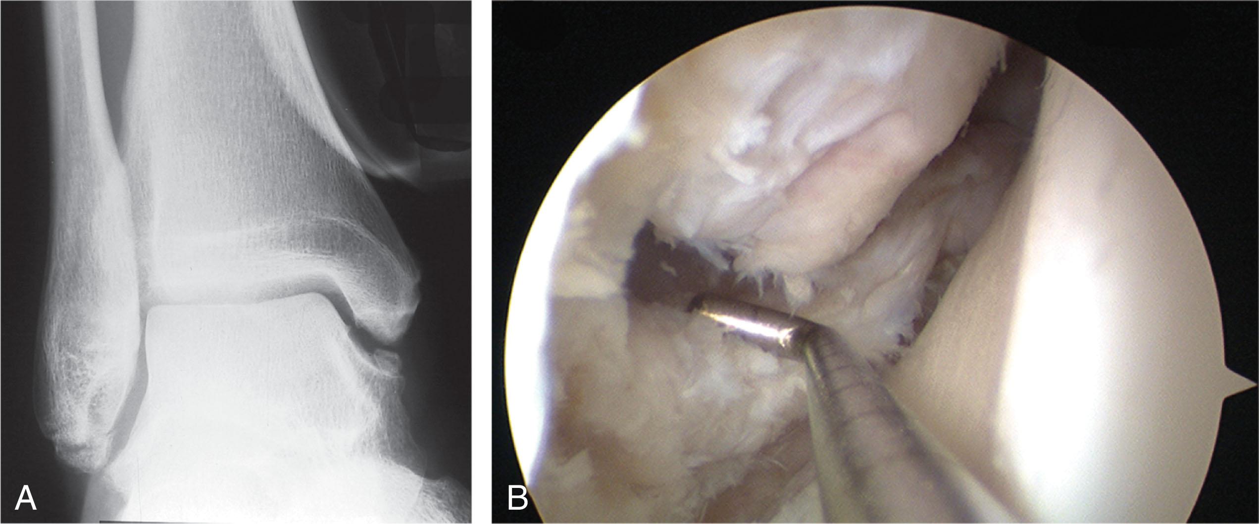
The ankle and hindfoot may be warm, swollen, and tender to examination and might have limited range of motion. Aspiration of the joint can yield a dark, serosanguineous fluid. Radiographs may be helpful in making the diagnosis of tenosynovial giant cell tumor by the characteristic periarticular erosions. An MRI can show nodular masses and reveal swollen synovial tissue ( Fig. 39-17A ).
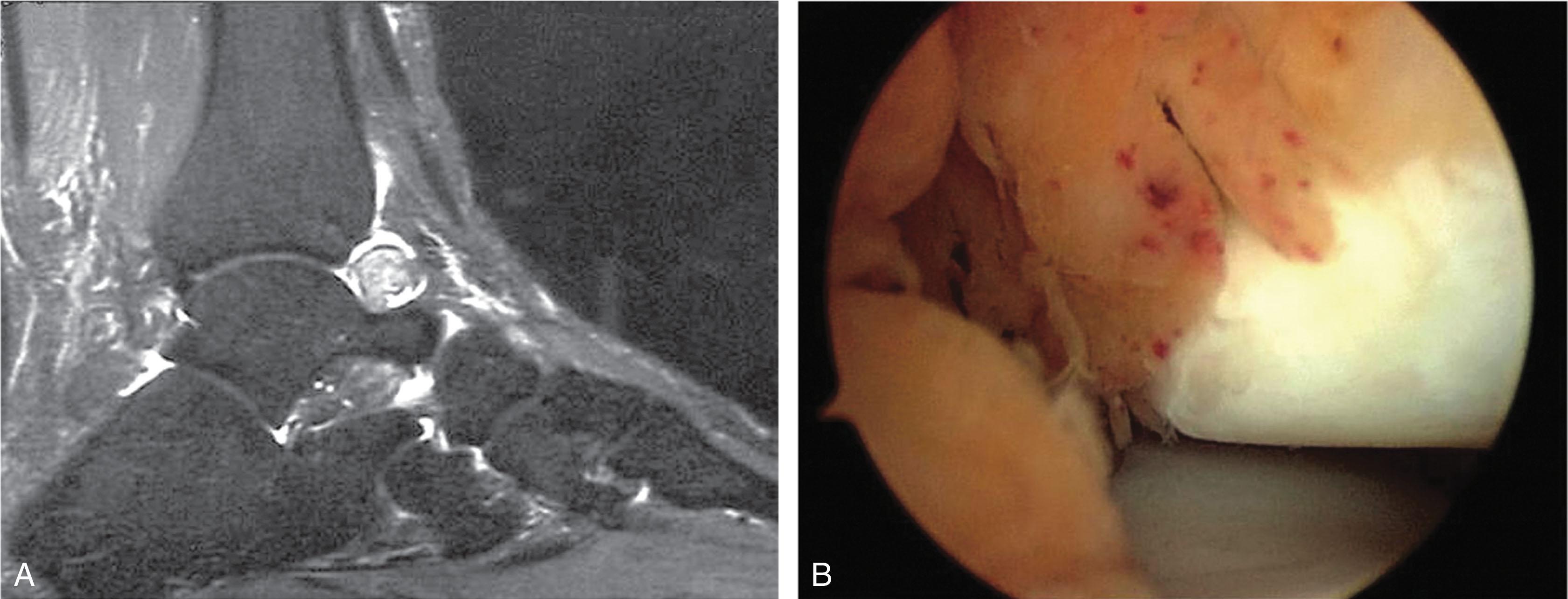
A 21-point examination is performed using a distraction device. Total synovectomy and debridement of hemosiderin deposits in all areas of the joint is important, using the same techniques and instrumentation as for rheumatoid arthritis ( Fig. 39-17B ). Arthroscopy of the subtalar joint may be beneficial in cases of hindfoot involvement. Preoperative adjuvant intraarticular radiation therapy before arthroscopy has been recommended by some people to lower the incidence of recurrence.
Arthroscopy offers several advantages over open synovectomy, including better visualization and surgical access to the ankle and hindfoot and decreased postoperative morbidity. Studies have shown successful treatment of tenosynovial giant cell tumor in the localized form, whereas the generalized form may not have lasting results, and recurrences are common.
Synovial osteochondromatosis (also known as synovial chondromatosis) is a rare condition of cartilaginous metaplasia of the subsynovial connective tissue. Pedunculated foci can become detached, resulting in multiple cartilaginous loose bodies within the joint. This condition occasionally afflicts the ankle and is most common in the monoarticular form. Three stages of the process have been described by Milgram ( Table 39-4 ).
| Stage | Description |
|---|---|
| 1 | Active intrasynovial phase, characterized by a lack of intraarticular loose bodies |
| 2 | Transitional phase, characterized by active synovial disease and chondral loose bodies |
| Early stage: No osseous development | |
| Later stage: Bone formation within synovial tissue and detached osteochondral fragments visible on radiographs | |
| 3 | Burned-out phase, characterized by no intrasynovial activity; however, the joint is filled with multiple osteochondral loose bodies. Radiographs demonstrate degenerative joint disease and loose bodies |
Arthroscopic treatment helps establish the diagnosis and alleviate pain and mechanical symptoms. Treatment may be contraindicated in patients with extensive disease, especially in cases of large osteochondral fragments that are more amenable to open treatment.
Patients can present with pain and symptoms of locking and giving way or with the sensation of loose bodies in the joint. Typically, there is limited range of motion as well as palpable synovium or nodules about the ankle joint. Synovial osteochondromatosis is most common in middle age. Radiographic studies can demonstrate multiple small intraarticular opacities and some calcification. The presence of intraarticular loose bodies does not establish the diagnosis of synovial chondromatosis. Loose bodies may be found in other diseases, such as degenerative joint disease, rheumatoid arthritis, neurotrophic arthritis, osteochondral fractures, osteochondritis dissecans, and tuberculosis.
In stages 1 and 2, arthroscopic surgery using joint distraction may be used to perform synovectomy and extraction of loose bodies ( Fig. 39-18 ). Debridement and loose body excision are performed in stage 3 because there is no intrasynovial activity to warrant synovectomy.
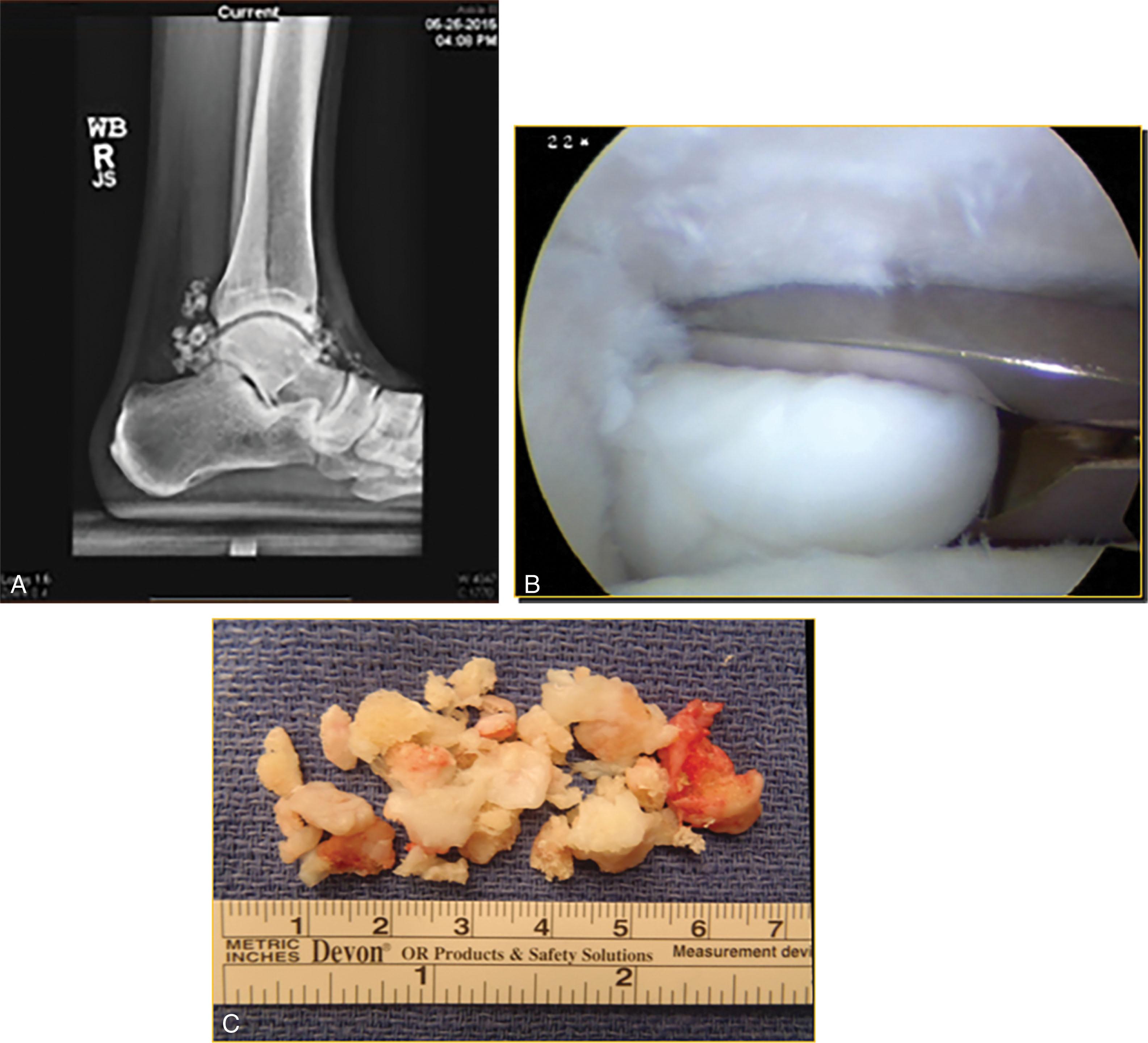
With open surgery, there is a 5% recurrence; Bojanic et al reported all good to excellent results in five patients with arthroscopic treatment. Recently, we have reported on results with synovial chondromatosis and found excellent outcome in patients as long as they did not have preexisting significant degenerative joint disease. An aggressive form of synovial chondromatosis has been described that tends to recur with occasional distal metastasis and should be considered a form of synovial chondrosarcoma. It is recommended to send all specimens for pathologic evaluation at the time of surgery and to follow the patients long term. Galat et al did open excision and found one of six cases (16%) to be later diagnosed with chondrosarcoma and needed below-the-knee amputation. Davis et al reported malignant transformation in 5% of cases.
Several hematologic conditions present with recurrent hemarthroses, which eventually may lead to cartilage and joint destruction.
Hemophilia is an uncommon inherited bleeding disorder involving reduced or absent levels of one of several clotting factors. Three types of hemophilia exist: A (factor VIII), B (factor IX), and C (factor XI). Hemophilic hemarthrosis often affects the ankle joint and usually occurs after minor trauma.
Von Willebrand disease is the most common inherited bleeding disorder. It involves a quantitative or qualitative defect in the von Willebrand factor, a protein critical to the normal clotting cascade. It most commonly presents with excessive bleeding after a surgical procedure but rarely may involve hemarthroses.
Arthroscopic synovectomy may be indicated in chronic forms of hemarthrosis. To avoid the postoperative complications and improve success, coagulation factors should be supplemented under the supervision of a hematologist.
In the acute stage, the joint may be swollen, red, and tender. Radiographs can demonstrate an extensive loss of joint space. The guidance of a hematologist is imperative, and an infection must be excluded. The chronic stage can show obliteration of the joint space and a chronic synovitis.
The technique of arthroscopic synovectomy is similar to that described for rheumatoid arthritis and tenosynovial giant cell tumor.
Arthroscopic synovectomy can help to reduce the frequency and severity of future hemarthroses. In a study of arthroscopic synovectomy of the ankle, elbow, knee, and shoulder, the median arc of motion was stable or improved in the year after surgery. The median follow-up was 79 months, and joints with sufficient follow-up data showed a significant decline in median bleeding frequency. Recently, Ferkel et al reviewed the orthopedic manifestations of van Willebrand disease and described successful treatment in a young athlete ( Fig. 39-19 ).
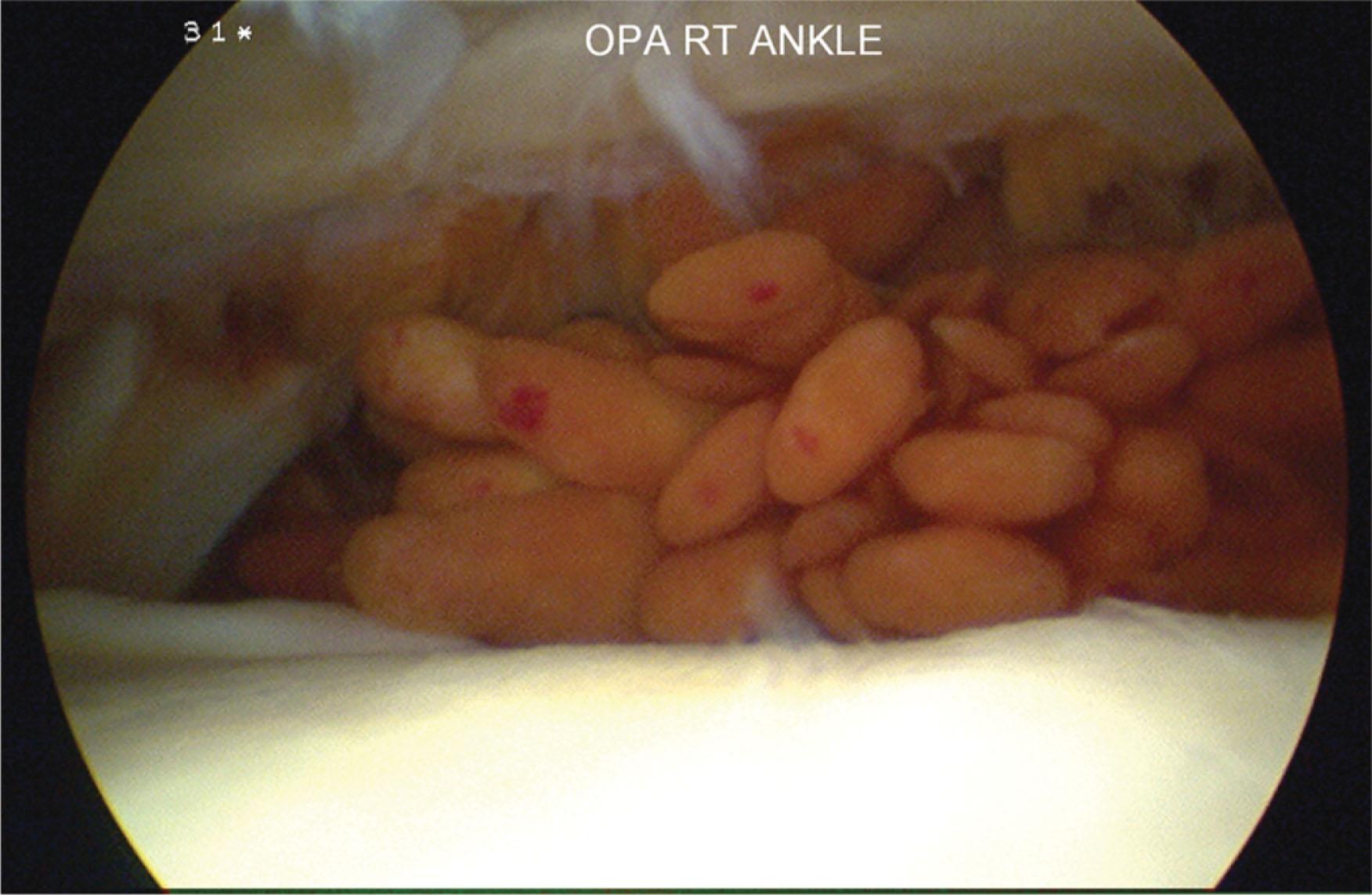
Bacterial, fungal, spirochete, mycobacterial, and other infections can afflict the ankle joint.
Acute ankle infection may be best diagnosed and treated arthroscopically. In the arthritic ankle of unknown diagnosis, arthroscopy can facilitate obtaining a biopsy and cultures as well as performing debridement. Infections involving the soft tissue envelope around the joint are best treated with open debridement.
A careful physical examination should be performed to determine the region of involvement. Diagnostic radiographic and laboratory studies are also helpful in making the diagnosis and assessing the extent of disease. Antibiotics should be withheld until a joint culture is performed to allow the use of appropriate antibiotics.
Arthroscopic treatment can be used to obtain cultures and perform a thorough debridement and synovectomy. Copious amounts of fluid are irrigated through the joint, and the debridement is performed using a shaver. Several series have reported successful results after arthroscopic management of intraarticular ankle infections caused by a variety of organisms.
Gonarthritis, Crohn disease, gout, chondrocalcinosis, and seronegative spondyloarthropathies can affect the ankle and subtalar joints. Usually, arthroscopy with debridement and occasionally synovectomy is very helpful in treatment of these conditions. Recently, Arroyo and Ferkel described crystal arthropathy with osteochondral lesions of the talus in eight patients. Six patients were found to have gout and two patients had chondrocalcinosis. Arthroscopic debridement resulted in a high percentage of good and excellent results, and all patients were able to resume sports at the same level postoperatively. Moreover, Wang et al showed improved outcomes in foot and ankle function and decreased repeat attacks of gouty arthritis after arthroscopic removal and debridement in the first metatarsophalangeal (MTP) joint.
Arthroscopic debridement for synovitis is recommended in the patient who fails to respond to prolonged conservative treatment after an adequate workup. The cause of ankle pain may be multifactorial, and careful evaluation is recommended. Contraindications to arthroscopic surgery were described earlier in the discussion of ankle arthroscopy.
After a traumatic injury to the ankle joint, a generalized or localized synovitis may occur. The patient with generalized nonspecific synovitis usually complains of swelling, aching, and soreness. The history of trauma may be significant or trivial. Sometimes, no specific inciting event can be recalled.
Ankle sprains and fractures are the most common causes of nonspecific generalized synovitis. Arthroscopic debridement with lysis of the adhesions and partial synovectomy is performed in this situation and may prove to be technically challenging. The key to success is a high-flow, gravity-assisted system using a posterolateral inflow cannula. The distractor may be very helpful in these cases to improve visualization and access to the joints.
After trauma, some patients develop a localized synovitis in the medial or lateral malleolar joints. Radiograph examinations are generally negative or might show small ossicles consistent with previous trauma. MRI might show a signal change in the area of pain with or without fluid. The arthroscopic examination of localized synovitis reveals mild or localized synovitis, papillary formation, and fibrosis.
The technique is as follows.
The patient positioning, equipment, and instrumentation used in synovectomy is the same as for ankle arthroscopy.
Distraction techniques are essential for proper visualization and synovectomy. Distraction should always be applied with caution.
A clinical pearl in successful arthroscopic synovectomy is to have a good inflow system into the ankle and suction out of the ankle. This can be accomplished by having a separate inflow portal posterolaterally or by using an arthroscopic pump. The authors do not advocate the use of mechanical pumps because of the risk of extravasation.
Interchangeable cannulas are important to minimize tissue trauma during instrumentation of the ankle.
Systematic evaluation of the entire ankle with the 21-point examination system should be performed. The ankle should be evaluated through both the anterior and posterior portals.
A 2.0-, 2.9-, or 3.5-mm full-radius blade is used for ankle synovectomy.
The entire joint should be debrided and freed from scar tissue, fibrotic material, and hemorrhagic synovitis. A systematic and thorough debridement is essential.
The surgeon is cautioned to avoid directing the shaver toward the anterior joint capsule to prevent neurovascular injury.
Debridement of associated chondromalacia and removal of loose bodies may be performed after improvement in visualization. In patients with rheumatoid arthritis or other synovial diseases, the capsule may be quite thin and friable.
The surgeon should use a whisker shaver tip when debriding anteriorly under the neurovascular structures, to prevent injury to these structures.
The portals should be closed postoperatively as was described for ankle arthroscopy. A drain may be placed if excessive bleeding is encountered after debridement.
A short-leg splint is applied for 5 to 7 days, after which the sutures are removed and a compression stocking is applied as well as a protective weight-bearing brace. The postoperative regimen should include early range of motion and weight bearing as tolerated, with physical therapy delayed for 2 to 3 weeks to allow the soft tissues to heal and the postoperative inflammation to resolve.
Chronic ankle pain after an ankle sprain is a well-documented problem and is seen in 20% to 40% of cases. The primary cause is soft tissue impingement, which can occur along the syndesmosis or in the syndesmotic interval, in the medial and/or lateral gutters, or posteriorly in the syndesmosis and posterior gutter. Anterolateral impingement is the most common type of soft tissue impingement about the ankle, although different types may coexist.
A list of differential diagnoses of chronic post-sprain pain include:
Osteochondral lesions of the talus
Calcific ossicles between the talus and the lateral or medial malleolus
Peroneal tears and/or instability
Tarsal coalition
Degenerative joint disease
Nerve entrapment
Occult fractures of the talus and/or calcaneus
Subtalar dysfunction
Sinus tarsi syndrome
Complex regional pain syndrome
Soft tissue impingement
Soft tissue impingement is most commonly seen anterolaterally because of the mechanism of most ankle sprains. In 1950, Wolin et al were the first to describe this entity in nine patients with persistent pain and swelling over the anterolateral aspect of the ankle. Patients reported pain for several weeks to months after an inversion sprain. Arthrotomy of the joint demonstrated a hyalinized connective tissue extending into the joint between the fibula and the talus likened to a “meniscoid” band. This mass was thought to originate from the talofibular joint capsule. It was thought that repeated pinching (or impingement) of this meniscoid tissue led to pain and swelling in the ankle. In 1982, Waller described the anterolateral corner compression syndrome. The pain in this syndrome was noted to be along the anteroinferior border of the fibula and the anterolateral talus and was thought to be a result of repetitive inversion injuries.
The authors think that the term “chronic ankle sprain” should be replaced by “soft tissue impingement of the ankle” as a more accurate description of this chronic condition around the ankle joint. The term “meniscoid” is misleading and should be avoided.
Patients with ankle pain that has failed a period of conservative management, including immobilization, progressive weight bearing, rehabilitation, nonsteroidal medications, and injections, may be candidates for arthroscopic debridement. The pain is often vague and activity related. Complex regional pain syndrome has been a well-described entity after ankle sprains and may be a contraindication to ankle arthroscopy.
There are some nomenclature changes for names of the ankle ligaments. In 1998, the Federative Committee on Anatomic Terminology stated that the term “inferior” should be dropped from both the anterior tibiofibular and posterior tibiofibular ligaments. In addition, the term “inferior transverse ligament” should be omitted and the ligament should be called the transverse ligament or deep component of the posterior tibiofibular ligament. The ankle pain is usually located anterolaterally but may also be anteromedially or in both areas. The borders of the lateral gutter of the ankle include the talus medially, fibula laterally, and tibia superiorly, bordered by the anterior and posterior tibiofibular (AtiFL and PtiFL) ligaments ( Figs. 39-20 and 39-21 ). The anteroinferior section of the lateral gutter is bordered by the anterior talofibular (ATFL), calcaneofibular (CFL), and anterior talocalcaneal ligaments. The posteroinferior borders are composed of the posterior talofibular (PTFL), calcaneofibular (CFL), and posterior talocalcaneal ligaments.
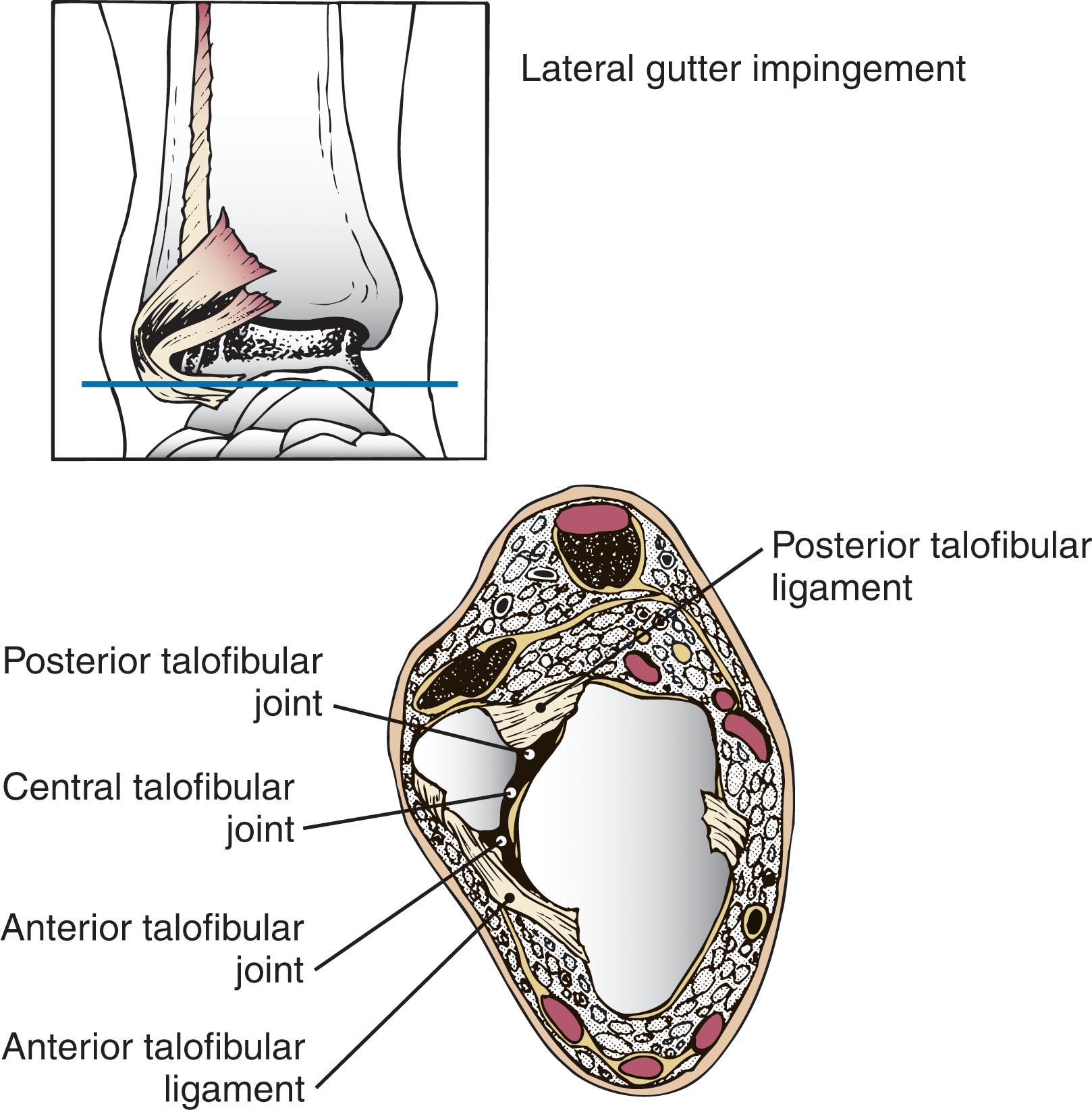
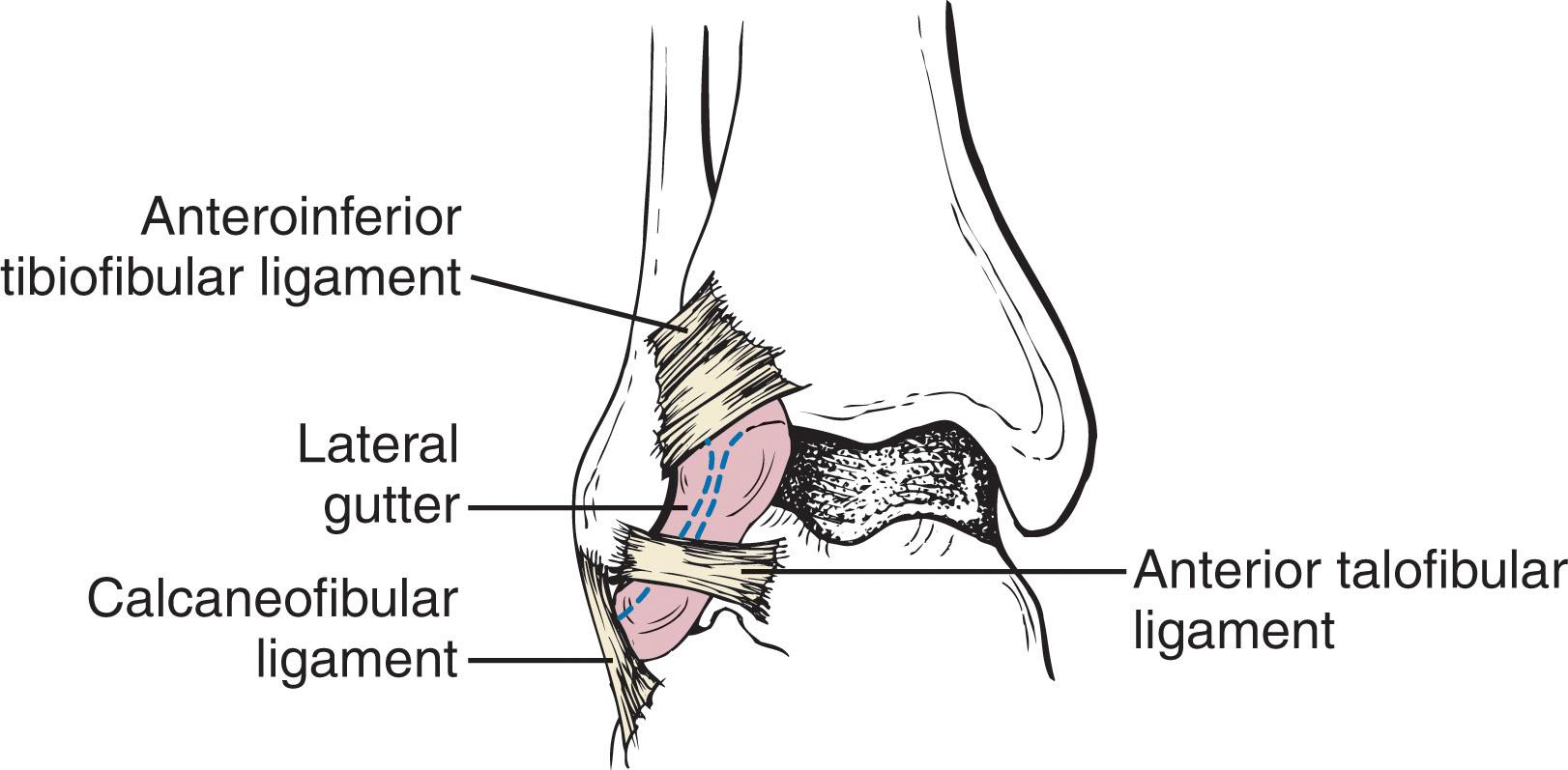
More recently, the anterior tibiotalar ligament has been reported as a possible contributing source of anterior soft tissue ankle impingement. Keller et al described the cadaveric anatomic and histologic features of the anterior tibiotalar ligament and postulated on its pathologic contributions to anterolateral soft tissue impingement. They found four different distinct types of ligaments. Types I and II are termed isolated ligaments and consist of a distinct band of organized fibers. Type I ligaments run over the talar neck more so than type II ligaments, which run over the lateral talar body. Types III and IV are termed widespread ligaments and are more diffuse, with no distinct thickening. Type III ligaments have parallel fibers, while type IV ligaments have crossing fibers.
The classic sequence associated with inversion injuries of the ankle is shown in Fig. 39-22 . There are three primary sites where anterolateral soft tissue impingement of the ankle occurs ( Fig. 39-23 ). The first site is the superior portion of the AtiFL ligament. The second site is the distal portion of the AtiFL ligament, which can involve a separate fascicle known as the Bassett ligament . The third and most common site is along the anterior talofibular ligament and lateral gutter near the area of the lateral talar dome.
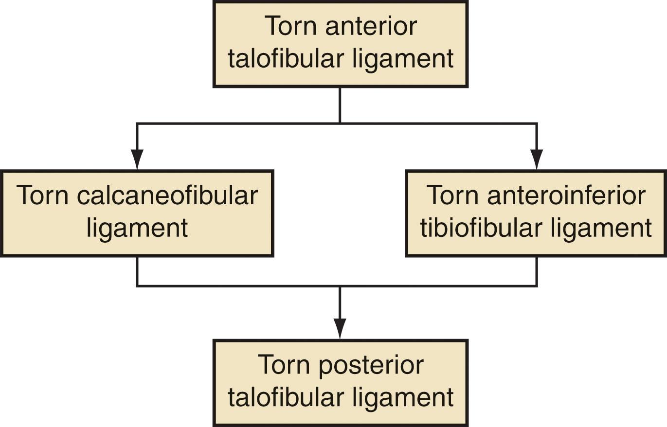
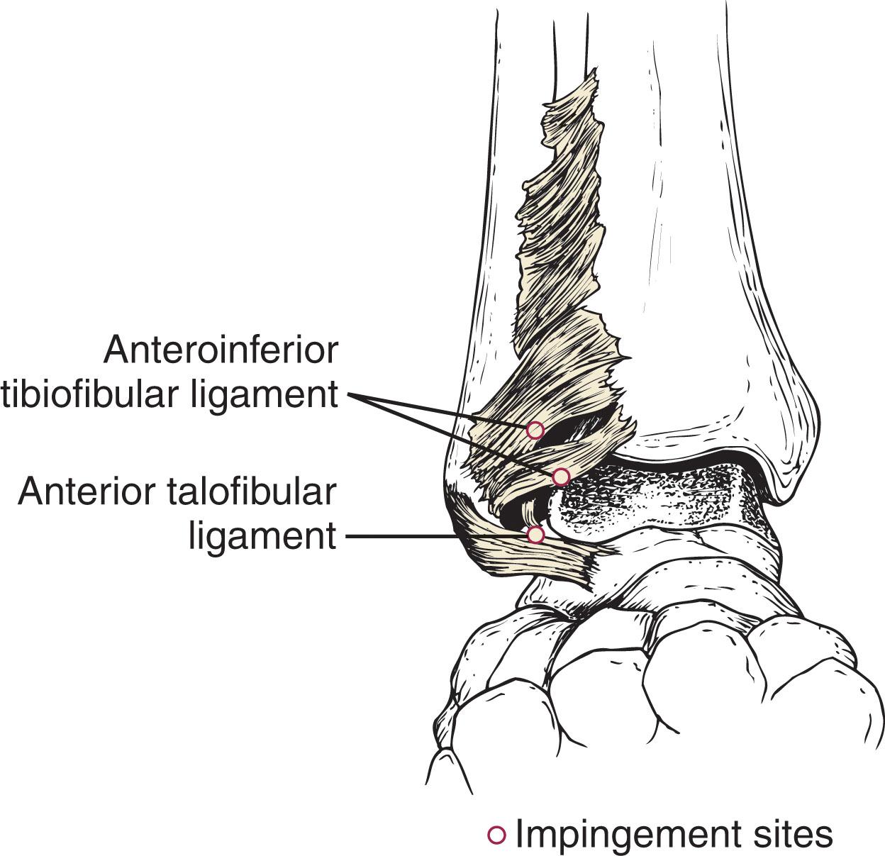
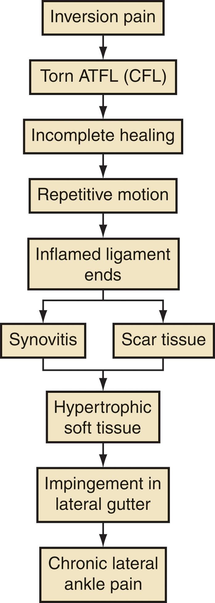
Figure 39-24 depicts the sequence of lateral ankle pain. Injury to the ligament initially results in synovitis and scar tissue. This in turn becomes hypertrophic and leads to impingement in the lateral gutter. Histologically, the impingement tissue is a layer of synovial membrane with subsynovial fat and collagen tissue anterior to the capsular tissues in the anterior joint space.
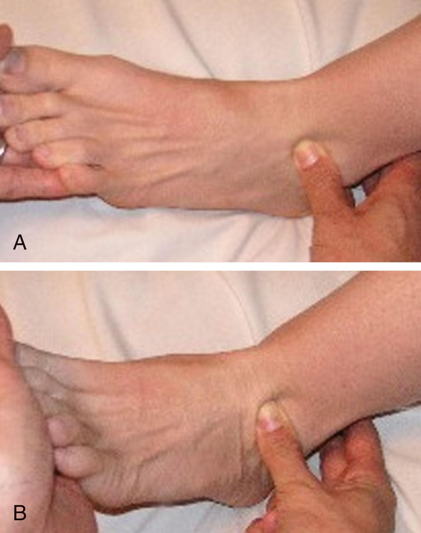
The diagnosis of soft tissue impingement can be made with a thorough physical examination and can be further confirmed with diagnostic studies. Careful palpation and stress testing of the ankle can assist in making the diagnosis ( Fig. 39-25 ). Attention should focus on the distal syndesmotic region, the area of the ATFL, CFL, and the anterior gutter. It is important to evaluate for subtalar pain as well because this presents in a similar manner and may coexist. Molloy et al reported a sensitivity of 94.8% and specificity of 88% on special physical examination testing for synovial impingement.
Radiographs can reveal calcifications or heterotopic bone in the interosseous space, indicating a previous injury to the distal tibiofibular syndesmosis. Ossicles along the tip of the fibula and the lateral talar dome are consistent with anterior talofibular ligament injuries. Stress view radiographs and ultrasonography have also been described in the literature for these conditions.
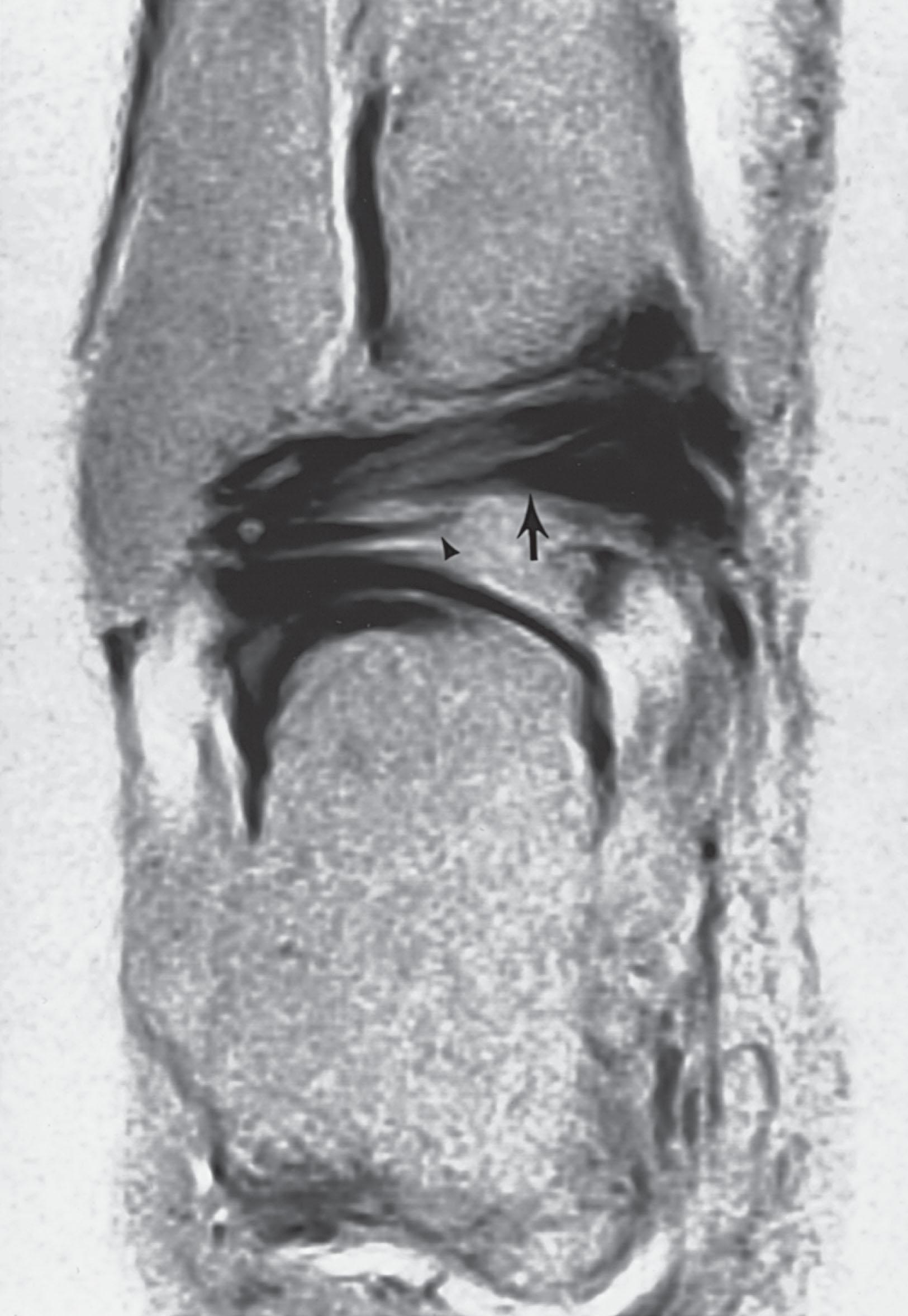
MRI has yielded a diagnostic accuracy of 78.8% and a sensitivity and specificity of 83.3% and 78.6%, respectively ( Fig. 39-26 ). Sagittal T1 and short tau inversion recovery (STIR) images help discern a collection of fluid (edema) or soft tissue that has caused the displacement of subcutaneous fat normally found immediately adjacent to the fibula anteriorly. Kinematic MRI has also been described to show impingement.
As with anterolateral soft tissue impingement, the patient with medial ankle pain that has failed conservative management may be a candidate for ankle arthroscopy.
Injury to the deltoid ligament or capsule can result in scarring and hyalinization or hypertrophy of the synovium. This can in turn lead to medial sided symptoms. Egol and Parisien have described a “medial meniscoid” lesion similar to that described in anterolateral impingement.
It is important to perform a careful physical examination to document medial ankle tenderness. Diagnostic studies can assist in making the diagnosis. Radiographs can reveal deep deltoid ligament avulsions, and stress radiographs can demonstrate medial joint space widening if there is associated ankle instability. An MRI can demonstrate inflammatory changes of scar tissue in the medial gutter or anteromedial joint.
Patients with chronic pain in the posterior ankle joint, usually worsened with plantar flexion, may be candidates for surgery. A trial of conservative management is advised.
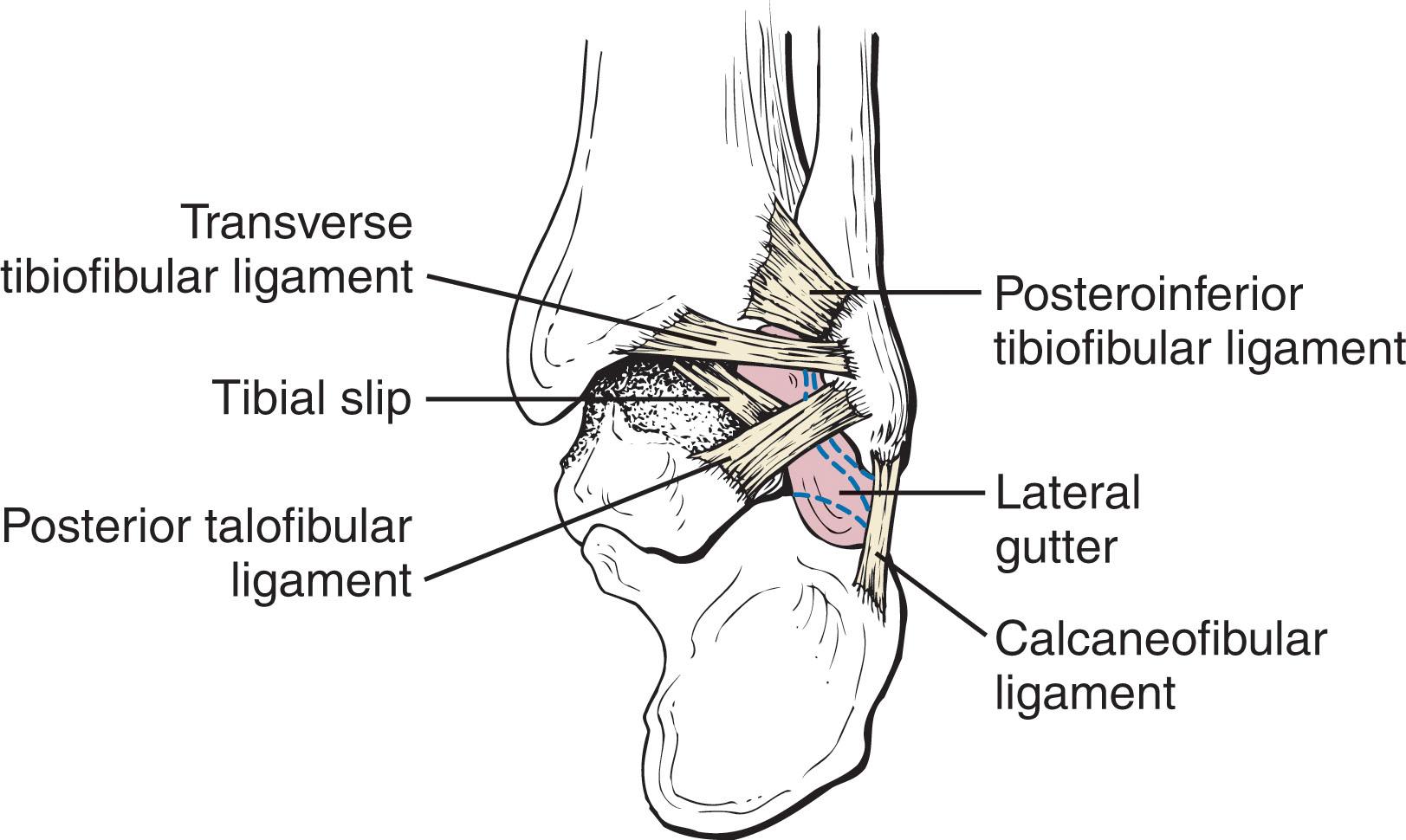
The posterior capsuloligamentous structures of the ankle have recently been classified in a review of 222 ankle arthroscopies. The posterolateral gutter structures include the posterior talofibular ligament, posterior tibiofibular ligament (PtiFL), transverse tibiofibular ligament (TTL), posterior intermalleolar ligament (tibial slip), FHL tendon, and a synovial nodule in the tibiofibular recess ( Fig. 39-27 ). The structures of the posteromedial gutter of the ankle include the FHL tendon and deep portion of the deltoid ligament.
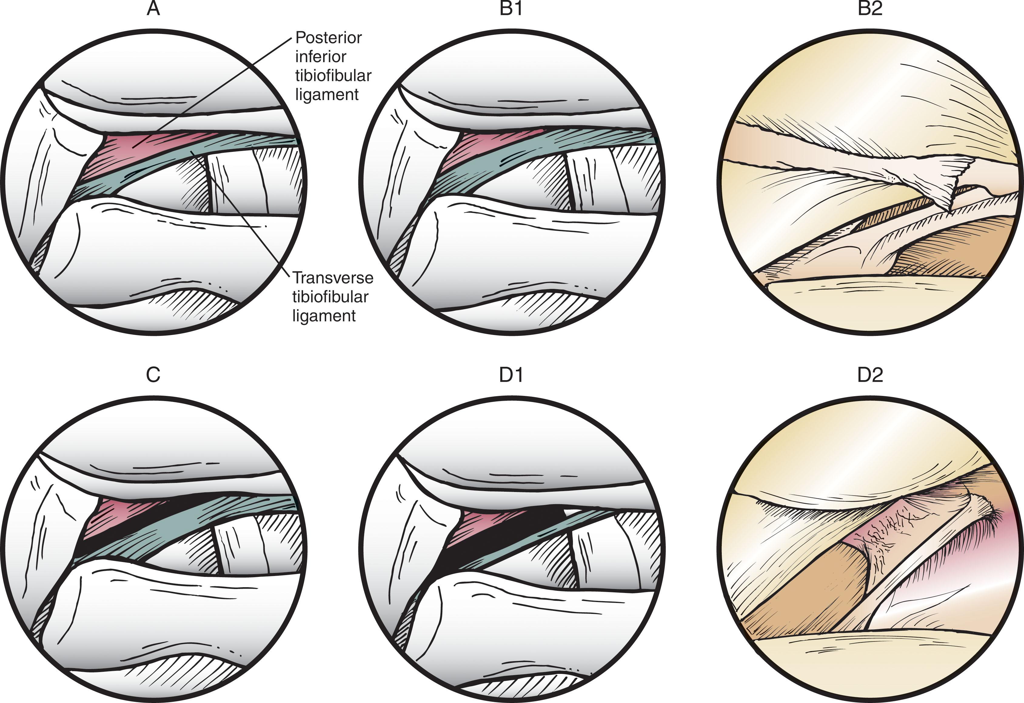
The configuration of the PtiFL and TTL remain controversial in the literature. Ferkel et al have classified these ligaments into four subtypes ( Fig. 39-28 ). Sarrafian describes the PtiFL as composed of a superficial and deep ligament. The deep ligament is also known as the TTL. Types II and III occurred most often at 33% and 35%, respectively, whereas types I and IV had an incidence of 26% and 9%, respectively. A normal variant bifid or three-limbed TTL has also been described, although it remains a rare finding and is most commonly found in type II and III configurations. In a review of 102 ankles, there were two tears of the TTL and one avulsion of the ligament from the posterior articular surface.
A posterior intermalleolar ligament (tibial slip) was noted in approximately 10% to 15% of ankles. This thin band lies in the floor of the posterior cul-de-sac and extends from the superior border of the posterior talofibular ligament laterally and courses medially and cephalad, eventually blending with the fibers of the TTL ( Fig. 39-29 ). Another posterior structure noted in 30% of ankles was a vertical band of tissue of varying thickness overlying the inferior aspect of the TTL.
Sarrafian found that the HL tendon and its sheath are in communication with the posterior capsule in 13% of ankles. Anatomic variation in capsule thickness or synovial proliferation can obstruct visualization of the flexor hallucis tendon in 60% of arthroscopies. In 20%, only a faint recess or shadow was evident, whereas in 11.8%, the recess was easily identified. Only 9.8% of patients had a discrete tendon ( Fig. 39-30 ).
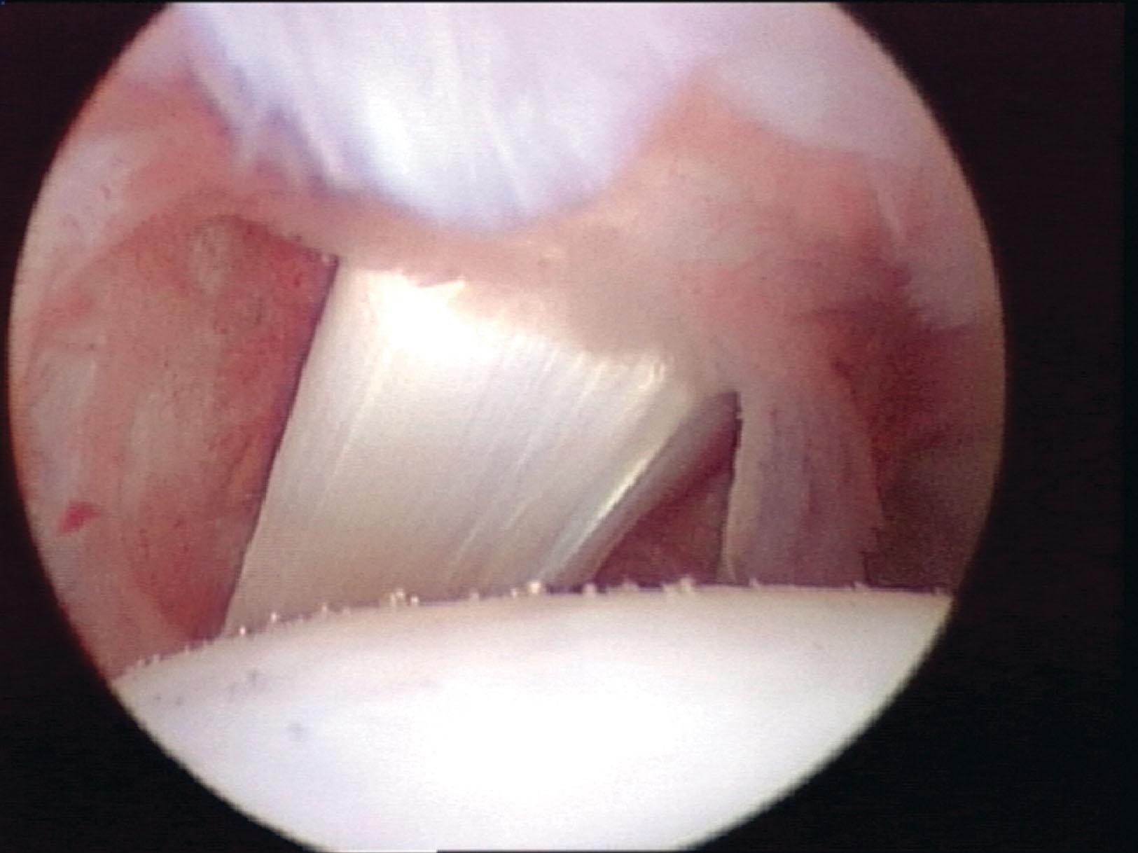
A synovial nodule is a normal finding of the tibiofibular synovial recess, located at the posterior margin of the distal fibula just anterior and superior to the PtiFL. The lesions ranged in size, apparently correlated to the amount of inflammation in the nodule. The majority of patients, 49%, had small discrete nodules without a significant inflammatory response, although a small subset of almost 12% had a seemingly large, noninflamed nodule. Twenty percent of ankles had small, inflamed nodules, whereas almost 20% had a large nodule with significant vascular injection.
Posterolateral soft tissue impingement may be an isolated entity; however, it is often found concomitantly with anterolateral soft tissue impingement of the ankle. In addition, generalized posterolateral soft tissue impingement can also occur with fibrosis, capsulitis, and synovial swelling. Although often normal, radiographs can reveal posterior osteophytes or loose bodies. An MRI study can show hypertrophy or tears of the posterior ligaments and might demonstrate synovitis.
Both localized and generalized posterolateral soft tissue impingement may be missed during ankle arthroscopy if visualization from the posterolateral portal is not performed. Furthermore, it is routinely difficult to see synovial disease in the posterolateral aspect of the ankle without the use of a distraction device.
Soft tissue posterior impingement can also be caused by hypertrophy or tears of the posterior tibiofibular ligament, transverse tibiofibular ligament, or posterior intermalleolar ligament (tibial slip) on the posterior ankle joint ( Fig. 39-31 ). The transverse tibiofibular ligament can be hypertrophied or become torn and pathologic in certain instances. Ferkel et al have reported 25 tears of the TTL tears, with an incidence of 1.56% in more than 1600 ankle arthroscopies ( Fig. 39-32 ).
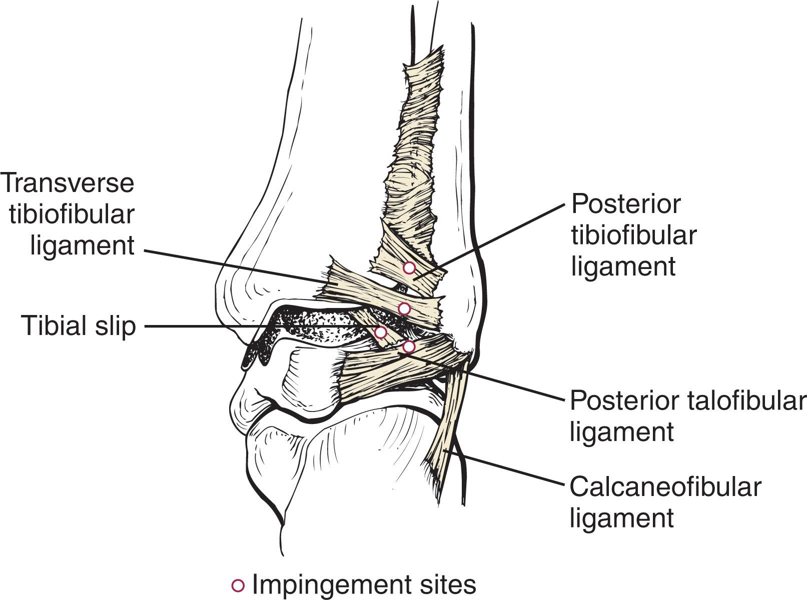
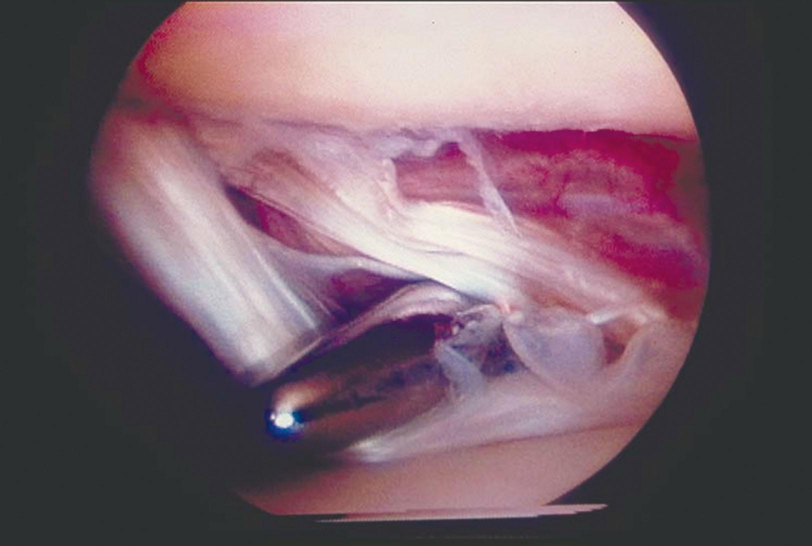
The posterior intermalleolar ligament (tibial slip) can also become hypertrophied and be a cause of posterior soft tissue impingement. The posterior intermalleolar ligament (tibial slip) runs between the posterior tibiofibular ligament and the transverse ligament (see Fig. 39-29 ). This ligament can develop fibrosis after trauma to the ankle, with subsequent pain and swelling. It can also be associated with injuries to the transverse ligament.
Hamilton (cited in Guhl) has described a tibial labrum on the posterior lip of the tibia. This can become pathologic, just like the superior labrum in the shoulder. The authors think that this disease is actually a tear of the transverse ligament or the posterior intermalleolar ligament (tibial slip). Hamilton has also described the “meniscus of the ankle” in the posterior ankle joint of ballet dancers. Tears in this region could displace into the joint, causing symptoms, and probably represent tears of the posterior structures.
Syndesmotic impingement may occur after injury to the syndesmotic complex. Injuries to the syndesmosis are vastly underestimated because they often occur with ankle sprains and fractures, and they frequently go undetected. Syndesmotic sprains have been estimated to occur in as many as 10% of all ankle injuries. Treatment of syndesmotic disruptions will be discussed later in this chapter. This section will focus on chronic pain after syndesmotic injury.
Surgery is indicated for patients with chronic pain after a “high ankle sprain” that fails conservative treatment. Those with syndesmotic instability are best treated with surgical stabilization.
The tibiofibular articulation is stabilized by the syndesmosis, which is composed of: the anterior tibiofibular ligament (AtiFL) and its fascicle; the posterior tibiofibular ligament (PtiFL), including its distal and deep component, the transverse ligament; and the interosseous ligament, and membrane.
There are three primary sites of syndesmotic impingement. These include the AtiFL, the tibiofibular joint, and the PtiFL ( Fig. 39-33 ).
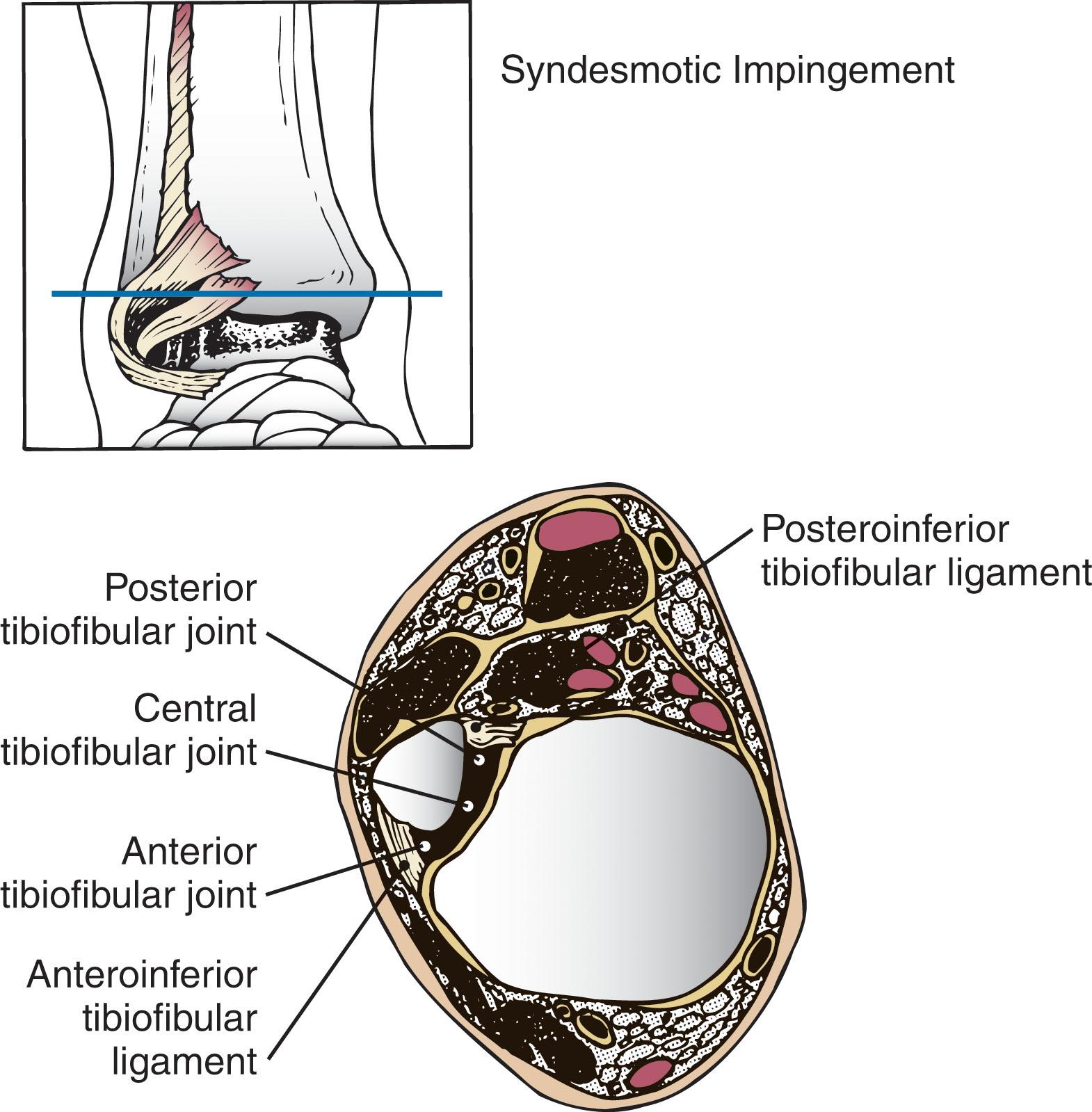
Injuries to the syndesmosis are among the most serious ligamentous injuries that occur in the ankle. Unfortunately, the incidence of these injuries has been underestimated in the past, particularly after an inversion injury. During an acute ankle sprain, if the talus inverts and the tibia rotates, stress is placed across the syndesmosis, and a tear of the syndesmosis may occur.
A careful examination includes assessment of the soft tissues, neurovascular status, and palpation over the syndesmosis and the interosseous membrane more proximally. The squeeze and external rotation tests are helpful in discerning syndesmotic injuries. Radiographs include anteroposterior (AP), mortise, and lateral views to discern ankle instability. Stress views under general anesthesia with fluoroscopy will verify an unstable ankle. Further studies may include MRI and weight-bearing CT scans.
After injury, synovitis and scarring can occur in the area of the AtiFL and corresponding joint. With increased activity, soft tissue impingement can develop around the area of the ligament and the undersurface of the joint. At surgery, a reddened synovium is often seen surrounding the AtiFL and extruding into the distal tibiofibular joint. Sometimes, the synovitis can extend to the posterior recess ( Fig. 39-34 ).
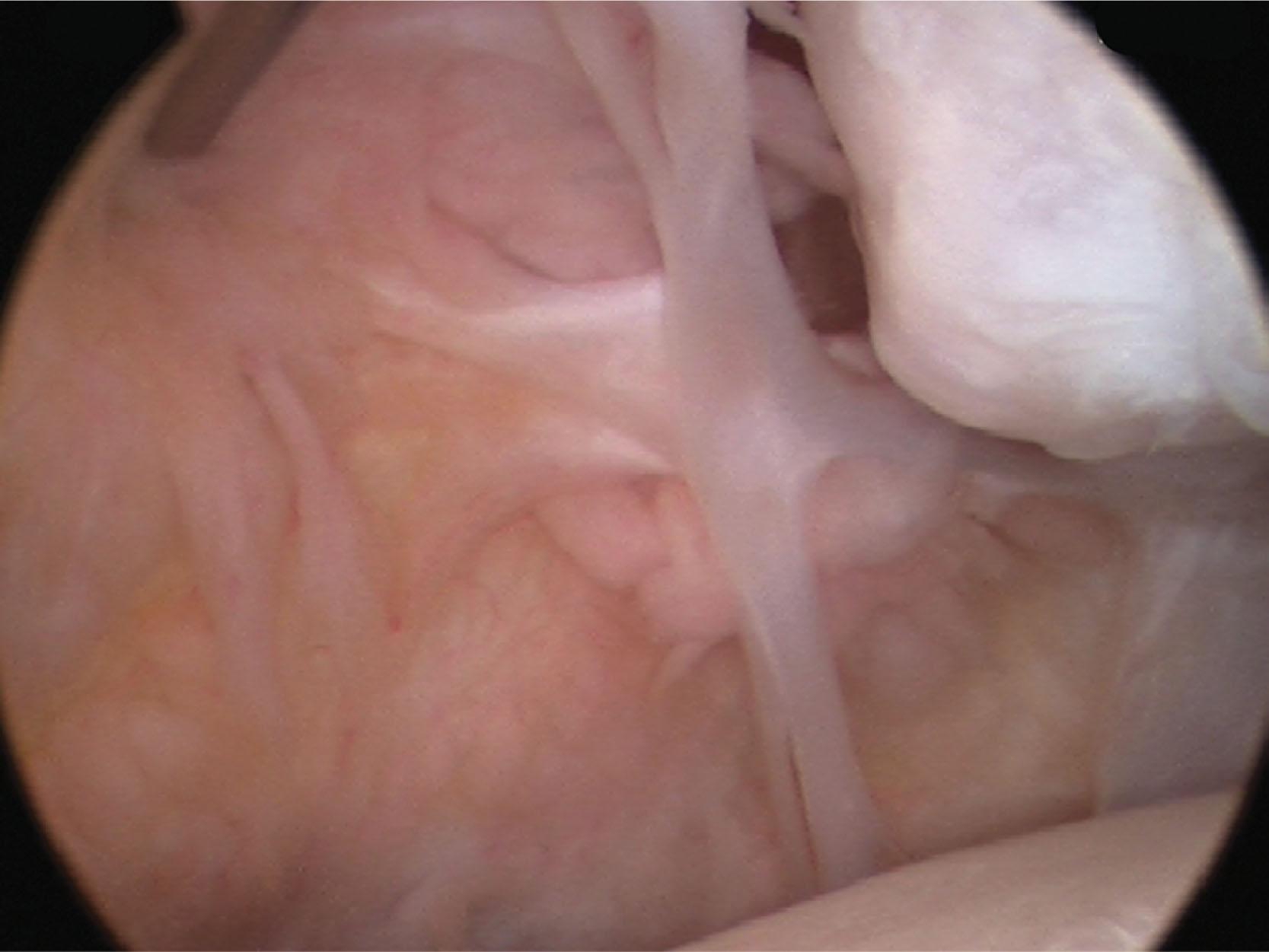
Several studies have reported impingement from a separate fascicle of the AtiFL. Bassett et al performed an anatomic study and found the fascicle to be present in 10 of 11 cadavers, a finding supported by Nikolopoulos et al in 22 of 24 cadaver specimens. The ligament is oriented distal and parallel to the AtiFL and separated by a fibrofatty septum. Bassett et al suggested that with a tear of the anterior talofibular ligament, the patient can develop increased laxity, and the talar dome can extrude anteriorly in dorsiflexion and cause soft tissue impingement. The authors think that the accessory fascicle rarely becomes symptomatic except after trauma.
A triad of pathologic findings were identified by Ogilvie-Harris in patients with chronic symptoms consistent with syndesmosis disruption: disruption of the PtiFL, interosseous ligament, and a posterolateral tibial plafond chondral fracture.
![]() The technique is as follows.
The technique is as follows.
Patient equipment, instrumentation, positioning, and anesthesia were described previously for ankle arthroscopy.
For syndesmotic impingement, before preparing and draping, intraoperative stress radiographs are taken while the patient is under anesthesia. Talar tilt and tibiotalar clear space widening are assessed. In case of tibiotalar instability, reduction and internal fixation is recommended and is performed after the arthroscopic syndesmotic evaluation and debridement.
A noninvasive ankle strap is used to for joint distraction; however, the anterolateral compartment of the ankle can become occluded with over-distraction, making visualization difficult. Visualization is best when the ankle is dorsiflexed with the distraction relaxed. In dorsiflexion, the anterior capsule becomes relaxed and the lateral malleolus lateralizes, rotates internally, and is pulled superiorly. Furthermore, the interosseous membrane, the anterior tibiofibular ligament, and the anterior talofibular ligament become more horizontal.
The anteromedial, anterolateral, and posterolateral portals are placed as described for ankle arthroscopy.
A 30-degree 2.7-mm arthroscope is used initially, and the ankle is visualized through all portals to perform the 21-point examination.
A 70-degree 2.7-mm arthroscope is routinely used to visualize the lateral and medial gutters. This arthroscope permits the surgeon to see over the talar dome from both the anterior and posterior portals. In severely tight ankles, a 30-degree, 1.9-mm arthroscope is recommended.
A posterolateral high-inflow cannula is placed to maintain fluid pressure and to remove shavings during the debridement.
A 2.9-mm full-radius shaver, baskets, and graspers are necessary for the debridement.
Debridement includes the removal of the inflamed synovium, thickened adhesive bands, inflamed capsular bands and ligamentous tissue, osteophytes, and loose bodies. Debridement is performed to expose the underlying cartilage, which often reveals chondromalacia of the fibula and talus.
For anterolateral soft tissue impingement, the inflamed tissue may extend superiorly into the syndesmosis region. Care must be taken to not excise any of the functional remnants of the anterior talofibular ligament ( Figs. 39-35 and 39-36 ). The lateral gutter can be approached with a 30- or 70-degree arthroscope in the anteromedial portal and the working instrument in the anterolateral portal. In addition, small ossicles or larger symptomatic nonunions may be present, and loose bodies may be hidden in the soft tissues at the tip of the fibula.
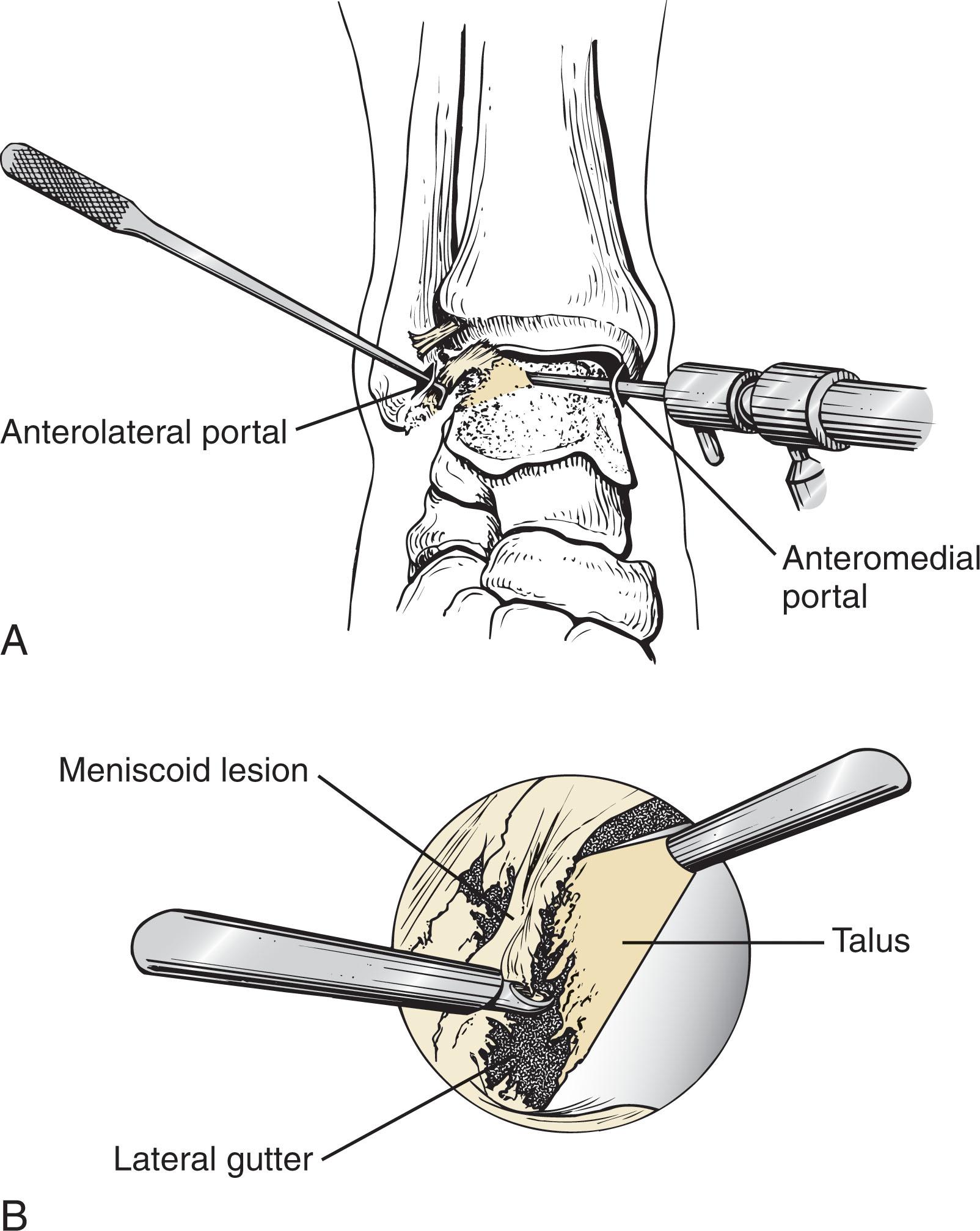
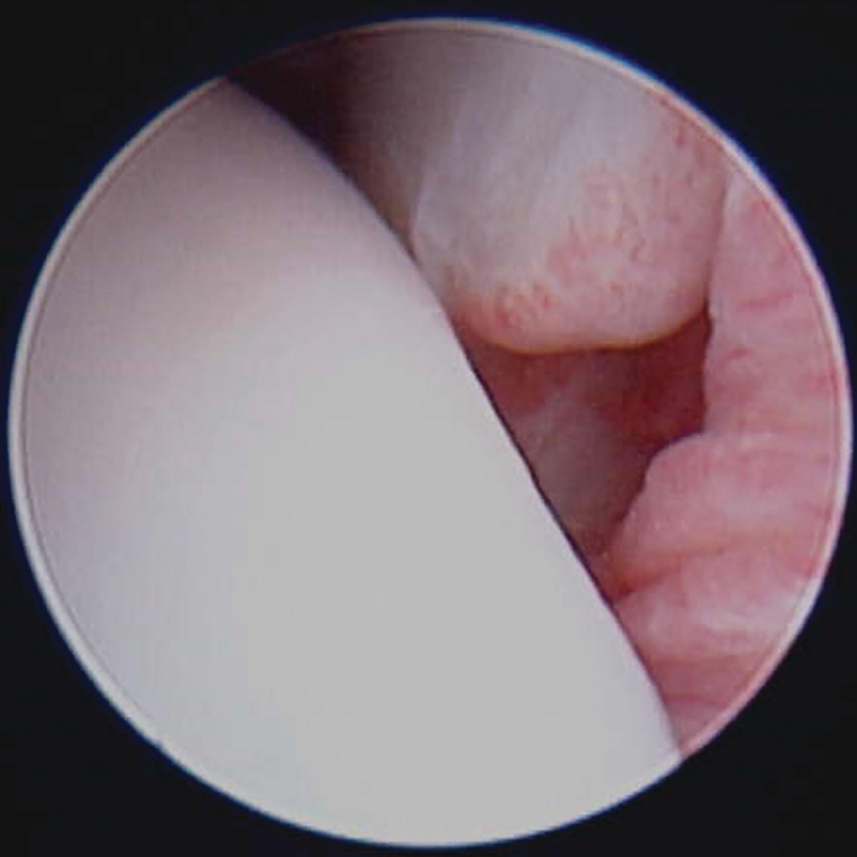
For medial soft tissue impingement, a 30- or 70-degree arthroscope can be placed in the anterolateral portal to view the medial gutter, with instrumentation in the anteromedial portal. The surgeon must be careful to preserve the deep deltoid ligament. For posterior impingement, the torn posterior ligaments can be visualized from the anterior portals as well as the posterior portal using 30- and 70-degree arthroscopes (see Fig. 39-32 ). The torn PtiFL, TTL, or posterior intermalleolar ligament (tibial slip) is debrided to its more normal appearing structure, with the shaver in the posterolateral portal. Visualization is best through the anteromedial portal through the notch of Harty. Alternatively, a suction basket may be used from the anterolateral portal.
For syndesmotic impingement, previous anatomic studies have shown that approximately 20% of the ATiFL syndesmotic ligament is intraarticular. Debridement of the ATiFL ligament and tibiofibular joint is performed using a full-radius 2.9-mm shaver or 2.5-mm suction basket. In addition, if a separate fascicle is seen, it should be removed. On rare occasions, the authors have excised this intraarticular portion of the ligament without any untoward effects to the patient at long-term follow-up ( Fig. 39-37 ).
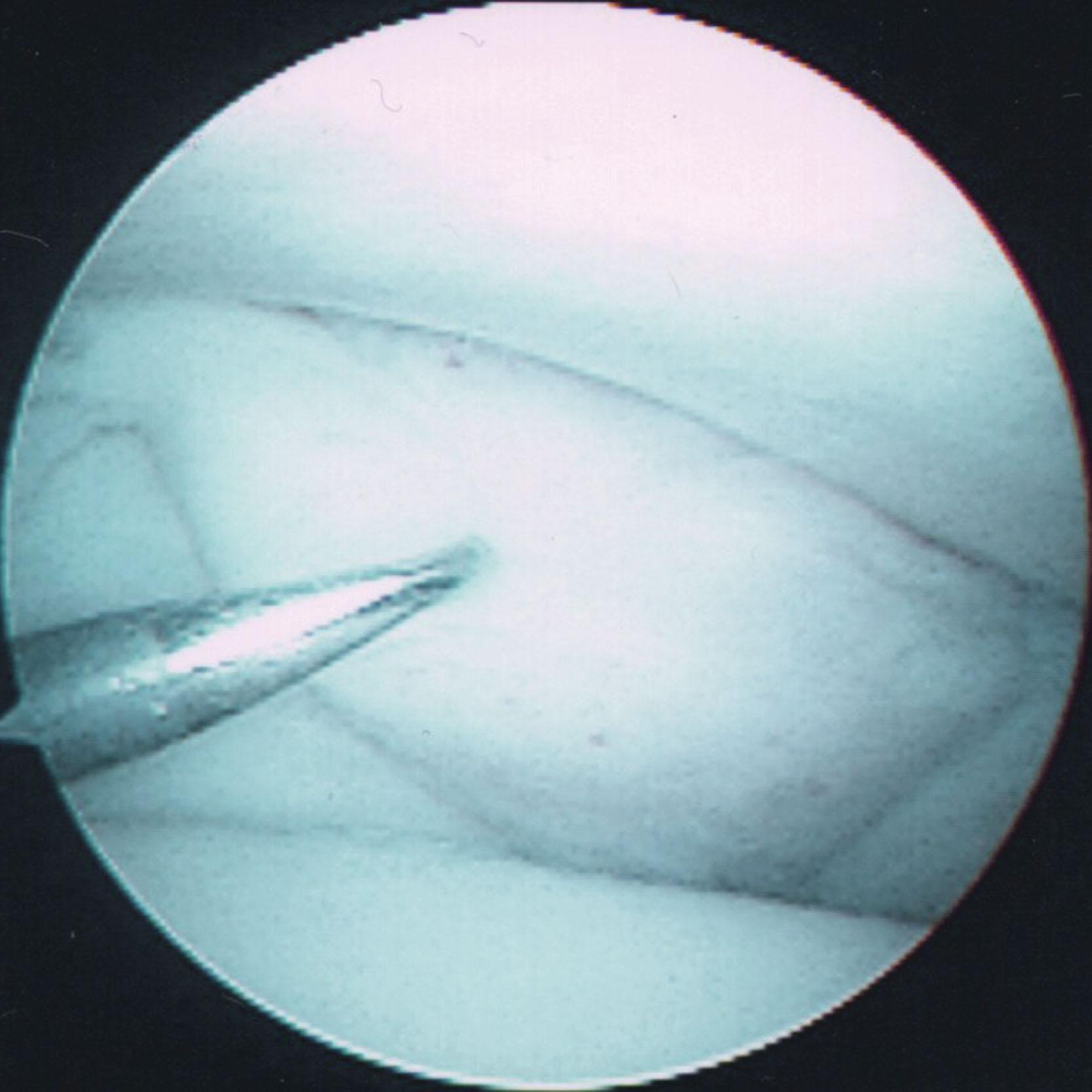
Postoperatively, patients are placed in a non–weight-bearing short-leg splint for 5 to 7 days. The stitches are removed, and the splint is discarded at 1 week, at which time a compression stocking and supportive brace are applied. Physical therapy is started at 2 to 3 weeks after surgery, and sports activities are permitted to begin 6 weeks postoperatively.
Since the 1980s, Ferkel has operated on numerous patients with pain in the anterolateral gutter of the ankle after an inversion stress injury. Initially, the results were analyzed in 30 patients with longer follow-up. The average patient age was 34 years, and average follow-up was 34 months. All patients had tenderness over the anterolateral corner of the talar dome and the lateral gutter. All patients had a history of a previous sprain, with an average interval of 24 months from injury to surgery. In every case, the patients had failed a complete course of conservative management, including nonsteroidal antiinflammatories (100%), physical therapy (100%), bracing (75%), casting (50%), and steroid injection (62%). Radiograph findings were normal in 50% of the patients; others had mild spurs, calcification, and joint space narrowing. Stress radiograph films of all patients showed no evidence of chronic ligamentous instability. MRI revealed synovial thickening consistent with soft tissue impingement in the anterolateral gutter.
At the time of surgery, all patients had synovitis and fibrosis of the anterolateral gutter, and some patients showed chondromalacia of the talus and fibula. In some patients, a thick band of tissue could be identified and excised, which probably corresponded to the meniscoid lesion Wolin described. Postoperative assessment found 84% of the patients rated good to excellent. Since then, numerous patients have been treated, with similar findings and results. Other authors have also found similar results with arthroscopic treatment of anterolateral soft tissue impingement.
Liu and Mirzayan initially reported a case of chronic posteromedial impingement after a lateral ankle injury that resulted in soft tissue impingement between the medial talus and tibial plafond. Medial impingement is believed to be predominately associated with lateral ankle instability, with secondary pathologic contact of medial structures. Since then, several authors have reported on small series of patients, showing promising results in most patients. An additional detailed review of evaluation and treatment of posterior ankle impingement is presented in the prone arthroscopy section.
Ogilvie-Harris and others have shown a significant improvement with arthroscopically treated syndesmotic injuries. Operative intervention requires a careful assessment of the soft tissue envelope, especially in grade III sprains with the potential for compartment syndrome. The surgeon is advised to assess the preoperative neurovascular status and be cognizant of complex regional pain syndromes occasionally associated with high-energy ankle sprains. Ankle stiffness, hardware loosening or breakage, loss of reduction, fibular stress fracture, and arthrosis are all possible complications.
This and the following sections discuss the arthroscopic management of chondral and osteochondral lesions of the ankle joint, excluding osteochondral lesions of the talus and the tibial plafond (OLTs and OLTPs). These include osteophytes, chondral and osteochondral loose bodies, protuberances secondary to fracture and trauma, as well as degenerative and posttraumatic arthritic changes.
A spectrum of symptoms can result from chondral and osteochondral lesions of the ankle, including pain, swelling, popping, locking, and limited range of motion. These lesions have often been a diagnostic challenge to orthopedic surgeons, particularly for chondral or soft tissue lesions. Advancement in arthroscopy of the ankle has allowed the orthopedic surgeon to directly visualize the ankle joint for evaluation of such disease and enables arthroscopic management of these lesions without an extensive surgical approach and ankle arthrotomy.
Loose bodies may be either chondral or osteochondral in origin. The presence of loose bodies is indicative of an underlying pathologic disorder of the ankle joint. The causes of loose bodies are many, and they range from a minor ankle sprain to synovial osteochondromatosis. Chondromatosis may be difficult to diagnose with radiography or CT because cartilaginous lesions are, in general, radiolucent, but some may be calcified. They are usually visible on MRI but may be missed because of the thickness of each imaging slice. Although this disorder is more common in the larger joints, it can certainly involve the ankle joint. Ossicles are most common in the subfibular region ( Fig. 39-38 ) and most likely represent avulsion fractures from an ankle sprain or injury. These chronic fractures may not heal, and a nonunion can develop. They may also represent a true accessory ossicle, referred to as an os subfibulare.
Patients with mechanical symptoms of locking, catching, and the feeling of a loose body in the joint may be candidates for surgery. A careful workup must be performed to determine the diagnosis and location of the loose body, and a trial of conservative management may be beneficial. With very large loose bodies, arthroscopic surgery occasionally may be contraindicated in favor of an open approach.
Loose bodies in the ankle joint can cause symptoms of catching and locking, with resultant pain, swelling, and limitation of range of motion. Symptoms may be intermittent because a loose body can become fixed in the synovial lining, therefore being asymptomatic until it is loosened and enters the ankle joint. The physical examination is generally unrevealing. Often, it is difficult to detect a specific area of tenderness. Rarely is a loose body palpable. It is important to rule out extraarticular pathology, which may mimic a loose body in the ankle joint.
Patients being evaluated for loose bodies and osteophytes should have weight-bearing AP, lateral, and mortise views of the ankle. Sometimes the lateral x-ray should be done in maximum dorsiflexion to detect bony impingement. The subtalar joint should also be carefully evaluated.
Scranton and McDermott, in 1992, proposed a radiographic classification system for anterior ankle osteophytes, based on the size of the anterior tibial osteophyte, the association with a talar osteophyte, and the presence of generalized degenerative changes in the ankle joint ( Table 39-5 ). In 1997, van Dijk et al developed a classification of osteoarthritis changes of the ankle joint that is commonly used to describe x-ray changes of degenerative joint disease on patients’ x-rays ( Table 39-6 ).
| Type | Characteristics |
|---|---|
| I | Synovial impingement; radiographs show inflammatory reaction with spurs as large as 3 mm |
| II | Osteochondral reaction exostosis; radiographs show osseous spur formation >3 mm but no talar spur is present |
| III | Severe exostosis with or without fragmentation, with secondary spur formation noted on the dorsum of the talus; also seen often is fragmentation of the osteophytes |
| IV | Pantalocrural osteoarthritic destruction; x-rays suggest degenerative osteoarthritic changes medially, laterally, or posteriorly |
| Grade | Characteristics |
|---|---|
| 0 | Normal joint or subchondral sclerosis |
| I | Osteophytes without joint space narrowing |
| II | Joint space narrowing with or without osteophytes |
| III | Total disappearance or deformation of the joint space |
Although plain radiographs generally reveal the presence of an osseous loose body, chondral loose bodies are not visible on plain radiograph examination ( Fig. 39-39 ). The CT arthrogram can effectively reveal the presence of chondral loose bodies, particularly in the presence of synovial chondromatosis.
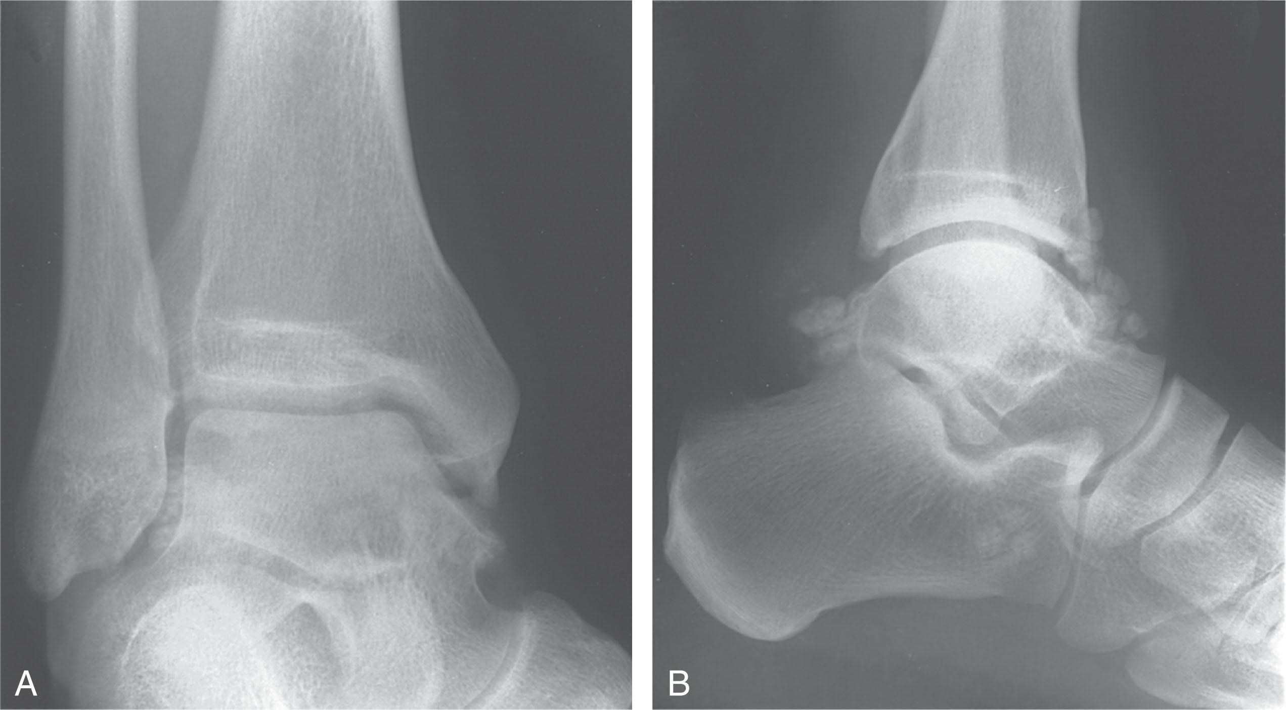
MRI appears to be the study of choice for revealing chondral lesions not otherwise visible on plain radiographs. Gadolinium used with the MRI helps to detect cartilage injury and chondral loose bodies. Therefore, a combination of diagnostic tests, including radiographs and CT or MRI scans should be used to effectively diagnose the presence and origin of the loose bodies preoperatively. Weight-bearing CT scans can be particularly helpful in evaluating loose bodies, osteophytes, osteochondral lesions, and numerous other conditions of the foot and ankle. Preoperative imaging should also be carefully studied to identify the source of the loose body (e.g., OLT). It is critically important to determine the general location of the loose body, whether it is intraarticular, intracapsular, or extraarticular, as part of the preoperative planning. This will aide in intraoperative localization of the lesion.
Treatment of loose bodies should entail the standard ankle arthroscopic technique, including the 21-point ankle examination protocol to systematically evaluate the entire ankle. This is particularly important when searching for loose bodies because they may be “hiding” in areas of the ankle not easily seen unless directly inspected. Distraction of the ankle joint may be particularly helpful, both to visualize the loose body and to obtain surgical access to the loose body in difficult-to-reach areas. After removing the loose body, the entire ankle joint surfaces should be examined again to detect any residual loose body and determine the origin of the lesion. Fluoroscopy may be used to determine the location of a radiopaque loose body and whether an adequate resection has been performed. If a substantial chondral or osteochondral defect (OCD) is detected, debridement and either burring, drilling, or microfracture should be performed. If a significant osteophyte was responsible for the loose body, the osteophyte should be aggressively debrided with an arthroscopic burr.
A loose body in the anterior compartment can usually be removed from the anterior portals ( Fig. 39-40 ). However, it may be necessary to place the arthroscope in the posterolateral portal and use the anteromedial or anterolateral portals for removing the loose body. A loose body in the posterior compartment of the ankle may be best removed with the arthroscope placed in either of the anterior portals and the loose body removed through instrumentation placed in the posterolateral portal or by pushing the loose body into the anterior compartment. Before removing the loose body, expand the portal adequately to prevent losing the loose body in the soft tissues. If the loose body is too large, break it up and remove it in smaller pieces. A strong grasping instrument or a large pituitary rongeur are very helpful in removing large loose bodies ( Fig. 39-41 ).
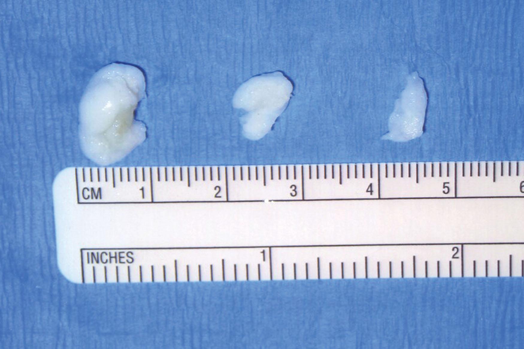
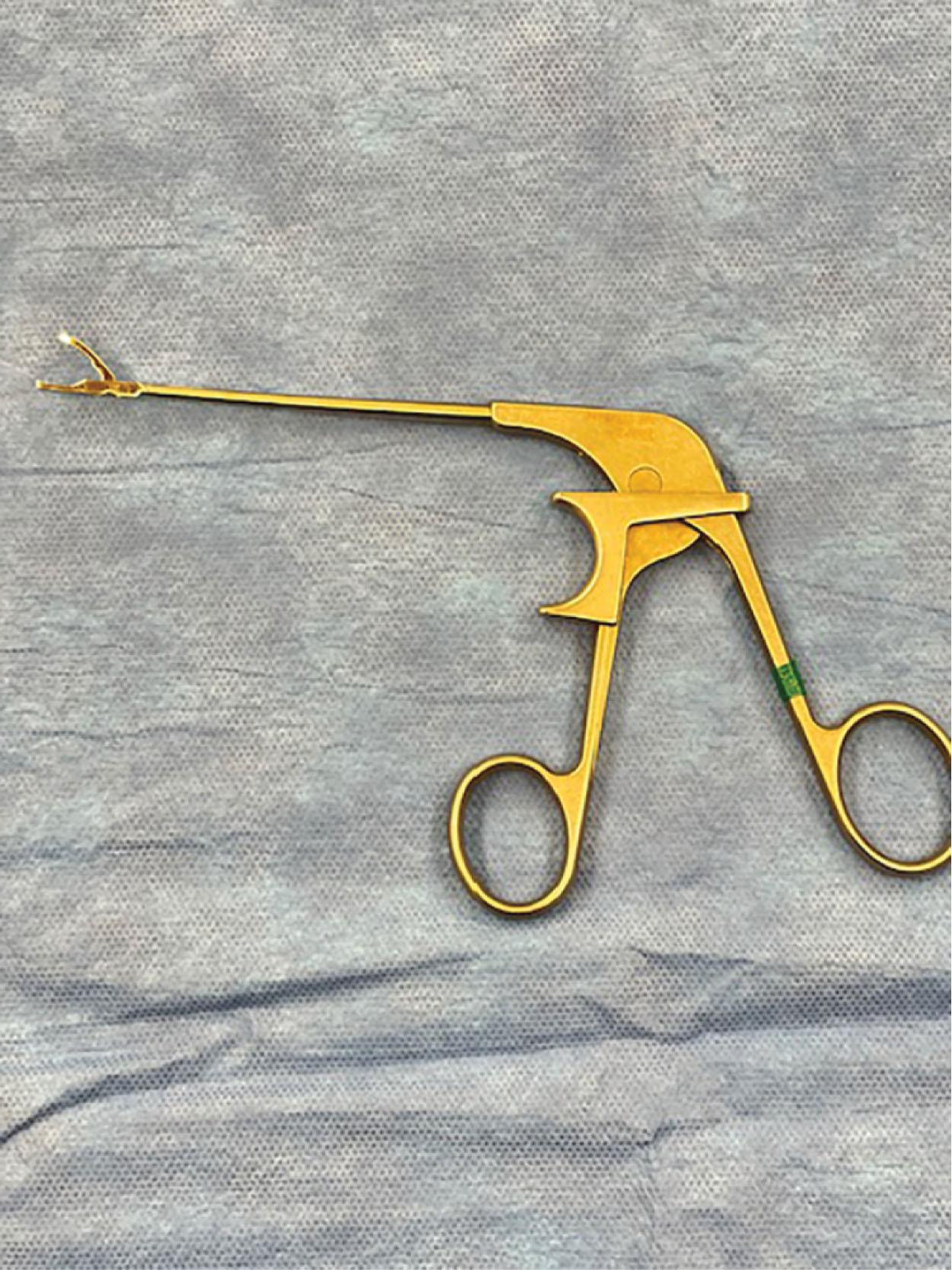
Ossicles are typically found within the lateral soft tissues, most commonly within the substance of the ATFL. Treatment should be aimed at removing the ossicle from within the substance of the ligament without disrupting the ligament; depending upon the size of the fragment, excessive damage to the ATFL may be done, necessitating repair of the ligament.
After surgery, a bulky compressive dressing with a posterior splint is applied. The patient is started on range-of-motion exercises at 5 to 7 days postoperatively, with a gradual increase in weight bearing. The exercise routine is then gradually advanced to include strengthening and increased range of motion.
The clinical results of loose body removal depend upon the underlying cause of the loose body and the presence of degenerative or posttraumatic arthritis in the ankle joint. In the presence of significant degenerative or posttraumatic arthritis, the results tend to be less predictable.
Osteophytes of the ankle joint are theorized to be the result of direct trauma after forced dorsiflexion injuries to the ankle, or they may be the result of forced plantar flexion causing capsular avulsion. One study, however, has shown that the anterior capsular attachments are proximal to the talotibial osteophyte formation sites, thereby possibly disproving this theory. Anterior impingement secondary to osteophyte formation is quite common after athletic injuries, with an incidence of 45% in football players and 59% in dancers.
Recently, the concept of cam-type impingement of the ankle has been reported as a likely etiology of bony ankle impingement. Amendola and colleagues found that in a consecutive series of 269 patients with ankle arthritis or anterior impingement syndrome, 34% were found to have some distortion of the anterior medial talar dome on lateral standing radiographs. The talar domes in these patients were not spherical, which the authors thought led to pathologic contact between the anterior medial distal tibia and talus. They specifically noted flattened talar domes that extrude into the anterior gutter space, or a thicker anterior neck just distal to the talar body articular surface. A comparative asymptomatic control group showed much lower rates of abnormal talar morphology. Studies are underway to further evaluate this concept.
Arthroscopic resection of ankle osteophytes may be performed in patients with progressive symptoms, pain well localized to the anterior aspect of the palpable bony prominence, and limitation of range of motion. Conservative management, including immobilization, should be exhausted before arthroscopic surgery. Severe degenerative joint changes are a contraindication to arthroscopic surgery and debridement. Other contraindications were discussed in the section on ankle arthroscopy.
Ankle joint osteophytes are most commonly seen at the anterior lip of the tibia. They usually appear as a beaklike prominence and are associated with a corresponding area over the neck of the talus. Talar lesions can be either a chondral defect or an opposing kissing osteophyte. CT scan evaluation has demonstrated a consistent anatomic location of osteophyte formation. Tibial and talar spurs typically do not overlap each other. Instead, the talar spur peak tends to lie medial to the midline, protruding off the medial side of the talar neck. The tibial spur peak, on the other hand, commonly lies lateral to the midline. In addition, the tibial spur is usually wider than the talar spur. On occasion, exostoses occur anterolaterally between the talus and the lateral malleolus and may be a source of symptoms and disability in the athlete. Osteophytes have also been noted on the anterior aspect of the medial malleolus and can be a source of impingement upon the talus, resulting in symptoms. Posterior ankle joint osteophytes can also form and cause clinical symptoms with plantar flexion of the ankle. Scranton and McDermott have developed a classification system to describe the different patterns of osteophyte formation.
Kim et al described a unique chondral lesion of the talar dome known as a “tram track” lesion. They were all found to be associated with variably sized anteromedial osteophytes of the distal tibia. In a more recent cadaveric and histologic study, these “tram track” lesions of the talar dome exactly articulated with the corresponding osteophytes of the distal tibia ( Fig. 39-42 ).
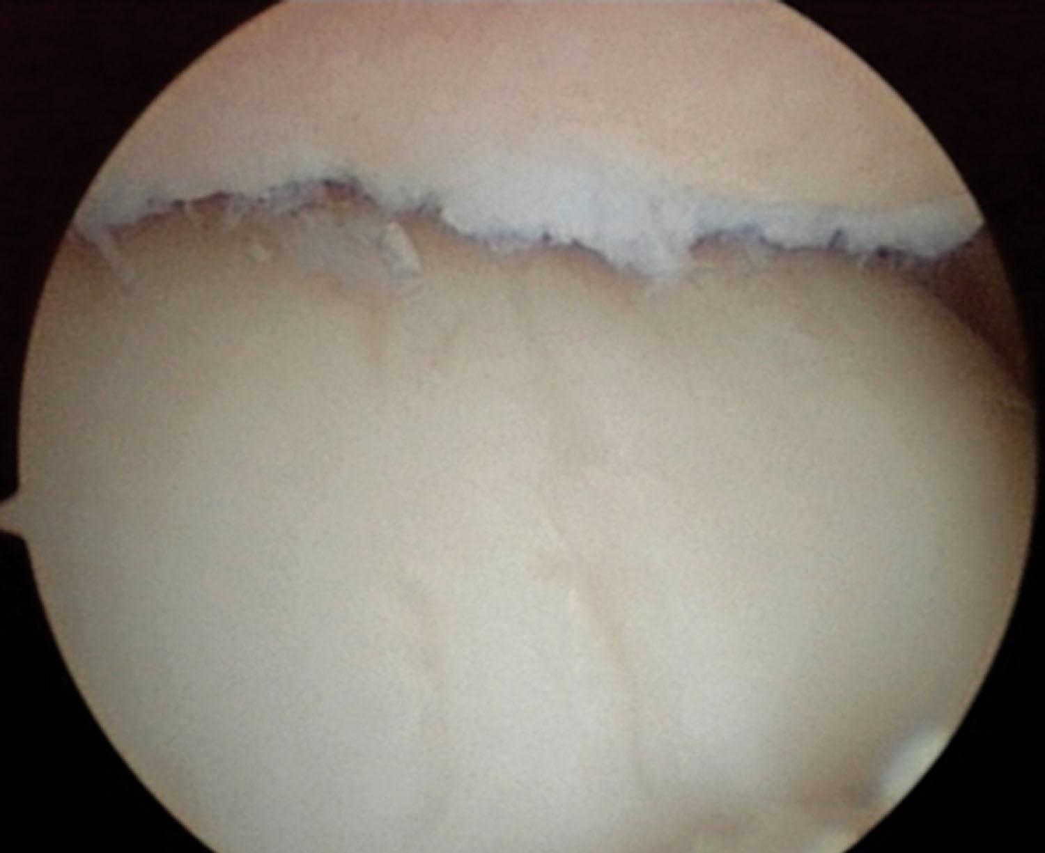
Amendola et al have described the concept of cam-type impingement in the ankle. In this situation, sagittal contour of the talar dome forms a noncircular arc with anterior flattening and causes pathologic contact with the anterior aspect of the tibial plafond in dorsiflexion.
It is important during preoperative evaluation to determine the anatomic location of the osteophytes because locating the osteophytes during surgery may be difficult. Preoperative AP, lateral, and dorsiflexion lateral views, and CT scans are essential. An oblique anteromedial radiograph has been proposed by van Dijk et al to locate anteromedial osteophytes ( Fig. 39-43 ). They demonstrated a dramatic increase in the sensitivity and specificity of radiographs in detecting anterior tibial and talar osteophytes. Normally, the angle between the distal end of the tibia and the talus is greater than 60 degrees ( Fig. 39-44A ). However, with the formation of osteophytes on the distal tibia and/or talar neck, this angle reduces to less than 60 degrees ( Fig. 39-44B ). A CT scan with sagittal and coronal reconstructions can also be helpful in localizing the size and the location of osteophytes. An MRI can also show evidence of osteochondral damage in the area of the osteophytes in symptomatic lesions, especially if the osteophyte is loose.
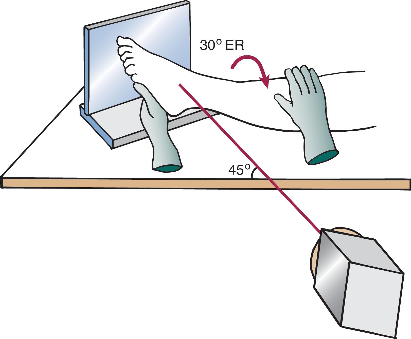
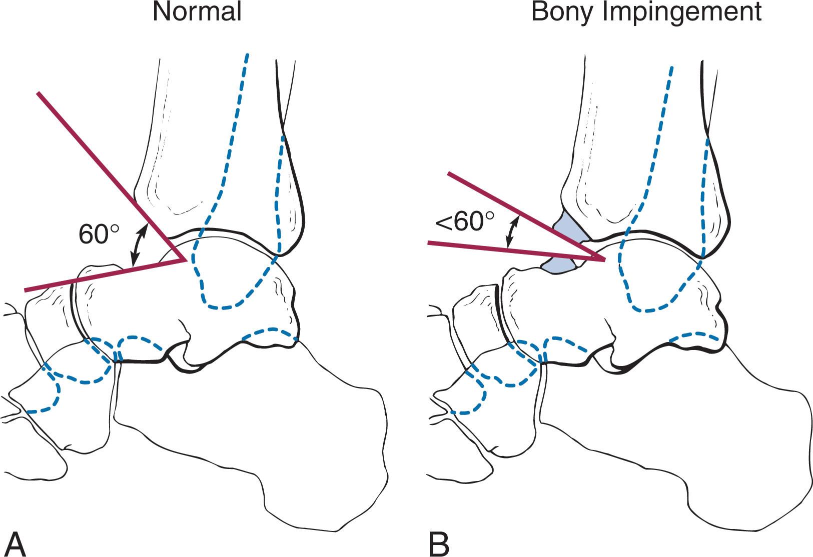
Clinically, a patient may have evidence of anterior ankle pain exacerbated by walking upstairs, squatting, or running. These osteophytes can cause impingement resulting in limited range of motion, pain, catching, and joint swelling. Tenderness may be localized over the bony prominence of the anterior ankle joint and exacerbated by passive ankle dorsiflexion. Range of motion may be limited, particularly in dorsiflexion. Posterior osteophytes can lead to impingement with ankle plantar flexion. Much of the pain associated with osteophytes appears to be due to synovial and capsular pinching associated with the osteophytes. The larger the osteophytes, the smaller the capsular space and the greater irritation that develops with motion.
![]() Preoperative radiographic assessment determines the amount and location of osteophyte resection. The goal of anterior osteophyte excision should be to restore the normal 60-degree angle between the distal tibia and talar neck. Positioning, equipment, instrumentation, distraction, and anesthesia were discussed previously for ankle arthroscopy. Ankle distraction can limit visualization of the anterior joint, particularly the superior and inferior confines of the osteophyte. Distraction should be decreased if this occurs.
Preoperative radiographic assessment determines the amount and location of osteophyte resection. The goal of anterior osteophyte excision should be to restore the normal 60-degree angle between the distal tibia and talar neck. Positioning, equipment, instrumentation, distraction, and anesthesia were discussed previously for ankle arthroscopy. Ankle distraction can limit visualization of the anterior joint, particularly the superior and inferior confines of the osteophyte. Distraction should be decreased if this occurs.
It is not imperative to completely resect both the talar and tibial osteophytes. As the talar lesion is typically more medial while the tibial osteophyte is more lateral, it is not always necessary to fully resect both lesions because they may not impinge. In addition, many of the talar osteophytes may be extraarticular. With that in mind, the authors recommend only safely resecting enough talar osteophyte to avoid any impingement and enough tibial osteophyte to restore the 60-degree angle.
The technique is as follows.
It is critical to identify the anterosuperior aspect of the tibial osteophyte and the inferior aspect of the talar osteophyte. This can require careful elevation of the soft tissues. Note that the neurovascular structures are at risk with this procedure.
The osteophyte can then be removed using a small burr, pituitary rongeur, or osteotome. The anterolateral and anteromedial portals must both be used for instrumentation and adequate resection.
Intraoperative fluoroscopy must be used to confirm the amount of resection.
On occasion, visualization through the posterolateral portal is important to assess the extent of resection and prevent overzealous resection. Mechanical distraction of the ankle is often necessary to visualize the anterior osteophytes through the posterior portals ( Fig. 39-45 ).
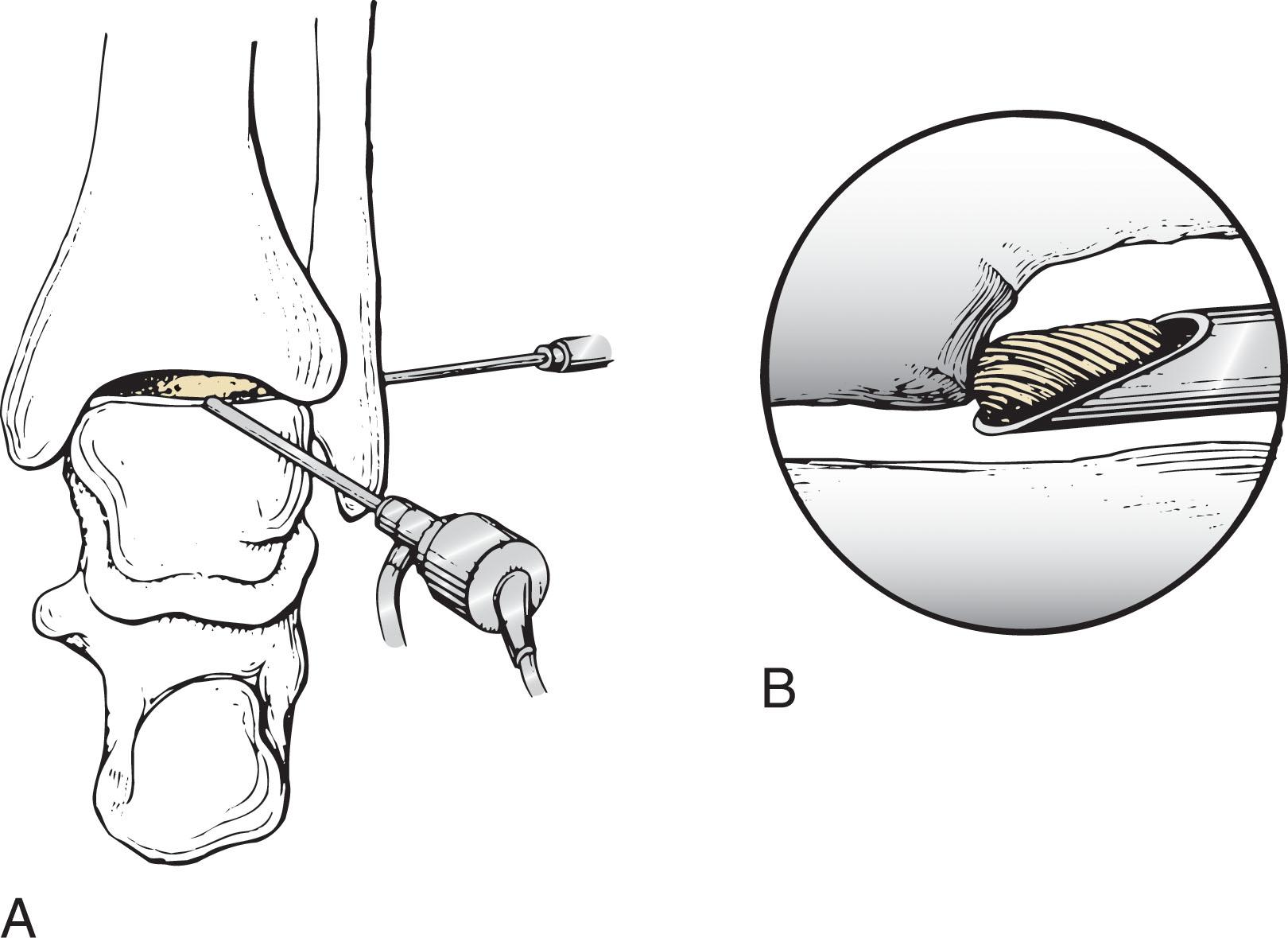
Posterior osteophytes are more challenging and can require the placement of a posteromedial portal. The posteromedial portal can be made using an outside-in technique. The posteromedial neurovascular structures and tendons must be avoided with this approach. Resection is performed using the same techniques as those described above. If posterior osteophytes are the primary source of pain, the surgeon should consider prone arthroscopy, using the posterolateral and posteromedial portals for excision (discussed later in the chapter).
The entire ankle joint should be systematically evaluated for the coexistence of other intraarticular disease or the presence of loose bodies. If significant synovitis is present as part of the inflammatory reaction to the presence of an osteophyte, synovectomy may be performed to improve visualization.
Lateral and oblique radiographs or fluoroscopic images may be used if any question remains about the completeness of the osteophyte excision.
The portals are closed with a nonabsorbable vertical mattress suture. A heavy, bulky dressing and a posterior splint are placed for 5 to 7 days. Range of motion and weight bearing are then encouraged.
Martin et al have suggested that patients with isolated osteophytes are more amenable to arthroscopic debridement than those with more diffuse disease. There have been several reports of good outcomes after arthroscopic resection for anterior osteophyte impingement, although outcomes have been correlated with the degree of arthritic joint changes. Patients undergoing arthroscopic excision recover faster compared with open excision. van Dijk et al studied 62 patients with ankle arthroscopy and found 90% of the patients had good and excellent results if they did not have joint space narrowing on their x-rays, while only 50% had good and excellent results if they had joint space narrowing. They felt that the stage of osteoarthritis was a better prognostic indicator to determine outcome than the size and location of spurs. Arthroscopic osteophyte excision may prove to be a challenging procedure, especially in cases of large anterior and posterior osteophytes. It is imperative that the surgeon has a thorough understanding of the ankle anatomy and that the location of the osteophyte is determined preoperatively.
Become a Clinical Tree membership for Full access and enjoy Unlimited articles
If you are a member. Log in here