Physical Address
304 North Cardinal St.
Dorchester Center, MA 02124

The role of advanced imaging techniques deserves special mention in abdominal trauma. Radiology has made a significant impact on the lives of traumatized patients by distinguishing those patients who can be managed conservatively from those who need surgical or other interventions and by helping to direct the most appropriate intervention for those who need it.
 Important Points
CT is the study of choice in abdominal trauma.
Intravenous contrast is always used (unless contraindicated) to identify devascularized areas, hematomas, active extravasation of blood or extraluminal urine (after contrast has passed through the kidneys) ( Box 25.1 ).
If a head CT is to be done as well, it should be done first before contrast is injected for the abdomen.
Intravenous contrast materials available today are nonionic, low osmolar solutions containing a high concentration of iodine that circulate through the bloodstream, opacify those tissues and organs with high blood flow, are absorbed by x-ray (and therefore appear whiter on images), and are finally excreted in the urine by the kidneys.
In some patients (e.g., those with a personal history of chronic kidney disease) who have an eGFR less than 30 mL/min/1.73 m 2 , the intravenous administration of iodinated contrast may be associated with acute kidney injury. Though usually reversible, in a small number of patients with underlying renal disease, renal dysfunction may permanently worsen. When the eGFR is lower than 30 mL/min/1.73m 2 and intravenous contrast is deemed necessary, prophylaxis may be indicated through the administration of isotonic volume expansion with normal saline.
Iodinated contrast agents can sometimes produce mild side effects, including a feeling of warmth, nausea, and vomiting, local irritation at the site of injection, and itching and hives; these side effects usually require no treatment. Occasional idiosyncratic reactions include itching, hives, and laryngeal irritation.
Asthmatics and those with a history of severe allergies or prior reactions to IV contrast have a higher likelihood of contrast reactions (but still very low overall) and may benefit from steroids or diphenhydramine (Benadryl™) administered prior and/or after injection. Prior shellfish allergy bears absolutely no relationship to iodinated contrast reactions.
In about 0.01% to 0.04% of all patients, severe and idiosyncratic reactions to contrast can occur that can produce intense bronchospasm, laryngeal edema, circulatory collapse, and, very rarely, death (1 in 200,000 to 300,000).
Oral contrast is usually not given. Rectal contrast is occasionally administered in penetrating trauma to search for a bowel laceration.
It is always best to consult with the radiologist so as to tailor the best study to fit the patient’s needs.
In some emergency settings, a quick abdominal ultrasound is used in unstable trauma patients to evaluate for hemoperitoneum (FAST scan, see Chapter 19 ).
The most commonly affected solid organs in blunt abdominal trauma (in order of decreasing frequency) are the spleen, liver, kidney, and urinary bladder. Traumatic injuries to each of them will be discussed under each organ.
The liver is discussed first because it is actually the most frequently injured organ if both penetrating and blunt trauma are included together. The liver is the largest intraabdominal organ and is fixed in position, making it especially susceptible to injury. Injuries to the liver account for the majority of deaths from abdominal trauma.
The posterior aspect of the right lobe is injured most frequently. Most hepatic injuries are associated with blood in the peritoneal cavity (hemoperitoneum).
Contrast-enhanced CT is the study of choice and, because of its ability to demonstrate both the nature and extent of the trauma, the overwhelming majority of patients with liver trauma are now managed conservatively and do not require surgery.
Important Points
CT findings in hepatic trauma:
Subcapsular hematoma. Lenticular fluid collections that conform to the shape of the outer contour of the liver but frequently flatten the adjacent liver parenchyma. Most occur anterolaterally over the right hepatic lobe ( Fig. 25.1A ).

Lacerations. Most common finding. Irregularly marginated, low attenuation, linear or branching defects, usually at the periphery . Fracture is a term that has been used to describe a laceration that avulses a section of the liver ( Fig. 25.1B ).
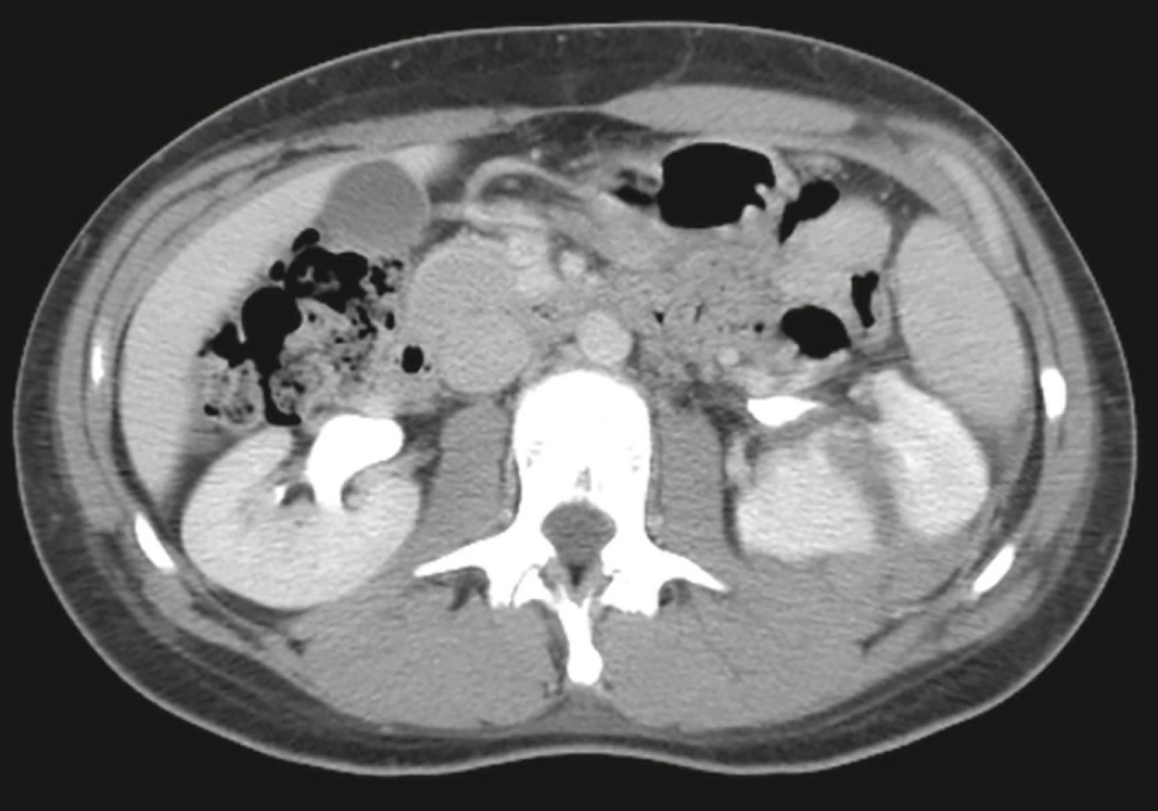
Intrahepatic hematomas. Focal, high-attenuation lesions first caused by blood, hematomas may progress to become low attenuation, mass-like lesions filled with serous fluid ( Fig. 25.1C ).
Wedge-shaped defects. Devascularized sections of liver parenchyma that do not contrast-enhance.
Contusions. A term used to describe an area of minimal parenchymal hemorrhage; they are lower in attenuation than the surrounding liver and have indistinct margins.
Pseudoaneurysms and acute hemorrhages. Irregular collections of high-attenuation, extravasated contrast that often require angiography with embolization and/or surgery.
Splenic trauma is usually caused by deceleration injuries in unrestrained occupants of motor vehicle collisions, by a fall from a height, or by being struck by a motor vehicle as a pedestrian. The spleen is affected in about 1/3 of patients with traumatic abdominal injuries.
Because the spleen is the most highly vascular intraabdominal organ , hemorrhage represents the most serious complication of trauma. Despite its vascular nature and the delayed presentation of many splenic injuries, most splenic trauma is treated conservatively (nonsurgically). However, the presence of hypotension in a patient with a suspected splenic injury can be a critical sign and a surgical emergency.
 Important Points
CT is the study of choice for evaluating splenic trauma. Findings include:
Subcapsular hematoma. Low-attenuation, crescent-shaped collection of fluid in the subcapsular space that frequently compresses the normal splenic parenchyma ( Fig. 25.2A ).

Laceration. Irregular, low-attenuation defect that typically transects the spleen ( Fig. 25.2B ).
Intraparenchymal hematoma . Lacerations filled with blood; they are intrasplenic, rounded areas of low attenuation that may have a mass effect and enlarge the spleen ( Fig. 25.2C ).
Contusion. Alterations in the normal homogeneous appearance of the spleen, including mottled areas of low attenuation ( Fig. 25.3 ).

Intraperitoneal fluid or blood. Hemoperitoneum occurs with almost all splenic injuries, also producing small amounts of blood in the pelvis. Its presence does not necessarily indicate active hemorrhage.
Become a Clinical Tree membership for Full access and enjoy Unlimited articles
If you are a member. Log in here