Physical Address
304 North Cardinal St.
Dorchester Center, MA 02124

A skull X-ray (SXR) continues to have an important role when there is suspicion of non-accidental injury (NAI) in an infant or a toddler . The primary indication for a SXR in these patients is forensic .
Be careful:
Accessory sutures are common.
Calling an accessory suture a fracture may lead to an incorrect suggestion of NAI.
Dismissing a fracture as an accessory suture can have serious clinical consequences.
To avoid mistakes:
Be aware of the positions of the common accessory sutures.
Assess and interpret an infant's or toddler's radiographs in a systematic step-by-step manner.
Lateral.
AP frontal view.
Towne's view.
The precise SXR views to be obtained will be specified by the local protocol for NAI assessment in infants and toddlers .
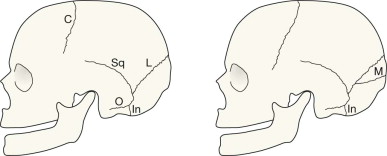
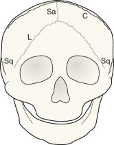
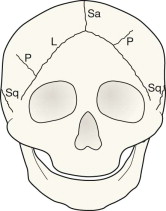
C, coronal;
In, innominate;
L, lambdoid;
M, mendosal;
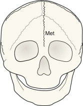
Met, metopic;
O, occipitomastoid;
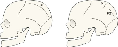
P1 and P2, accessory parietal;
Sa, sagittal;
Sq, squamosal.
NAI, non-accidental injury.
Evaluating the SXR in an infant or toddler presents unique problems. Diagnostic confusion between sutures and fractures may have serious consequences. A basic understanding of the locations and variable appearances of these sutures will help to reduce the likelihood of misdiagnosis .
| Grouping | Notes | Sutures |
|---|---|---|
| The normal sutures | Visible on the SXR in all infants and toddlers – persisting in all adults | Sagittal, coronal, lambdoid, squamosal, and smaller sutures around the mastoid |
| A normal developmental suture | Visible on the SXR in all infants and many toddlers – but not in adults | Innominate |
| The most common accessory sutures | Visible on the SXR in some infants and toddlers – occasionally persisting to adulthood | Metopic, accessory parietal, mendosal |
| Suture | Most commonly seen on | Notes |
|---|---|---|
| Metopic suture | Frontal √ √ Towne's √ |
The commonest accessory suture. It is also the one that most commonly persists in older children, and even in a few adults. |
| Accessory parietal suture | Towne's √ √ Frontal √ Lateral √ |
May be complete or incomplete. Occurs in vertical, horizontal or oblique orientations. Most commonly vertical. |
| Mendosal suture | Lateral √ √ Towne's √ |
Extends posteriorly from the lambdoid suture on the lateral view. Passes medially on Towne's view. |
| Innominate suture | Lateral √ √ | Sometimes classified as an accessory suture but best regarded as a normal developmental suture because it is always present in infants. As the child matures this suture disappears. |
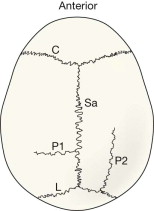
Accessory parietal sutures vary in position. This drawing does not correspond to any radiographic projection. It shows the general positions and direction of the more common incomplete accessory parietal sutures (P1 and P2) when looking down from above the cranium.
L = lambdoid suture;
C = coronal suture;
Sa = sagittal suture;
P = accessory parietal sutures.
Become a Clinical Tree membership for Full access and enjoy Unlimited articles
If you are a member. Log in here