Physical Address
304 North Cardinal St.
Dorchester Center, MA 02124

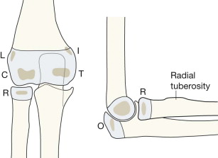
Undisplaced supracondylar fracture.
Fracture, lateral condyle of humerus.
Monteggia injury .
AP in full extension.
Lateral with 90 degrees of flexion.
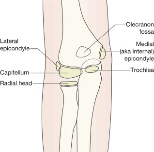
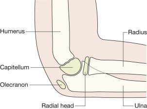
CRITOL: C apitellum, R adial head, I nternal epicondyle, T rochlea, O lecranon, L ateral epicondyle.
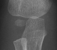
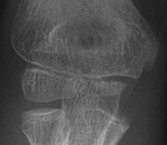
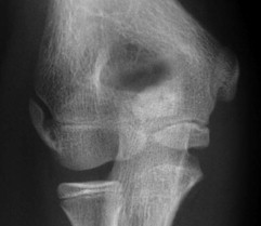
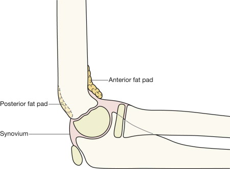
There are pads of fat close to the distal humerus, anteriorly and posteriorly. They are extrasynovial but intracapsular.
Look for the fat pads on the lateral. They are not seen on the AP view.
The fat is visualised as a dark streak amongst the surrounding grey soft tissues.
The anterior fat pad is seen in most (but not all) normal elbows. It is closely applied to the humerus, as shown below.
The posterior fat pad is not visible on a normal radiograph because it is situated deep within the olecranon fossa and hidden by the overlying bone.
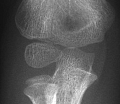
At birth the ends of the radius, ulna and humerus are lumps of cartilage, and not visible on a radiograph. The large, seemingly empty, cartilage filled gap between the distal humerus and the radius and the ulna is normal.
From 6 months to 12 years the cartilaginous secondary centres begin to ossify. There are six ossification centres. Four belong to the humerus, one to the radius, and one to the ulna. Gradually the humeral centres ossify, enlarge, and coalesce. Eventually each of the fully ossified epiphyses fuses to the shaft of its particular bone.
Exceptions are an occasional normal variant .
A 2011 survey of 500 paediatric elbow radiographs found:
97% followed the CRITOL order.
3% showed a slightly different order.
But: there were no instances in which the trochlear ossification centre appeared before the medial (internal) epicondylar centre.
CRITOL is a really helpful tool when analysing a child's injured elbow.
Occasionally a minor variation in the sequence may occur.
Use the rule: “I always appears before T”. So, if you see the ossified T before the I then the internal epicondyle has almost certainly been avulsed and is lying within the joint… ie it is masquerading as the trochlear ossification centre (see p. 105 ).
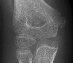
Is the medial epicondyle slightly displaced/avulsed? A common dilemma.
The rule to apply:
On the AP radiograph a normally positioned epicondyle will be partly covered by some of the humeral metaphysis.
A caveat:
Occasionally a child in pain will hold the forearm in a position of slight internal rotation. Rotation will project the metaphysis of the humerus away from a normally positioned epicondyle.
Conclusions:
When checking the position of the internal epicondyle on the AP radiograph:
If part of the epicondyle is covered by part of the humeral metaphysis then an avulsion has not occurred.
A completely uncovered epicondyle indicates an avulsion… unless the forearm bones are slightly rotated.
The ossification centre for the internal (ie medial) epicondyle is the point of attachment of the forearm flexor muscles. Vigorous muscle contraction may avulse this centre (see p. 105 ). The most common injury mechanism is a fall on an outstretched hand. Avulsions also occur in children who are involved in throwing sports, hence the term “little leaguer's elbow”.
When a major displacement of the internal epicondyle occurs the bone can become trapped within the elbow joint. This is a well recognised complication of a dislocated elbow, occurring in 50% of cases following an elbow subluxation or dislocation. A major avulsion is easy to overlook when an elbow has been transiently dislocated and then reduces spontaneously because the detached epicondyle may, on the AP radiograph, be mistaken for the normally positioned trochlear ossification centre ( p. 105 ).
I before T. Though the CRITOL sequence may vary slightly there is a constant: the trochlear (T) centre always ossifies after the internal epicondyle. Therefore apply this rule: if the trochlear centre (T) is visible then there must be an ossified internal epicondyle (I) visible somewhere on the radiograph. If the internal epicondyle is not seen in its normal position then suspect that it is trapped within the joint.
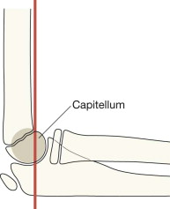
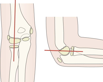
Normal alignment : when drawn along the anterior cortex of the humerus, in most normal patients at least one third of the ossifying capitellum lies anterior to this line.
Be carefu l: in very young children the ossification within the cartilage of the capitellum might be minimal (ie normal and age related), and so is insufficiently calcified and does not allow application of the above rule.
This line helps you to detect a supracondylar fracture with posterior displacement ( pp. 106–108 ).
Become a Clinical Tree membership for Full access and enjoy Unlimited articles
If you are a member. Log in here