Notes on staining techniques


Histochemical Staining Techniques

Most histological stains are dyes which, when applied to tissues, form a chemical bond with proteins within the tissues. Staining tissue sections is essential to differentiate between the different cells and tissues on the slide, permitting examination by light microscopy. Many histological and pathological features can be identified using routine histological stains and by far the most commonly used is haematoxylin and eosin or ‘H&E’. This is regarded as the ‘bread and butter’ stain of both histology and pathology. Additional stains, as detailed below, can help to illustrate particular features within tissues such as collagen or mucin production and to identify infective organisms. If these routine stains do not provide an answer, there are further ancillary stains which can be applied such as immunohistochemistry and immunofluorescence. Further details are provided in the boxes below.

  • Haematoxylin and eosin (H&E)

    • This is the most commonly used stain in histology and pathology. Haematoxylin is a basic dye which stains acidic structures a purplish-blue. In contrast, eosin is an acidic dye which stains basic structures pinkish-red. In the mammalian cell, most of the acidic structures reside in the nucleus due to the presence of nuclear DNA, whereas most of the cytoplasmic structures are basic. Thus, nuclei stain purplish-blue and the cytoplasm a pinkish-red. If a cell’s cytoplasm contains abundant RNA, then the cytoplasm may have a purplish tint. In the first micrograph, the serous cells of the parotid gland show dense granular purple cytoplasm which is full of zymogen granules.

    • The intensity of the staining by H&E depends upon many factors including tissue processing and fixation, the thickness of the tissue section and the formulation of the stain. The thickness of the section has the most impact upon the staining intensity as can be seen in some thicker, low-power images throughout this text.

    • The second micrograph shows early secretory endometrium with subnuclear vacuolation in the glands which are easily demarcated from the background endometrial stroma.

  • Periodic acid–Schiff (PAS)

    • The periodic acid–Schiff (PAS) reaction demonstrates the presence of complex carbohydrates by staining them a deep magenta colour. This is referred to as ‘ PAS positive’ . PAS will stain pure complex carbohydrates, such as glycogen in hepatocytes and muscle cells, but will also stain carbohydrates when they are bound to lipids and proteins. Basement membranes and the brush borders of kidney tubules and the small and large intestines are PAS positive, as is cartilage and some collagen.

  • Diastase periodic acid–Schiff (D-PAS, PASD, PASF)

    • Many of these complex carbohydrate–lipid or protein compounds are present in basement membranes, fungal cell walls and some neutral mucin. In order to differentiate between pure glycogen and these other compounds, glycogen can be eradicated in the tissue section by pre-treatment with the enzyme diastase (amylase) prior to the PAS reaction.

  • Alcian blue (acid mucin)

    • Epithelial mucins secreted by epithelial cells are either acidic or neutral. Acidic mucins are present in goblet cells and oesophageal submucosal glands. Most invasive adenocarcinomas also produce acidic mucin. Alcian blue stains acidic mucin a vivid blue colour. In contrast, neutral mucins, e.g. those present in gastric foveolar cells and prostate glands, are PAS positive. Alcian blue can also be used in combination with other stains such as H&E or van Gieson.

  • Masson trichrome

    • Masson trichrome is a connective tissue stain as it is used to demonstrate supporting tissue elements such as collagen. As the name implies, this staining method produces three colours in tissue sections. Nuclei and other basophilic structures are stained black, collagen is stained green or blue depending on which variant of the staining protocol is used, and muscle, erythrocytes and keratin are stained bright red. In pathology, it is used routinely in liver and kidney biopsies.

  • Martius Scarlet Blue (MSB)

    • MSB is another connective tissue stain. This three-colour staining method stains fibrin red, erythrocytes yellow and collagen blue. It is also useful for the examination of thrombi in histological sections, particularly in the autopsy setting.

  • Miller’s elastic van Gieson (EVG)

    • This stains collagen red and elastic fibres black. It is particularly useful in staining the elastic laminae of arteries (black), the smooth muscle of the media and erythrocytes (yellow) and collagenous tissue (red).

  • Phosphotungstic acid haematoxylin

    • This stain demonstrates muscle cross-striations such as in the myocytes of the heart. It may also be used to demonstrate the presence of mitochondria in particular types of tumours.

  • Gordon & Sweet’s reticulin

    • This is a silver-based stain that demonstrates reticulin fibres of supporting tissues which are stained blue/black by this technique. It is particularly useful in the interpretation of liver architecture (illustrated in the micrograph), demonstrating the liver cell plates. Background tissues may be counterstained with a red dye (neutral red).

  • Sirius red

    • This stain is most commonly used to detect amyloid deposits. These are acidophilic (eosinophilic) deposits of abnormal protein which can occur in many different tissue sites. Amyloid is characterised by beta pleated sheets on electron microscopy. Congo red is a similar stain to Sirius red. Both stain amyloid red and display apple-green birefringence under polarised light.

  • Giemsa stain

    • This stain is mainly used to stain smears of blood cells and other cells e.g. in bone marrow and in neuropathology. It can also be used to demonstrate some microorganisms. Nuclei are stained dark blue to violet, background cytoplasm pale blue and erythrocytes pale pink.

  • Gram stain

    • This method is mainly used in smears of bacteria in diagnostic microbiology, but can also be used in histological sections to show whether any bacteria are present and, if so, whether they are Gram-positive or Gram-negative. Gram-positive bacteria stain dark blue as illustrated in the micrograph opposite.

  • Perls’ stain

    • This stain is used to demonstrate the presence of ferric iron in tissues, usually at the site of old bleeding where the iron-containing pigment, haemosiderin (derived from local haemoglobin breakdown), accumulates. It can also be used to demonstrate accumulation of iron in various tissues in the primary iron storage disease haemochromatosis and can be used for the identification of ferruginous asbestos bodies in lung tissue sections.

  • Masson-Fontana

    • This stain is used to demonstrate melanin pigment in tiss ues but can also identify argentaffin granules and some neurosecretory granules. This stain can be used in the diagnosis of malignant melanoma as well as some melanin-producing microorganisms, e.g. Cryptococcus . Melanin pigment stains black and the background tissue stains pink.

  • Wade–Fite (modified Ziehl–Neelsen stain)

    • This stain is used for identification of Mycobacterium leprae and atypical Mycobacteria and is a modification of the Ziehl–Neelsen stain for Mycobacterium tuberculosis organisms which are more acid and alcohol fast. The organisms stain red whilst the background tissues stain blue.

  • Ziehl–Neelsen

    • This stain can be used to demonstrate Mycobacterium tuberculosis . This bacterium possesses a protective capsule containing lipids that affects the rate at which dyes move into and out of the organism during staining. Two dyes are used, basic fuchsin mixed with phenol and methylene blue. The first dye (which is red) is forced into all the tissues in the section (including any Mycobacteria ) by heating. The section is then exposed to acid and alcohol which wash the red dye out of everything except the Mycobacteria , which hold on to it because of the lipid capsule. All the other tissues are free to take up the contrasting blue dye, but the Mycobacteria remain red against a blue background.

  • Goldner’s trichrome stain

    • This method is applied to acrylic resin sections of undecalcified bone to distinguish between mineralised bone and unmineralised osteoid. It has a haematoxylin component which also stains the nuclei of osteoblasts, osteocytes, osteoclasts and marrow cells.

    • The von Kossa silver method stains calcium deposits black. It can be used in a variety of scenarios, including distinguishing between mineralised bone and osteoid and in identifying dystrophic calcium in tissues and in conditions such as pseudogout and malakoplakia.

  • Cresyl fast violet

    • This is a basic dye commonly used in staining tissues of the brain and spinal cord. The neuropil stains purplish/blue and the Nissl substance of neurones stains dark blue due to the abundance of RNA. A modified version of this stain is also used to stain Helicobacter pylori organisms in gastric biopsies as illustrated in the micrograph.

Immunological Techniques

A variety of immunohistochemical techniques are vital for diagnostic purposes in pathology as well as for research. The basis of the technique and some examples are given below. Further examples are also illustrated throughout this book to highlight specific histological features.

Immunohistochemistry (IHC; also known as immunocytochemistry) is a staining method based on the antigen–antibody reaction. IHC markers are antibodies that are produced commercially and target particular antigens (e.g. nuclear proteins, cytoplasmic proteins, cell adhesion proteins) in cells and tissues. There are hundreds of commercially available IHC markers which may highlight different components of tissues, e.g. smooth muscle markers (SMA, desmin and H-caldesmon) stain normal smooth muscle and smooth muscle tumours. IHC can provide useful information in normal histology to identify particular cells or identify the function of a cell/tissue and therefore this technique is often utilised in research laboratories. IHC is also extensively used by diagnostic pathologists for a variety of purposes such as identifying the site of origin of primary tumours, as prognostic and predictive markers in cancer and to screen for genetic diseases. Interpretation of IHC staining is complex and outwith the scope of this book, however some basic points are discussed below and throughout the organ system chapters. There is also more detailed information in Chapter 1 of Wheater’s Pathology (6th edition).

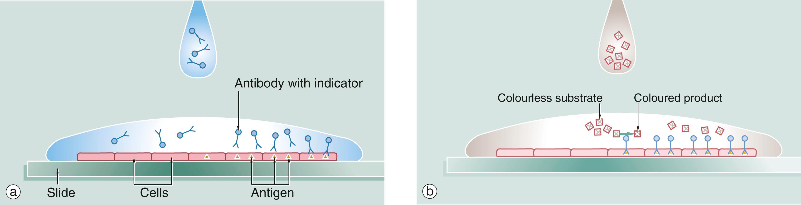
The basic technique of immunostaining is as follows: a section is placed on a glass slide (a) and a solution of antibody is laid over the issue. The antibody binds to the antigen of interest in the tissue and excess antibody is washed away so that only cells with the particular antigen have antibody bound to them. The antibody is pre-linked to an indicator substance (b). This is transformed by an enzyme from a colourless substrate to a coloured product which can be viewed by light microscopy. An enzyme commonly used for this purpose is horseradish peroxidase and the technique is known as the immunoperoxidase reaction. The substrate may also be a fluorescent substance such as fluorescein, in which the technique is known as immunofluorescence , and the position of the antibody can be viewed by a fluorescence microscope. A further modification of this principle for electron microscopy uses antibodies linked to gold which is electron dense and can be detected by EM (immunogold labelling). Further information on EM is given in Appendix 1 .

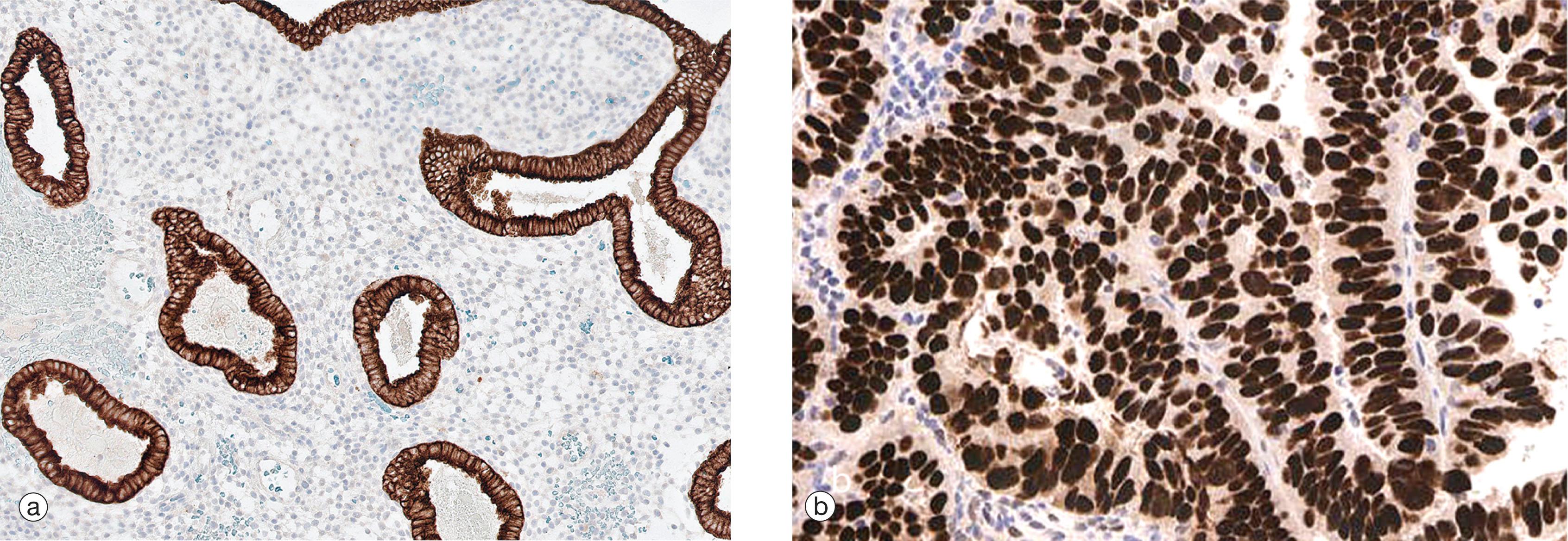
Immunostains can produce a variety of patterns of staining within the cell (nuclear, cytoplasmic, membranous or a combination). Image (a) shows endometrial tissue which has been stained with an epithelial marker (cytokeratin). The positive cytoplasmic brown staining of the glands contrasts to the lack of staining in the endometrial stroma where only the blue counterstain is seen. Image (b) shows a nuclear pattern of staining for the biomarker oestrogen receptor (ER) in a breast carcinoma.

Online questions

You're Reading a Preview

Become a Clinical Tree membership for Full access and enjoy Unlimited articles

Become membership

If you are a member. Log in here