Physical Address
304 North Cardinal St.
Dorchester Center, MA 02124

Lisfranc subluxations.
Fatigue fractures involving the 2nd or 3rd metatarsals.
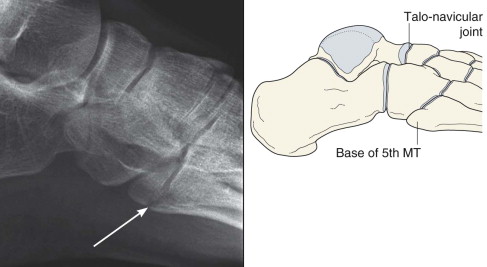
Avulsion fracture of the base of the 5th metatarsal—overlooked on ankle radiographs.
Protocols vary. In the UK a two view series is commonplace: AP and Oblique. Elsewhere, and in the USA, a three view series is common practice : AP, Oblique , and a Lateral .
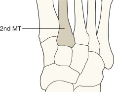
AP, anterior-posterior; MT, metatarsal.
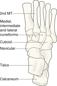
The bones of the midfoot form an arch. As a consequence several of the tarsal bones, specifically the three cuneiform bones and the bases of the metatarsals, overlap on both the AP and oblique projections. The individual bones can be separated from one another when the AP and oblique radiographs are examined as a complementary pair.
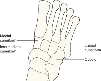
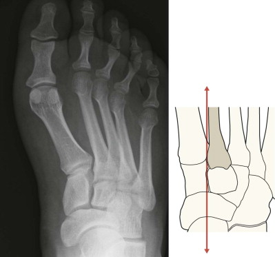
The base of the 2nd metatarsal is held in a mortice created by the three cuneiform bones. This mortice helps to prevent lateral slip of the bases of the metatarsals during weight bearing.
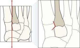
Alignment of 2nd metatarsal and the intermediate cuneiform. May appear “notched”.
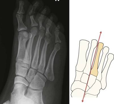
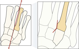
Alignment of 3rd metatarsal and the lateral cuneiform. May appear “notched”.
Analysis of the images will be influenced by the clinical findings such as the precise site of swelling, bruising, tenderness and pain.
Check:
Metatarsal and phalangeal shafts.
The Lisfranc joints. Always ask yourself … does the medial margin of the base of the 2nd metatarsal align with the medial margin of the cuneiform bone?
Check:
Metatarsal shafts.
The Lisfranc joints. Does the medial margin of the base of the 3rd metatarsal align with the adjacent cuneiform bone?
The hindfoot bones and the hindfoot–midfoot articulations.
Check:
Base of the 5th metatarsal.
The hindfoot bones and the midfoot articulations.
Become a Clinical Tree membership for Full access and enjoy Unlimited articles
If you are a member. Log in here