Physical Address
304 North Cardinal St.
Dorchester Center, MA 02124

A 56-year-old man with no significant medical history had a left medial unicondylar arthroplasty done at an outside institution for isolated medial knee osteoarthritis. He presented to our institution with several months of chronic knee pain and feelings of instability. He had no new trauma and no fevers or other constitutional symptoms suggestive of infection. On physical examination, he had a well-healed midline incision with a large effusion but no surrounding erythema. His active range of motion was 15 to 90 degrees with 10 mm varus and valgus laxity. The erythrocyte sedimentation rate (ESR) and C-reactive protein level (CRP) were within normal limits; aspiration of the knee revealed only 137 white blood cells with no bacteria. Standing radiographs ( Fig. 21.1 , A and B ) showed an unstable medial unicondylar arthroplasty with severe subsidence of the tibial tray and a large bony defect in the medial plateau. The overall alignment of the knee was valgus, which suggested a lateral plateau bony deficiency as well. Intraoperatively, the femoral and tibial components were both found to be grossly loose. On the tibial side, there was a large contained defect on the medial plateau and an uncontained defect on the lateral plateau. After all bony cuts were made and components were trialed, the defects were addressed. Because the medial defect was contained, morselized bone graft was packed into the defect. However, a wedge augment was used on the lateral side because the bony defect was uncontained there. A constrained condylar knee implant was used because of the lack of stability. The soft tissue envelope closed well, and the patient was made weight bearing as tolerated, with therapy focusing on increasing the range of motion. Postoperative radiographs are shown in Figure 21.1 , C and D .
 view of bilateral knees. B , Lateral view of the left knee. C , Postoperative AP radiograph shows a well-positioned prosthesis with bone graft used in the contained defect medially and a wedge in the uncontained defect laterally. D , Postoperative lateral radiograph.")
An algorithm for the management of bone defects in revision total knee arthroplasty (TKA) is presented.
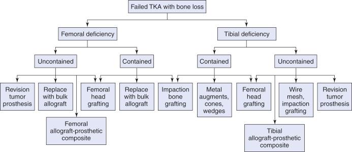
This chapter describes an overview and surgical technique of using allograft for the management of bone defects in revision total knee replacement. A classification system is described and may be used to help guide treatment. Surgical complications, outcomes, and postoperative care are also discussed.
Preoperative planning is critical. The more a surgeon knows preoperatively about potential bone defects, the more prepared he or she can be for the revision. It is important to know that there is not one right way of performing a revision, and the use of structural bone graft is just one tool of several available to the arthroplasty surgeon.
Preoperative radiography and computed tomography should be used to help determine the extent of bony defects.
Care should be taken in removing the implant to keep as much native bone stock as possible.
If screws or Steinmann pins are used, they should be secured to native bone so that the hardware is buried within the allograft and will not touch the final implanted prosthesis.
Cement should be used to secure a stemmed prosthesis to a bulk allograft, because the allograft cannot be relied on for bony ingrowth.
Revision knee arthroplasty in the setting of an active or latent infection with the use of allograft will result in failure.
Use of allograft or implants that are too large may result in problems with wound healing.
Use of allografts without addressing incompetent collateral ligaments will result in failure.
Become a Clinical Tree membership for Full access and enjoy Unlimited articles
If you are a member. Log in here