Lesional Morphology and Assessment


Introduction

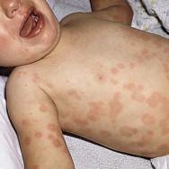
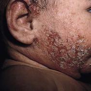
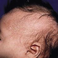
Newborn infant skin can manifest with an extraordinary array of conditions. Neonatal cutaneous findings may indicate transitory, benign processes such as erythema toxicum neonatorum, or may represent important harbingers of internal disease or genetic alteration, as might be observed in patients with herpes simplex virus infection or incontinentia pigmenti. Dermatologic manifestations are readily visible to the clinician, and it is often more efficient to first assess lesional morphology and then focus history-taking on the basis of the observed clinical findings. The timely identification and accurate diagnosis of skin findings in the newborn infant therefore relies on combining a comprehensive history with a meticulous physical examination, as well as on a proper understanding of physiologic differences between neonatal, pediatric, and adult skin that will influence both the diagnosis and the management of skin conditions appropriate to the neonate. This chapter reviews the principles of morphologic assessment in the term and preterm infant.

Reaction patterns

An understanding of the specialized reaction patterns is outlined in Tables 3.1–3.3 and Box 3.1 and, in conjunction with a comprehensive history and assessment of cutaneous morphology, will aid the clinician in making the proper dermatologic diagnosis.

TABLE 3.1
Primary lesions
Drawings reproduced with permission from Bolognia JL, Jorizzo JL, Rapini RP, eds. Dermatology. 3rd ed. St. Louis: Saunders; 2012.
Primary lesions are defined as lesions that arise de novo and are therefore most characteristic of the disease process. The graphic representations are intended to demonstrate three-dimensional and topographic relationships and not necessarily the histology of the example shown.
MACULE EXAMPLES

Café-au-lait macules

A circumscribed, flat lesion with color change, up to 1 cm in size, although the term is often used for lesions >1 cm. By definition, they are not palpable Ash leaf macules, café-au-lait macules, capillary malformations
PATCH EXAMPLES

Hemangioma precursor

A circumscribed, flat lesion with color change, >1 cm in size Nevus depigmentosus, Mongolian spots, nevus simplex
PAPULE EXAMPLES

Umbilical granuloma

A circumscribed, elevated, solid lesion, up to 1 cm in size. Elevation may be accentuated with oblique lighting Verrucae, milia, and juvenile xanthogranuloma
PLAQUE EXAMPLES

Nevus sebaceus

A circumscribed, elevated, plateau-like, solid lesion, >1 cm in size Mastocytoma, nevus sebaceus
NODULE EXAMPLES

Juvenile xanthogranuloma

A circumscribed, elevated, solid lesion with depth, up to 2 cm in size Dermoid cysts, neuroblastoma
VESICLE EXAMPLES

Acropustulosis of infancy

A circumscribed, elevated, fluid-filled lesion up to 1 cm in size Herpes simplex, varicella, miliaria crystallina
BULLA EXAMPLES

Insect bite reaction

A circumscribed, elevated, fluid-filled lesion >1 cm in size Sucking blisters, epidermolysis bullosa, bullous impetigo
PUSTULE EXAMPLES

Transient neonatal pustular melanosis

A circumscribed, elevated lesion filled with purulent fluid, <1 cm in size. Pustules can be primary skin lesions or can initially be a vesicle that then becomes filled with cells or debris. Transient neonatal pustular melanosis, erythema toxicum neonatorum, infantile acropustulosis
WHEAL EXAMPLES

Drug eruption

A circumscribed, elevated, edematous, often evanescent lesion, caused by accumulation of fluid within the dermis Urticaria, bite reactions, drug eruptions
ABSCESS EXAMPLE

Abscess

A circumscribed, elevated lesion filled with purulent fluid, >1 cm in size. Pyodermas

TABLE 3.2
Secondary lesions
Drawings reproduced with permission from Bolognia JL, Jorizzo JL, Rapini RP, eds. Dermatology. 3rd ed. St. Louis: Saunders; 2012.
Secondary lesions are characteristically brought about by modification of primary lesions, either by the individual or through the natural evolution of the lesion in the environment. The graphic representations are intended to demonstrate three-dimensional and topographic relationships and not necessarily the histology of the example shown.
CRUST EXAMPLES

Infected atopic dermatitis

Results from dried exudate overlying an impaired epidermis. Can be composed of serum, blood, or pus Epidermolysis bullosa, impetigo
SCALE EXAMPLES

Seborrheic dermatitis

Results from increased shedding or accumulation of stratum corneum as a result of abnormal keratinization and exfoliation. Can be subdivided further into pityriasiform (branny, delicate), psoriasiform (thick, white, and adherent), and ichthyosiform (fish scale-like) Ichthyoses, postmaturity desquamation, seborrheic dermatitis
EROSION EXAMPLES

Epidermolysis bullosa

Intraepithelial loss of epidermis. Heals without scarring Herpes simplex, certain types of epidermolysis bullosa
ULCER EXAMPLES

Aplasia cutis congenita

Full-thickness loss of the epidermis, with damage into the dermis. Will heal with scarring Ulcerated hemangiomas, aplasia cutis congenita
FISSURE EXAMPLES

Atopic dermatitis

Linear, often painful break within the skin surface, as a result of excessive xerosis Inherited keratodermas, hand and foot eczema
LICHENIFICATION EXAMPLES

Atopic dermatitis

Thickening of the epidermis with exaggeration of normal skin markings caused by chronic scratching or rubbing Sucking callus, atopic dermatitis
ATROPHY EXAMPLES

Focal dermal hypoplasia

Localized diminution of skin. Epidermal atrophy results in a translucent epidermis with increased wrinkling, whereas dermal atrophy results in depression of the skin with retained skin markings. Use of topical steroids can result in epidermal atrophy, whereas intralesional steroids may result in dermal atrophy Aplasia cutis congenita, intrauterine scarring, and focal dermal hypoplasia
SCAR EXAMPLES

Aplasia cutis congenita

Permanent fibrotic skin changes that develop as a consequence of tissue injury. In utero, scarring can occur as a result of certain infections or amniocentesis, or postnatally from a variety of external factors Congenital varicella, aplasia cutis congenita

TABLE 3.3
Borders, configuration, and distribution of lesions
BORDER EXAMPLES

Note the characteristically well-demarcated border in these lesions of acrodermatitis enteropathica

The border of a cutaneous lesion may also help in the differential diagnosis. Some lesions, such as acrodermatitis enteropathica, ichthyosis linearis circumflexa, and erysipelas, have distinct borders Lesions with indistinct borders include cellulitis and atopic dermatitis. The borders of the lesion may be raised and indurated, as in granuloma annulare and neonatal lupus
Configuration
LINEAR
Several lesions follow a linear pattern. Linear lesions can be subdivided (see below).
BLASCHKO (LINEAR) EXAMPLES

Linear epidermal nevus

These linear V- and S-shaped lines are believed to represent patterns of neuroectodermal migration, and skin lesions in this distribution indicate areas of cutaneous mosaicism. They do not follow any known vascular, nervous, or lymphatic pattern Linear epidermal nevus, incontinentia pigmenti
DERMATOMAL/ZOSTERIFORM (LINEAR) EXAMPLES

Herpes zoster

Lines demarcating a dermatome supplied by one dorsal root ganglia Herpes zoster
KOEBNERIZATION (LINEAR) EXAMPLES

Lichen nitidus

Certain skin conditions tend to recapitulate at sites of skin injury, which may give them a linear configuration. Classic examples include: psoriasis, lichen planus, and lichen nitidus Lichen nitidus, psoriasis
ANATOMIC LESIONS (LINEAR) EXAMPLES

Iatrogenic calcinosis cutis resulting from extravasation of calcium from an intravenous catheter

Skin lesions that occur in a linear configuration may indicate underlying involvement of a vascular structure, as might be encountered with infiltration of an intravenous cannulation site. Infectious organisms, such as Aspergillus, Rhizopus , or mycobacteria, may also spread along a vascular or ‘sporotrichoid’ distribution Sporotrichosis, atypical mycobacterial infection
EXOGENOUS (LINEAR) EXAMPLES

Sockline hyperpigmentation

If lesions are found to be discordant with normal lines of demarcation, one should search for an external insult Allergic contact dermatitis, pressure-induced injury, non-accidental trauma
SEGMENTAL PATTERNS EXAMPLES

Segmental infantile hemangioma

The configuration of segmental lesions is thought to be determined by the location of embryonic placodes or other embryonic territories, as can be seen in PHACE(S) syndrome Infantile hemangioma
Nevus of Ota
SESSILE EXAMPLES

Sessile juvenile xanthogranuloma

Papules, nodules, or tumors having a broad base Molluscum, dermatofibroma, dermal nevus, juvenile xanthogranuloma
PEDUNCULATED (POLYPOID) EXAMPLES

Pedunculated lobular capillary hemangioma

Papules, nodules, or tumors having a narrow, stalk-like base Skin tags (fibroepithelial polyps), lobular capillary hemangioma, condyloma
ANNULAR EXAMPLES

Annular lesions of neonatal lupus

A round, ring-shaped lesion, where the periphery is distinct from the center Tinea corporis, neonatal lupus, syphilis, annular erythema of infancy
NUMMULAR EXAMPLES

Nummular eczema

A coin-shaped lesion, with homogenous character throughout Nummular eczema, discoid lesions of neonatal lupus
GYRATE/POLYCYCLIC/ARCIFORM/SERPIGINOUS EXAMPLES

Urticarial drug eruption

Variations in the spectrum of annular lesions Neonatal lupus erythematosus, urticaria
TARGETOID/IRIS EXAMPLES

Early lesions of erythema multiforme

Concentric ringed lesions, often with a dusky or bullous center. This is characteristic of erythema multiforme Erythema multiforme
HERPETIFORM EXAMPLES

Herpes simplex infection

Clustered, similar to herpes simplex Herpes simplex
CORYMBIFORM EXAMPLES

Verrucae (‘ring warts’)

Defined as a central cluster of lesions surrounded by scattered individual lesions Verrucae
RETIFORM/RETICULATE EXAMPLES

Cutis marmorata telangiectatica congenita

A net-like pattern of lesions Cutis marmorata, cutis marmorata telangiectatica congenita

Box 3.1
Color of lesions

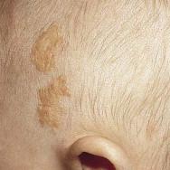
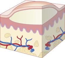
To the untrained eye, the appreciation of subtle variations in color is often the most difficult concept to grasp. Fortunately, this assessment does not carry the diagnostic weight of the primary or secondary lesions. When evaluating the color, one must take into account the background pigmentation of the patient. In infants with darker skin type, subtle erythema or jaundice may be difficult to appreciate. Likewise, pigment dilution is more difficult to evaluate in lighter skin. The most prominent colors seen in cutaneous pathologic processes are described below.

Red

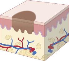
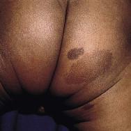
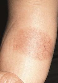
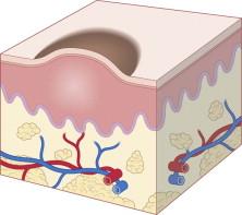
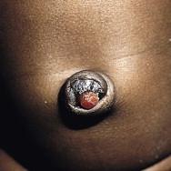
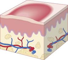
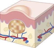
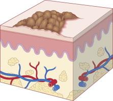
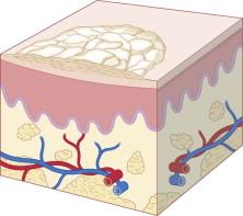
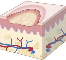
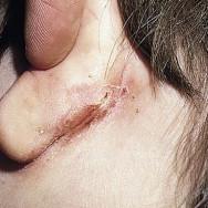
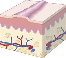
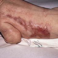
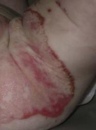
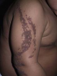
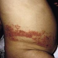
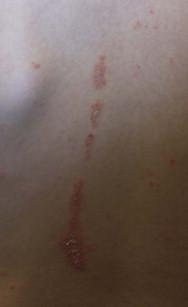
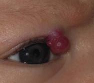
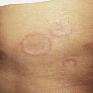
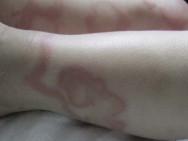
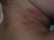
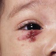
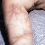
Red color can be the result of vasodilation or hyperemia caused by inflammation. Deeper red or purple hues suggest extravasation of red blood cells. Diascopy is a diagnostic maneuver to help differentiate these possibilities. By applying pressure to the lesion, one can see whether the lesion blanches, which suggests rubor due to vasodilation or inflammation. Conversely, nonblanching lesions suggest vascular damage, with consequent extravasation of blood into the dermis.

White

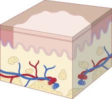
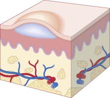
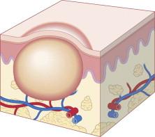
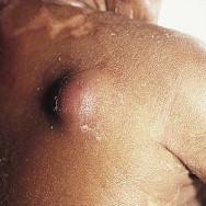
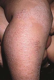
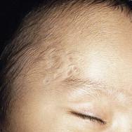
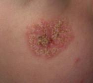
White color can be the result of loss of pigment within the epidermis or the accumulation of white material such as purulent exudate or keratinous material. One should not use the term white to describe skin-colored lesions.

You're Reading a Preview

Become a Clinical Tree membership for Full access and enjoy Unlimited articles

Become membership

If you are a member. Log in here