Physical Address
304 North Cardinal St.
Dorchester Center, MA 02124

Patients with traumatic injuries can be placed into one of three major groups. The imaging approach will differ between these groups.
Imaging:
Strict local protocols and algorithms utilising early ultrasound (US) and/or multidetector computed tomography (CT). The use of plain film radiology in the Emergency Department (ED) is generally limited .
Imaging:
Plain film radiology is utilised in the ED.
Imaging:
Plain film radiology is the principal imaging investigation.
This book describes the assessment and interpretation of the plain radiographs that are customarily obtained in patients who have not sustained a life threatening injury.
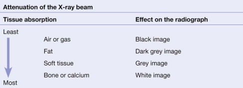
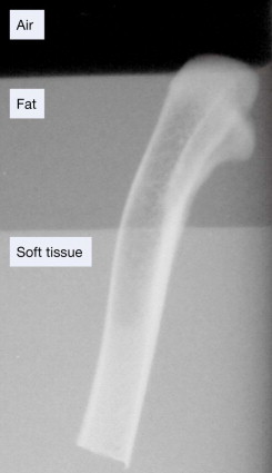
The tissues that lie in the path of the X-ray beam absorb (ie attenuate) X-rays to differing degrees. These differences account for the radiographic image.
When a fracture results in separation of bone fragments, the X-ray beam that passes through the gap is not absorbed by bone. This results in a black (ie lucent) line on the radiograph.
On the other hand, bone fragments may overlap or impact into each other. The resultant increased thickness of bone absorbs more of the X-ray beam and so results in a white (ie sclerotic or denser) area on the radiograph.
There are radiological soft tissue signs which can provide a clue that a fracture is likely. These include displacement of the elbow fat pads (see pp. 97 and 102 ), or the presence of a fat–fluid level at the knee joint (see pp. 248–249 ).
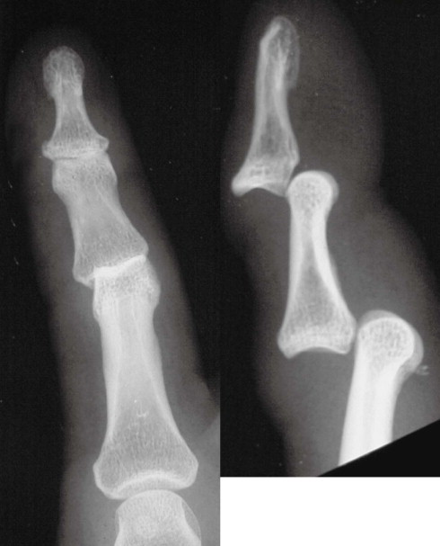
‘One view only is one view too few’
Many fractures and dislocations are not detectable on a single view. Consequently, it is normal practice to obtain two standard projections, usually at right angles to each other. The example below shows two views of an injured finger.
At sites where fractures are known to be exceptionally difficult to detect (for example a suspected scaphoid fracture), it is routine practice to obtain more than two views.
Become a Clinical Tree membership for Full access and enjoy Unlimited articles
If you are a member. Log in here