Infections of histological importance


Introduction

A number of infections may be diagnosed from the characteristic appearances seen in biopsy specimens. In some cases, the diagnosis may first be suspected when a biopsy is examined by light microscopy. In other cases, the clinician suspects an infective agent and seeks confirmation by biopsy as well as by other means. Although histological appearances are often characteristic, it is usually necessary to confirm the presence of the infective agent by other techniques such as microbiological cultures or serology. In addition, the pathologist may employ a wide variety of techniques such as electron microscopy, immunohistochemical staining with specific monoclonal antibodies and a range of special stains to confirm the diagnosis.

Many infections that are otherwise difficult to diagnose arise in immunosuppressed patients, for example patients with HIV/AIDS and organ transplant recipients and, as the number of such patients increases, so the role of histopathology in diagnosis of infection expands. This chapter aims to give an overview of infections that are important in routine diagnostic pathological practice, to illustrate the appearances of these organisms in the tissues and to consider the patterns of tissue damage they cause. As prion disease specifically affects the central nervous system, this is discussed in Ch. 23 .

Bacterial infections

Most bacteria cause disease by exciting an acute exudative inflammatory response (pyogenic bacteria) . This inflammatory exudate is responsible for many of the clinical features of the disease (e.g. lobar pneumonia, see Fig. 3.3 ; bronchopneumonia, see Fig. 12.7 ). The pattern of tissue damage is similar irrespective of the pyogenic bacteria causing it and the organism can only be identified by microbiological methods. More complex mechanisms are involved in the tissue damage caused by infection with bacteria such as Helicobacter pylori , which cause inflammation by interfering with the normal physiological regulation of gastric acid secretion (see Figs 4.2 and 13.9 ). Other bacteria cause disease by producing toxins, which induce necrosis of cells and tissues (e.g. Clostridium difficile toxins destroy the surface epithelium of the colon ( E-Fig. 5.1 G ) ( Fig. 5.1 ), and some bacteria initiate a type IV hypersensitivity reaction (e.g. Mycobacteria and some Treponema organisms) and so produce characteristic changes.

Fig. 5.1, Pseudomembranous colitis.

Key to Figures

A Actinomyces C colonic crypts E inflammatory exudate FB filamentous bacteria P pus

Fig. 5.2, Actinomyces organisms.

Mycobacterial infections

Infections caused by various Mycobacteria can frequently be diagnosed histologically because of the tissue reaction to their presence. The organisms are difficult and slow to grow in microbiological culture and therefore biopsy often plays an important role in early diagnosis. Mycobacterium tuberculosis causes tuberculosis , a disease that is increasing in incidence because of the emergence of drug-resistant strains. Mycobacterium leprae is the cause of leprosy ( Fig. 5.9 ). There are other pathogenic Mycobacteria (called atypical Mycobacteria ) that cause a range of disorders.

Important points to note about the histology of mycobacterial infections are:

  • Most show a granulomatous pattern of chronic inflammation (see Ch. 4 ) due to a delayed type hypersensitivity reaction.

  • Caseous necrosis ( Fig. 5.3 ) is particularly associated with M. tuberculosis infection.

  • Suppurating granulomas (with neutrophils in the central necrotic area of the granuloma) may occur in infections by atypical Mycobacteria ( Fig. 5.8 ). These organisms are also referred to as Mycobacteria other than tuberculosis (MOTT) or non-tuberculous Mycobacteria (NTM).

  • The causative organism can sometimes be identified in histological sections by the use of special stains (Ziehl–Neelsen, Wade–Fite) as shown in Appendix 1 .

Key to Figures

CN caseous necrosis F fibroblasts L Langhans’ giant cell M epithelioid macrophages W fibrous wall

Fig. 5.3, Early pulmonary tuberculosis. (A) Early tubercle (MP); (B) early tubercle (HP); (C) later tubercle (MP); (D) later tubercle (HP).

Fig. 5.4, Fibrocaseous tuberculous nodule (LP).

Fig. 5.5, Tuberculous lymph node (LP).

Some Other Aspects of Tuberculosis

In all cases of suspected tuberculosis, material should be submitted for culture. Culture of M. tuberculosis is difficult and time-consuming and may take 6 weeks or more. When the organisms have been cultured successfully, this allows assessment of antimicrobial resistance and sensitivity. It is often necessary to begin treatment empirically whilst awaiting microbiological confirmation and so initial histological findings supportive of this diagnosis can be very valuable. Histological identification of the organisms using special staining techniques is dependent upon the nature of the mycobacterial cell wall. This lipid-rich coating allows the organisms to retain certain dyes, even upon heating with acid and alcohol solutions, and therefore mycobacteria are often called acid and alcohol fast bacilli (see Fig. 5.10 ). Another name for M. tuberculosis which is sometimes used in clinical practice is the eponymous designation Koch’s bacillus .

Key to Figures

B bronchiole Bo bony trabeculae C caseous necrosis D destroyed wall G granuloma L Langhans’ giant cell T tubercle

Fig. 5.6, Tuberculous bronchopneumonia (MP).

Fig. 5.7
Disseminated tuberculosis. (A) Liver (MP); (B) kidney (MP); (C) bone (MP); (D) tuberculous meningitis (MP).
If a ruptured tuberculous lymph node (or a rapidly enlarging focus of post-primary tuberculosis) erodes a blood vessel wall, masses of mycobacteria are discharged into the circulation and lodge in the microvasculature. When the eroded vessel is a branch of the pulmonary artery, the organisms pass to other areas of the lung, but when a pulmonary venous tributary is involved, they are spread in the systemic circulation to many organs, notably the liver, kidney and spleen. In this way, vast numbers of new tubercles may be produced throughout the body. Such multiple lesions rarely attain any great size because this occurrence usually produces rapid clinical deterioration and death. Because the gross appearance of individual lesions resembles millet seeds, this condition is known as miliary tuberculosis .
Fig. 5.7A shows several miliary tubercles (T) in the liver ( E-Fig. 5.4 H ). One of the tubercles exhibits Langhans’ giant cells (L) and the larger tubercle shows early central caseous necrosis.
It seems that a relatively small number of organisms can be disseminated by the bloodstream to various organs without causing overt disease, only to become reactivated at a later date when the host’s immune status is impaired. These organisms remain viable but quiescent and active tuberculosis may then reappear in tissues remote from the original lesion many years later. This phenomenon is known as metastatic or isolated organ tuberculosis and most commonly involves the kidneys, adrenals, meninges, bone, Fallopian tubes, endometrium and epididymis.
Fig. 5.7B illustrates renal involvement ( E-Fig. 5.5 H ) with the formation of small tubercles (T) in the renal cortex. Continuation of this process results in destruction of much of the renal cortex and medulla, with eventual rupture of large confluent tubercles into the pelvicalyceal system, which becomes distended with caseous material. This condition is known as tuberculous pyonephrosis . In more advanced cases, infection spreads to involve the ureter and bladder. Renal tuberculosis is frequently bilateral and may result in renal failure.
Bone tuberculosis (tuberculous osteomyelitis) most frequently affects the long bones, associated joints and the vertebrae (Pott’s disease) . In long bones, infection may produce a localised, painful swelling, which may drain to the skin to form a chronic sinus. Joint involvement (tuberculous arthritis) is most common in children and often affects the hips or joints associated with the vertebrae (tuberculous spondylitis) as part of Pott’s disease of the spine. As in other tissues, Fig. 5.7C shows the characteristic caseating granulomas (G), which cause progressive destruction of the bony trabeculae (Bo) . The infection tends to spread extensively in the cancellous medullary bone, leading to necrosis of surrounding cortical bone ( E-Fig. 5.6 H ).
Tuberculous meningitis is an uncommon but frequently fatal complication of tuberculosis. Most often, it affects the meninges around the base of the brain and the spinal cord. As shown in Fig. 5.7D , tuberculous granulomas (G) , with characteristic central areas of caseation, develop in the leptomeninges and adjacent brain tissue ( E-Fig. 5.7 H ) where they may damage cranial and spinal nerves. The presence of numerous lymphocytes in CSF obtained from lumbar puncture is useful in distinguishing tuberculous meningitis from purulent (bacterial) meningitis. In the latter, neutrophils are the major cell type present.

Fig. 5.8
Atypical mycobacterial infection involving lymph node (MP).
Infection with non-tuberculous mycobacteria (also known as atypical mycobacteria or mycobacteria other than tuberculosis, MOTT) is not uncommon in the cervical lymph nodes of young children. Another group at risk from these organisms are patients with AIDS who, because of severe immunosuppression, commonly develop widespread infections involving the liver, lymph nodes and spleen. In these patients, there is usually a deficient immune response and the histological appearance may simply be of aggregates of epithelioid cells or even foamy macrophages in the tissue with no lymphocytic component.
Fig. 5.8 shows a lymph node from an otherwise healthy child with MOTT infection. The lymphoid tissue is replaced with a granulomatous inflammatory response, consisting of confluent sheets of epithelioid macrophages (E) . In contrast to M. tuberculosis , the granulomas in MOTT infection show suppurative necrosis with aggregates of neutrophils (N) , rather than caseous necrosis. As in this example, giant cells may be sparse.

Key to Figures

C carbon pigment DN dermal nerve E epithelioid macrophages G granuloma N neutrophils

Fig. 5.9
Leprosy. (A) Dermal infiltration (LP); (B) nerve involvement (HP).
Leprosy is a disease caused by infection with M. leprae . The tissue reaction to the bacillus depends on the immune response of the infected person. In the tuberculoid form of the disease, there is an active cell-mediated immune response and granulomas are formed that are similar to those seen in tuberculosis, but without evidence of caseation. In the lepromatous form, there is no effective cell-mediated immune response and tissues are infiltrated with macrophages colonised by large numbers of bacteria. Intermediate forms of leprosy exist with both tuberculoid and lepromatous features.
Clinically, people with the lepromatous form of the disease have nodular dermal and subcutaneous deposits of macrophages filled with bacteria and lipid. The disease affects the face, ears, arms, knees and buttocks, as the leprosy bacilli require cooler areas of the body for proliferation. In contrast, the tuberculoid form of the disease gives rise to macular or plaque-like skin lesions and, in addition, causes extensive inflammatory destruction of peripheral nerves. This typically gives rise to anaesthesia (loss of sensation) in the limbs, which then become prone to damage through repeated non-perceived injury.
Fig. 5.9A is a micrograph of the skin ( E-Fig. 5.8 H ) of a person with tuberculoid leprosy. Histiocytic granulomas (G) with a heavy surrounding lymphocytic infiltrate are present at all layers throughout the dermis, but particularly in relation to small nerves. This is illustrated at higher magnification in Fig. 5.9B . Here, a small dermal nerve (DN) is shown surrounded by a cuff of lymphocytes. There is an associated granuloma (G) .

Fig. 5.10
Special staining for mycobacteria. (A) Ziehl–Neelsen stain in tuberculosis (HP); (B) Wade–Fite stain in lepromatous leprosy (HP).
M. tuberculosis and atypical mycobacteria are not visible on routine H&E stains. To demonstrate the presence of mycobacteria in tissue, the Ziehl–Neelsen (ZN) staining method is employed. The stain is taken up by the cell walls of the mycobacteria and remains despite treatment with acid and alcohol. This is the origin of the term acid and alcohol fast bacilli , which is used in describing mycobacteria. In a very high-power micrograph ( Fig. 5.10A ), scattered magenta-coloured, rod-shaped organisms can be seen in the lung of an immunocompromised patient. Some carbon pigment (C) can be identified within alveolar macrophages. In an immunocompetent individual, these organisms may be very sparse and difficult to find, but even a single organism in the appropriate background of a caseating granuloma is diagnostic. In immunosuppressed individuals (as shown here) and also in MOTT infection, the organisms are often much more numerous.
The micrograph of lepromatous leprosy ( Fig. 5.10B ) shows the Wade–Fite stain, which is a modified version of the ZN stain. Again, it stains the organisms red. The similarity in appearance between M. tuberculosis and M. leprae can be readily appreciated.

Spirochaete infections

The major form of spirochaete infection worldwide is syphilis , due to Treponema pallidum . Yaws and pinta are further important treponemal infections, but these are rare except in specific tropical regions. Although now relatively uncommon, late-stage syphilis is still regarded as one of the classical examples of specific chronic inflammation. The infecting organism, the spiral-shaped spirochaete T. pallidum , resists usual tissue defences and excites a progression of fascinating pathological and clinical phenomena, which represent typical chronic inflammatory responses with superimposed hypersensitivity reactions mounted by the immune system. The condition usually proceeds through three stages over a long period.

In brief, the organism usually gains access to the body by penetrating the genital mucosa where it produces a single, small primary lesion known as a chancre . The chancre is a raised, reddened nodule caused by an intense local accumulation of plasma cells and lymphocytes in the subepithelial connective tissue. The chancre may ulcerate at this stage, but it is often painless and may easily pass unnoticed. By the time the chancre has developed, the organism has multiplied extensively and has been disseminated to regional lymph nodes and thence into the bloodstream, causing a generalised bacteraemia. The chancre and concomitant bacteraemia (primary syphilis) are followed some weeks or months later by a transient secondary stage . This is characterised by a widespread, variable skin rash, often with moist, warty, genital lesions and ulceration of the oral mucosa. These various mucosal lesions are histologically similar to the primary chancre and are full of treponemal organisms. The disease is now at its most contagious, yet the patient usually feels well and the only other evidence of a generalised infection is a widespread lymphadenopathy and positive serological findings.

In most untreated cases, the infection is effectively resolved by body defences and, in many of these patients, even serological evidence of previous infection disappears. Unfortunately, a proportion of untreated cases proceed from the secondary stage to develop tertiary syphilis , after a variable interval from one to many years. The lesions of tertiary syphilis may be either focal or diffuse and it is the focal lesion of tertiary syphilis, known as the gumma , which exhibits many of the features of a granulomatous inflammation. Tertiary lesions may occur in almost any organ or tissue and the clinical consequences vary enormously. The diffuse form of tertiary syphilis most notably involves the cardiovascular system, particularly the ascending aorta and, less commonly, the central nervous system; the well-known tabes dorsalis and general paresis of the insane (GPI) are two of the manifestations of neurosyphilis. In the focal form of tertiary syphilis, gummas may develop in the liver, bone, testes and other sites. The clinical outcome depends upon the nature and extent of local tissue destruction.

Fig. 5.11, Syphilis. (A) Syphilitic gumma of the liver (LP); (B) syphilitic aortitis (MP).

Viral infections

Viruses cause disease in three main ways:

  • By causing death of the cell they infect, either by a direct effect or by modifying the genome such that the host cell is recognised as foreign and is destroyed by the host immune system.

  • By causing excessive proliferation of the infected cell line, which may result in the development of malignant tumours. Human papillomavirus (HPV) is particularly important in this respect.

  • By integrating themselves in the cell nucleus where they produce latent infection.

Although viral culture and the demonstration of rising titres of antibodies against the virus remain the mainstays of diagnosis, some viral infections can be diagnosed by characteristic histological appearances. Individual viruses are too small to be seen by light microscopy, but when congregating together in enormous numbers within the host cell, they are visible as viral inclusion bodies and may be either intranuclear, intracytoplasmic or both. Inclusion bodies provide a histological clue to the causative virus and this can be confirmed by electron microscopy and immunohistochemistry.

One viral infection of particular importance is the human immunodeficiency virus (HIV),which is the cause of the acquired immune deficiency syndrome (AIDS). As well as producing certain characteristic histological changes, particularly affecting lymph nodes, HIV compromises the host immune system and so a range of other unusual infections may result. HIV-1 is a lymphotropic virus that gains access to cells by way of the CD4 surface protein, normally found on T helper cells as well as on most monocytes and other macrophages. Infection with HIV-1 is associated with several clinical and pathological syndromes. Some patients develop fever, weight loss, diarrhoea and generalised lymph node enlargement (lymphadenopathy) in which there is generalised follicular hyperplasia. In patients with the full-blown immunodeficiency state of AIDS, lymph nodes show loss of follicles, lymphocyte depletion, vascular proliferation and fibrosis.

Key to Figures

E elastin I inclusion body L lymphocytes around vessels N necrosis S syncytial cells V vesicle

The two main consequences of the immunodeficiency state seen in AIDS are:

  • Predisposition to opportunistic infections, particularly

  • Predisposition to certain tumours, particularly

    • Kaposi’s sarcoma ( Fig. 11.11 )

    • Non-Hodgkin’s lymphoma, especially diffuse large B-cell lymphoma (see Ch. 16 ).

Clinical Features of Herpes Virus Infection

Herpes simplex virus types 1 and 2 (HSV-1 and HSV-2) cause the common skin eruptions known as cold sores and genital herpes, whilst the chicken pox virus (Herpes zoster) is responsible for shingles . In immunocompetent individuals, Herpes viruses may remain latent in nerve cells with intermittent episodes of reactivation when the virus replicates within and destroys either epidermal cells or the epithelial cells of mucous membranes. Clinically, this gives rise to the typical appearance of groups of vesicles on an erythematous background. Most lesions heal spontaneously although the latent viral DNA remains in the nerve cells and can emerge in subsequent clinical episodes. In immunosuppressed patients, such as those with AIDS or those on immunosuppressive treatment, herpetic infection may result in widespread skin or visceral infections, rather than the self-limiting rash seen in the immunocompetent.

Fig. 5.12, Herpes virus infection. (A) Skin (LP); (B) oesophagus (HP).

Fig. 5.13, Cytomegalovirus infection (HP).

Fig. 5.14, Human papillomavirus (HP).

Fungal infections

Fungal infections range from minor localised skin infections to life-threatening systemic diseases in immunosuppressed patients. The inflammatory reaction in fungal infections may have one of three patterns. The classical appearance is of a granulomatous inflammation, which may exhibit central suppurative necrosis. A second pattern is of acute inflammation with an infiltrate consisting primarily of neutrophils. This pattern is seen in Candida infection of the oesophagus. The third is a very minimal inflammatory response, as in superficial infections of the skin by dermatophytic fungi.

Fungi are not usually obvious on routine H&E staining, but the thick cell walls are highlighted by special stains such as periodic–acid Schiff (PAS) or silver stains. Some fungi are easily recognised histologically because of the characteristic shape and structure of the hyphae and the pattern of budding of the yeast forms. Despite this, culture techniques are preferable for definitive identification of fungal species. An important diagnostic point is that the presence of fungal yeast forms, such as Candida at a mucosal surface or on the skin, does not necessarily indicate active infection, as these agents are common commensals. Evidence of active invasion must be demonstrated and, in most cases, there is an appropriate inflammatory response.

Clinical Aspects of Fungal Infection

Many superficial forms of fungal infection are very indolent and slow growing and, as a result, treatment with anti-fungal drugs may need to extend over a prolonged period. It may be important to identify the precise type of fungus that is causing infection in order to ensure that the organism is sensitive to the form of treatment chosen. As fungi tend to have different growth requirements from bacteria, the microbiology laboratory must be informed if fungal infection is suspected so that appropriate cultures can be undertaken.

Key to Figures

B binucleate cell D dyskeratotic cell G giant cell H hyphae I inclusion body K koilocyte ND necrotic debris Y yeast forms

Fig. 5.15, Candidiasis. (A) Oral candidiasis (PAS) (HP); (B) oesophageal candidiasis (PAS) (HP); (C) myocardial candidiasis (PAS) (HP).

Key to Figures

A Aspergillus hyphae E exudate F fruiting bodies H hyphae I inflammatory cells L lymphocytes N necrotic tissue S septa

Other Fungal Infections – Zygomycosis

Zygomycosis (also termed mucormycosis ) is a form of opportunistic fungal infection caused by one of a range of environmental fungi of the class Zygomycetes . These organisms pose no threat to immunocompetent individuals but can cause very serious and often life-threatening infections in those who are immunocompromised, particularly in patients with diabetes in whom the organisms may infect the nose and paranasal sinuses, invading from this site to involve the orbits and cerebrum (rhinocerebral mucormycosis) . In cases of mucormycosis, the hyphae are broad and irregular, branching at near right angles (in contrast to the acute angles seen in Aspergillus infection). Also, the hyphae typically appear open and non-septate in routine preparations. Often, the fungi invade into artery walls and may then give rise to thrombosis and tissue necrosis.

Fig. 5.16, Aspergillus . (A) MP; (B) HP; (C) HP; (D) silver stain (HP); (E) Aspergillus niger (MP).

Fig. 5.17
Pneumocystis in the lung. (A) H&E (MP); (B) silver stain (HP).
The organism Pneumocystis jirovecii (formerly known as Pneumocystis carinii ) was previously classified as a protozoan, but more recently it has become clear that it is a member of the fungal family. It is a ubiquitous organism and can be demonstrated in the lungs of most normal individuals where it causes no disease. Pneumocystis has, however, been brought into the spotlight by AIDS, although it may infect patients with immunosuppression from other causes. In patients with AIDS, Pneumocystis causes a diffuse patchy pneumonia, which may be the presenting feature of full-blown AIDS and is often fatal. Diagnosis may be difficult and transbronchial lung biopsy is often required. With routine H&E staining, as in Fig. 5.17A , the organisms may not be apparent ( E-Fig. 5.11 H ).
The alveoli are often filled by a foamy, acellular exudate (E) with an interstitial infiltrate of lymphocytes (L) in the alveolar wall. The inflammatory infiltrate may be minimal or more severe, showing features of diffuse alveolar damage (see Fig. 12.12 ) with hyaline membranes, capillary dilatation and exudation of red cells. Demonstration of the organisms requires a silver stain as shown in Fig. 5.17B . The organisms are cup-shaped and measure 4–6 μm.
Giemsa and toluidine blue stains may also be used. The above stains may also be carried out on sputum samples and bronchial washings. Patients with AIDS frequently have concurrent infections with other organisms such as cytomegalovirus ( Fig. 5.13 ).

Fig. 5.18
Cryptococcus. (A) Brain H&E (MP); (B) alcian blue stain (HP); (C) mucicarmine stain (HP).
Cryptococcus neoformans is another yeast that causes serious infections in many tissues, mainly in immunosuppressed individuals such as patients with AIDS, haematological malignancy and transplant recipients. Occasionally, however, it may cause meningitis, meningoencephalitis or lung infection in an otherwise well individual.
Fig. 5.18A shows the typical appearance of Cryptococcus in the brain. The organisms are seen forming a cyst in a Virchow–Robin perivascular space. These lesions are known as ‘soap bubble’ lesions. The cryptococci (C) have a thick surrounding capsule that appears as a clear space. This capsule is an important virulence factor as it inhibits phagocytosis of the organisms and reduces normal leukocyte migration. Typically, there is a minimal inflammatory response, which may be due to the immunosuppressed state of the patient. However, in chronic infections in non-immunosuppressed individuals, the organisms may incite a granulomatous response.
Techniques employed in demonstrating cryptococci are highly dependent upon the thick capsule of the organisms. One simple method uses India ink to create a negative image, showing the organism with a clear capsular halo against a dark background.
Figs 5.18B and C show other special staining techniques that are useful in highlighting cryptococci. The organism’s thick polysaccharide capsule (Cap) can be stained using Alcian blue as illustrated in Fig. 5.18B . Here, the nuclei of surrounding cells are counter-stained red. Fig. 5.18C shows brain tissue stained using the mucicarmine method. The cryptococci organisms are surrounded by a thick, magenta-coloured capsule (Cap) .
In the lung, Cryptococcus may cause a diffuse infection or it may form a solitary mass lesion in previously healthy individuals. Focal lesions such as this may lead to suspicion of other pathologies, such as bronchial carcinoma. In this case, the lesion is actually a large granuloma with a jelly-like centre, formed by a mass of organisms surrounded by a thick layer of macrophages, lymphocytes and giant cells.

Protozoa and helminths

Of the wide range of protozoa that are of pathological importance, only a few are usually diagnosed histologically. Many of these organisms are major causes of morbidity and mortality in particular geographic regions in the developing world. Increasingly, such infections may be seen elsewhere as a result of more widespread travel and migration. Protozoa are unicellular organisms that may reproduce asexually or sexually. Many have a complex life cycle involving one or more animal hosts. Giardia lamblia is a common protozoan infecting the small intestine in communities worldwide ( Fig. 5.19 ). Trichomonas vaginalis is another protozoan parasite that is a common cause of female genital tract infection ( Fig. 5.20 ). This organism is frequently encountered in cervical cytology screening. Malaria and amoebiasis are also described in this chapter ( Figs 5.21 and 5.22 ). A wide variety of helminths infect humans, many primarily infecting the gut, although they may pass through other anatomical sites en route. Some helminths, such as the nematodes Enterobius vermicularis and Toxocara canis , are found worldwide, while others, such as Wuchereria bancrofti , have a more limited tropical distribution. Enterobiasis is fairly commonly seen in routine practice ( Fig. 5.24 ) and Schistosomiasis is of particular histological importance ( Fig. 5.23 ). These infections are illustrated.

Table 5.1
Chapter review.
Disorder Main features Figure
Bacterial infections
Clostridium difficile
E-Fig.5.1 G
Pseudomembranous colitis with focal epithelial necrosis due to toxin. ‘Volcano’ pattern with inflammatory exudate 5.1
Actinomyces Filamentous bacteria forming ‘sulphur granule’ colonies with encrusted proteinaceous material 5.2
Mycobacterium tuberculosis
E-Fig. 5.3 G
Granulomatous inflammation with caseation and Langhans’ giant cells Ghon complex. May progress to miliary disease if immunosuppressed 5.3–5.7,
5.10A
Atypical mycobacteria (MOTT, NTM) Often in immunocompromised patients. Suppurative granulomas 5.8
Mycobacterium leprae Pattern depends upon immune status (tuberculoid/lepromatous). Granulomatous inflammation of skin and dermal nerves 5.9, 5.10B
Treponema pallidum Primary, secondary and tertiary syphilis. Chancre, gumma, endarteritis obliterans 5.11
Viral infections
Herpes simplex virus Types 1 & 2. Oral and genital herpes. Disseminated in immunosuppressed. Vesiculation with syncytial giant cells and intranuclear inclusions 5.12
Varicella zoster virus Cause of chickenpox and shingles. Morphology similar to HSV Like 5.12
Cytomegalovirus Non-specific symptoms normally. Disseminated in immunosuppressed. Very large cells with intranuclear and intracytoplasmic inclusions 5.13
Human papillomavirus Many types, causes common wart. Oncogenic forms cause cervical cancer Koilocytosis, binucleation, dyskeratosis and dysplasia (CIN) 5.14, 17.6, 17.7
Fungal infections
Candida Common, often affects mucous membranes. Typically neutrophil response. Yeast forms and branching pseudohyphae 5.15
Aspergillus Mostly lung disease. Allergic, colonising and invasive forms Narrowing septate hyphae with branching at acute angles 5.16
Zygomyces Mucormycosis. Typically sinonasal disease in diabetic patients.
Invades vessels. Broad and non-septate hyphae with right-angle branching
Pneumocystis Environmental fungus, pathogenic in immunocompromised, e.g. AIDS. Foamy exudate in alveoli, cup-shaped organisms on silver stains 5.17
Cryptococcus Mostly in immunocompromised patients. Can cause lung disease in normal people. Yeasts with thick outer capsule 5.18
Protozoa and helminths
Giardia lamblia Cause of infective diarrhoea. Protozoan parasite infecting small intestine. Small sickle-shaped organisms on mucosal surface 5.19
Trichomonas vaginalis Common cause of vaginal discharge and irritation, often seen in cervical smear tests. Pear-shaped protozoan parasites with flagellae 5.20
Plasmodium sp. Cause of malaria. Intracellular parasites with complex life cycle, spread by mosquito. Pigmented organisms in erythrocytes, vascular complications 5.21
Entamoeba histolytica Very common cause of dysentery worldwide. Invades colorectal mucosa. Flask-shaped ulcers and organisms larger than macrophages, PAS positive 5.22
Schistosoma sp. Important worldwide. Complex lifecycle involving snail.
Eggs lodge in various tissues and cause granulomatous inflammation
5.23
Enterobius vermicularis Threadworm or pinworm. Common, colonises ileocolic area. Worms do not invade. Eggs cause perianal irritation. Often seen in appendix 5.24

Key to Figures

C Cryptococci Cap capsule E erythrocytes G Giardia O Trichomonas organism S squamous cell V villus

Fig. 5.19, Giardiasis (HP).

Fig. 5.20
Trichomonas vaginalis (Papanicolaou stain) (HP).
This flagellated protozoan parasite commonly infects the female genital tract, presenting with itching and a frothy greenish vaginal discharge. The organism is motile and can be demonstrated in wet preparations but is most commonly noted at the time of examination of a routine cervical smear test (Pap test). In Fig. 5.20 , the organisms (O) are pear-shaped or ovoid and are somewhat smaller than the surrounding squamous epithelial cells (S) . The organisms stain a smudgy grey-green colour. Indistinct nuclei and red cytoplasmic granules may be seen. Flagellae are not usually identifiable on routine preparations. The surrounding epithelial cells may show reactive changes and scattered neutrophil polymorphs may be present.

Fig. 5.21
Cerebral malaria (HP).
Malaria, which is caused by four species of the protozoan Plasmodium , is common in the tropics and subtropics and is a major cause of mortality. After entering the blood via the proboscis of a mosquito, the merozoites enter erythrocytes. Further cycles of division occur within the erythrocytes, which rupture, allowing further cycles of infection. The sexual phase of the life cycle occurs within the mosquito. Plasmodium falciparum is the most virulent form of malaria, with cerebral involvement being a major cause of death. The cerebral tissue is characteristically congested, as shown in Fig. 5.21 , where a dilated small blood vessel is packed with erythrocytes within which the malarial parasites can be seen as dark brown dots. Falciparum malaria has the property of causing erythrocytes to adhere to endothelium, thus obstructing blood flow. The congested vessels often rupture to cause ‘ring haemorrhages’ (not shown here). Without specific treatment, cerebral malaria is almost always fatal and the increasing incidence of drug resistance makes it difficult to treat. Malarial parasites can be seen within erythrocytes in any other tissues and the diagnosis is usually made by examination of a blood smear. The different species can be identified by their characteristic appearance. The other species that cause malaria, namely P. vivax, P. ovale and P. malariae , cause a much milder illness that may be recurrent. P. falciparum may cause severe anaemia, pulmonary oedema, renal failure, shock, hypoglycaemia and cerebral disease.

Fig. 5.22
Amoebiasis (HP).
Infections with Entamoeba histolytica occur worldwide. The organism is restricted to humans and it is estimated that about 10% of the world’s population may carry the organism in the colon. The most common form of infection is amoebic dysentery , where the Entamoebae invade the mucosa of the colon and rectum, causing painful, bloody diarrhoea. Pathologically, the mucosa is extensively undermined, producing typical flask-shaped ulcers, which may perforate ( E-Fig. 5.2 H ). Fig. 5.22 shows organisms (A) adherent to colonic mucosa (M) . The Entamoebae are slightly larger than a macrophage and characteristically phagocytose erythrocytes (visible within the organisms). The organisms may invade and obstruct colonic arteries, causing superimposed ischaemic necrosis. The organism may spread to the liver, causing an amoebic abscess , and thence to the lung or pleural, peritoneal or pericardial cavities. Venereal infections of the cervix and penis also occasionally occur.

Key to Figures

A amoebae AP alar projection C cuticle E epithelioid cells G giant cell L longitudinal section through worm M mucosa SE schistosome eggs W transverse section adult worm

Fig. 5.23
Schistosomiasis. (A) MP; (B) HP.
Schistosomiasis is a systemic parasitic infection caused by the organism Schistosoma , a genus of trematode worms (flukes) . Three species are of pathological importance. Their life cycle includes water snails (the intermediate host), with humans (the definitive host) becoming infected by bathing or working in water containing the larvae or cercaria released from snails. The cercaria penetrate the skin, in the process converting to schistosomules , and then migrate via the venous system to the pulmonary vessels where they mature for 4 weeks before entering the systemic circulation. From here, they migrate to the hepatic branches of the portal vein ( Schistosoma mansoni and Schistosoma japonicum ) or the pelvic veins ( Schistosoma haematobium ) where they mature and may persist for several years or even more. In their chosen location, the adult worms mate with the females, producing up to 3000 eggs per day. Some eggs leave the body in urine or faeces and, on reaching still or gently flowing fresh water, hatch into a ciliated form called miracidia and thus reach their intermediate snail hosts. Other eggs lodge in the tissues ( S. mansoni and S. japonicum in the small and large intestine and thence to the liver; S. haematobium in the bladder and rectum), exciting a florid granulomatous reaction progressing to extensive fibrosis. In the liver, there is severe fibrosis of the portal tracts where the eggs lodge. Schistosomiasis is the major cause of portal hypertension worldwide. S. haematobium causes a similar reaction in the bladder, with a number of manifestations including papillomas, ulcers and bladder contractures. Chronic inflammation predisposes to dysplasia and malignancy. Eggs may also be found at many other sites such as the lungs and CNS.
Fig. 5.23A shows a low-power view of schistosome eggs (SE) in the small intestine. Unusually, part of the adult worm (W) is seen. Fig. 5.23B shows two schistosome eggs (SE) at high power. The groups of eggs are surrounded by epithelioid cells (E) , giant cells (G) and eosinophils. The eggs of each species can be identified by their size and the position of the spine, which in this case is terminally located and therefore likely to come from S. haematobium .

Fig. 5.24
Enterobius vermicularis in appendix. (A) LP; (B) MP; (C) HP.
Enterobius vermicularis is also known as pinworm or threadworm. The organism is spread by the faecal-oral route and is commonly present within the lumen of the bowel, particularly in children. The organisms do not invade the tissues. The female worm lays eggs in the peri-anal area and symptoms, if any, tend to be of perineal irritation. The adult worms inhabit the ileocaecal area and are most commonly identified in the appendix.
Fig. 5.24A shows the tip of the appendix, sectioned longitudinally ( E-Fig. 5.12 H ). Adult worms are visible within the lumen, one shown in longitudinal section (L) . Note the normal reactive lymphoid tissue within the wall of the appendix. Typically, there is no tissue reaction associated with the presence of the parasites and the normal appendiceal mucosa with its associated lymphoid tissue can been seen at higher magnification in Fig. 5.24B .
Fig. 5.24C shows a transverse section through an adult worm, lying within the lumen of the appendix. Again, note that there is no invasion of the mucosa (M) . The thick cuticle (C) of the worm has a typical alar projection (AP) on each side and the oesophagus and other internal structures of the worm can also be seen.

E-Fig. 5.1 G
Pseudomembranous colitis. F/68. Colectomy was performed because the patient developed toxic megacolon. She was taking ampicillin and this allowed overgrowth of the Clostridium difficile which produced the multiple, discrete white plaques of purulent exudate on the mucosal surface.

Reproduced from Cooke, R., Stewart, B., Colour Atlas of Anatomical Pathology, 3rd edition. Copyright 2004, Elsevier Ltd.

E-Fig. 5.2 H
Colorectal type absorptive/protective mucosa, H&E (MP).
Four basic mucosal types are found lining the gastrointestinal tract and these can be classified according to their main function: Absorptive/protective. This form lines the entire large intestine and is shown in micrograph. The mucosa is arranged into closely packed, straight tubular glands consisting of cells specialised for water absorption, as well as mucus-secreting goblet cells to lubricate the passage of faeces.

Reproduced from Young, B., O’Dowd, G., Woodford, P., Wheater’s Functional Histology, 6th edition. Copyright 2014, Elsevier Ltd.

E-Fig. 5.3 G
Vertical section of the left lung and mediastinum. This shows multiple creamy nodules – the military tubercles, which were present throughout both lungs. There is also a large, round white focus of tuberculous granulation tissue in the left upper lobe, just beneath the pleura. This has the appearance of what is called a Ghon focus of primary tuberculosis. The child was moribund on admission and died soon afterwards.

Reproduced from Cooke, R., Stewart, B., Colour Atlas of Anatomical Pathology, 3rd edition. Copyright 2004, Elsevier Ltd.

E-Fig. 5.4 H
Liver. (A) Capsule and parenchyma, H&E (MP); (B) architecture, H&E (LP).
Micrograph (A) shows the structure of the liver, which is a solid organ composed of tightly packed pink-staining plates of hepatocytes . The outer surface of the liver is covered by a capsule composed of collagenous tissue C called Glisson’s capsule , covered by a layer of mesothelial cells M from the peritoneum. The sinusoids can just be seen as pale-stained spaces between the plates of liver cells. The hepatic sinusoids form a very low-resistance system of vascular channels that allows blood to come into contact with the hepatocytes over a huge surface area. Micrograph (B) shows the overall architecture of the liver at a slightly lower magnification. The liver does not contain much in the way of connective tissue. Most of the collagenous connective tissue in the liver is found in the portal tracts P which contain the main blood vessels running into the liver. Larger vessels can be seen containing bright red blood, even at this low magnification. The other structures that run in the portal tracts are branches of the bile ducts B . Less conspicuous than the portal tracts are the centrilobular venules (hepatic venules) V that drain the liver. These are tributaries of the hepatic vein and take blood away from the liver. The very close association of the sinusoidal vasculature of the liver with the hepatocytes is essential for normal function. Certain diseases of the liver cause obliteration of the normal sinusoidal arrangement and this then causes impairment of liver function.

Reproduced from Young, B., O’Dowd, G., Woodford, P., Wheater’s Functional Histology, 6th edition. Copyright 2014, Elsevier Ltd.

E-Fig. 5.5 H
Renal cortex H&E (MP).
At higher magnification, the renal corpuscles are dense rounded structures, the glomeruli G , surrounded by narrow Bowman’s spaces, normally filled with plasma ultrafiltrate and only just visible at this magnification The tubules T fill the bulk of the parenchyma between the corpuscles. The cortex consists mainly of proximal convoluted tubules lined by more eosinophilic epithelial cells, with smaller numbers of distal convoluted tubules and collecting tubules. At the left side of the micrograph, part of a medullary ray MR composed of collecting tubules is easily identified. An interlobular artery IA and vein V are also easily identified.

Reproduced from Young, B., O’Dowd, G., Woodford, P., Wheater’s Functional Histology, 6th edition. Copyright 2014, Elsevier Ltd.

E-Fig. 5.6 H
Bone, cortical and trabecular H&E (LP).
This micrograph shows bone from the head of the femur. It illustrates the origin of the trabecular (cancellous) bone T from the compact cortical bone CB . As this end of the bone forms part of a synovial joint, the outer cortical plate consists of articular hyaline cartilage AC . On the shaft of this long bone, the outer layer would be formed from fibrous periosteum. Between the bony trabeculae, there are intervening spaces. Note that these marrow spaces FM are filled with adipose tissue (fatty or yellow marrow).

Reproduced from Young, B., O’Dowd, G., Woodford, P., Wheater’s Functional Histology, 6th edition. Copyright 2014, Elsevier Ltd.

E-Fig. 5.7 H
Meninges H&E (LP).
The pia and arachnoid layers of the brain meninges are illustrated in this micrograph. Pia mater P is attached to the surface of the brain and continues into the suclus S and around the penetrating vessels. The arachnoid mater A appears to be a completely separate layer and bridges the sulcus. Meningeal vessels lie in the subarachnoid space.

Reproduced from Young, B., O’Dowd, G., Woodford, P., Wheater’s Functional Histology, 6th edition. Copyright 2014, Elsevier Ltd.

E-Fig. 5.8 H
Skin architecture H&E (LP).
This shows the basic structure of the skin, with the three component layers: epidermis, dermis and subcutis.The surface layer in contact with the exterior is the epidermis E , a highly specialised self-regenerating stratified squamous epithelium which produces a non-living surface rich in a protein, keratin K , that is tough and protective and is also partially water resistant. The epidermis also contains non-epithelial cells: melanocytes produce melanin pigment to protect against UV light, Langerhans cells act as antigen-presenting cells and induce immune responses to new antigens and Merkel cells act as touch receptors. The epidermis is tightly bound to the underlying dermis by a specialised basement membrane. Additional resistance to frictional shearing force is provided by a series of epidermal downgrowths ( rete ridges ) which extend into the superficial dermis, with their papillary dermal mirror images projecting upwards ( dermal papillae ) to provide stronger tethering. These are most developed where exposure to shearing forces is almost constant (e.g. sole, palm).The dermis immediately adjacent to the epidermis is called the papillary dermis PD ; it has relatively fine collagen fibres and contains numerous small blood vessels, sensory nerve endings and sensory structures. The reticular dermis RD is the deeper tough layer of horizontally arranged collagen and elastin fibres with fibroblasts.The deepest layer is the subcutis SC , also called the panniculus or hypodermis . It is a layer of adipose tissue often compartmentalised by fibrous septa, extending downwards from dermis to the underlying structural connective tissue fascia. The subcutis acts as a shock absorber and thermal insulator as well as a fat store.The dermis and subcutis contain an assortment of skin adnexa (appendages) such as hair follicles, sebaceous glands, eccrine (sweat) glands EG and ducts ED and, in some areas, apocrine glands .

Reproduced from Young, B., O’Dowd, G., Woodford, P., Wheater’s Functional Histology, 6th edition. Copyright 2014, Elsevier Ltd.

E-Fig. 5.9 H
Uterine cervix.
The uterine cervix protrudes into the upper vagina and contains the endocervical canal, linking the uterine cavity with the vagina. The function of the cervix is to admit spermatozoa to the genital tract at the time when fertilisation is possible, i.e. around the time of ovulation. At other times, including pregnancy, its function is to protect the uterus and upper tract from bacterial invasion. In addition, the cervix must be capable of great dilatation to permit the passage of the fetus during parturition.As seen in this micrograph, the endocervical canal EC is lined by a single layer of tall columnar mucus-secreting epithelial cells. Where the cervix is exposed to the more hostile environment of the vagina V , the ectocervix , it is lined by thick stratified squamous epithelium as in the vagina and the vulva. The cells of the ectocervix often have clear cytoplasm due to their high glycogen content (not apparent in this specimen).The junction J between the ecto- and endocervical epithelium is quite abrupt and is normally located at the external os, the point at which the endocervical canal opens into the vagina.The main bulk of the cervix is composed of tough collagenous tissue containing a little smooth muscle. At the squamocolumnar junction, the cervical stroma is often infiltrated with leucocytes, forming part of the defence against ingress of microorganisms.

Reproduced from Young, B., O’Dowd, G., Woodford, P., Wheater’s Functional Histology, 6th edition. Copyright 2014, Elsevier Ltd.

E-Fig. 5.10 H
Myocardium.
(A) H&E, LS (HP); (B) H&E, TS (HP). In longitudinal section (A) , cardiac muscle fibres form an interconnecting network, joined to each other by intercalated discs ID . These specialised intercellular junctions provide both mechanical and electrophysiological coupling, allowing the cardiac myocytes to act as a functional syncytium. The cells possess central nuclei and regular cytoplasmic cross-striations. The intercalated discs and cross-striations can be clearly seen using special methods such as the immunohistochemical technique for α-B crystallin and in thin resin sections stained with toluidine blue. In transverse section in micrograph (B) , the extensive and intimate capillary network C between the myocardial fibres is easily seen. The vessels in this section are distended with red blood cells. This high level of vascularity is a reflection of the high and constant oxygen demand of the myocardium, particularly in the left ventricle which is shown in these two pictures. Further structural details of the cardiac muscle of the myocardium are given in.

Reproduced from Young, B., O’Dowd, G., Woodford, P., Wheater’s Functional Histology, 6th edition. Copyright 2014, Elsevier Ltd.

E-Fig. 5.11 H
Terminal portion of the respiratory tree.
H&E (LP) Terminal bronchioles T are the smallest diameter passages of the purely conducting portion of the respiratory tree. Beyond this, branches become increasingly involved in gaseous exchange. Each terminal bronchiole divides to form short, thinner walled branches called respiratory bronchioles R which contain a small number of single alveoli A in their walls. The epithelium of the respiratory bronchioles is devoid of goblet cells and largely consists of ciliated cuboidal cells and smaller numbers of non-ciliated cells called Clara cells . In the most distal part of the respiratory bronchioles. Clara cells become the predominant cell type. Clara cells have three functions:• They produce one of the components of surfactant .• They act as stem cells , i.e. they are able to divide, differentiate and replace other damaged cell types.• They contain enzyme systems which can detoxify noxious substances.Each respiratory bronchiole divides further into several alveolar ducts AD which have numerous alveoli A opening along their length. The alveolar ducts end in an alveolar sac AS , which in turn opens into several alveoli.In histological sections, all that can be seen of the walls of the alveolar ducts are small aggregations of smooth muscle cells, collagen and elastin fibres which form alveolar rings AR surrounding the alveolar ducts and the openings of the alveolar sacs and alveoli. The smooth muscle of the respiratory bronchioles and alveolar ducts regulates alveolar air movements. Each alveolus consists of a pocket, open at one side, lined by flattened epithelial cells ( pneumocytes ). The alveolar septa contain occasional small openings about 8 µm diameter, the alveolar pores (of Kohn ), which allow some movement of air between adjacent alveoli. The collagen and elastic fibres of the septum condense around the openings of the alveoli and form a supporting meshwork for the lung parenchyma.

Reproduced from Young, B., O’Dowd, G., Woodford, P., Wheater’s Functional Histology, 6th edition. Copyright 2014, Elsevier Ltd.

E-Fig. 5.12 H
Appendix.
(A) H&E, LP; (B) H&E (MP) (C) H&E (MP).The appendix is a small, blind-ended, tubular sac extending from the caecum just distal to the ileocaecal junction. The general structure of the appendix conforms to that of the rest of the large intestine. In some mammals, the appendix is capacious and is involved in prolonged digestion of cellulose, but in humans its function is unknown. Micrograph (A) illustrates the suspensory mesentery or mesoappendix M , in continuity with the outer serosal layer S . The serosa contains extravasated blood due to haemorrhage during surgical removal. The mesenteries of the gastrointestinal tract conduct blood vessels, lymphatics and nerves to and from the gastrointestinal tract.The most characteristic feature of the appendix, particularly in the young, is the presence of masses of lymphoid tissue in the mucosa and submucosa. As seen in micrographs (B) and (C) , the lamina propria LP and upper submucosa SM are diffusely infiltrated with lymphocytes. Note that the mucosal glands are much less closely packed than in the large intestine. As seen in micrographs (A) and (B) , the lymphoid tissue also forms follicles F , often containing germinal centres. These follicles bulge into the lumen of the appendix and, like the follicles of Peyer’s patches in the small intestine, are invested by a simple epithelium of M cells , which presumably facilitates sampling of antigen in the lumen. The most common disorder affecting the appendix is acute appendicitis (inflammation of the appendix). This typically presents with severe abdominal pain, initially centred in the middle of the abdomen and then later localising to the right iliac fossa. Appendicitis is a fairly common acute surgical emergency. If it is left untreated, the appendix may rupture and discharge infected pus into the peritoneal cavity, resulting in acute peritonitis.

Reproduced from Young, B., O’Dowd, G., Woodford, P., Wheater’s Functional Histology, 6th edition. Copyright 2014, Elsevier Ltd.

Questions

Chapter 5 Question 1

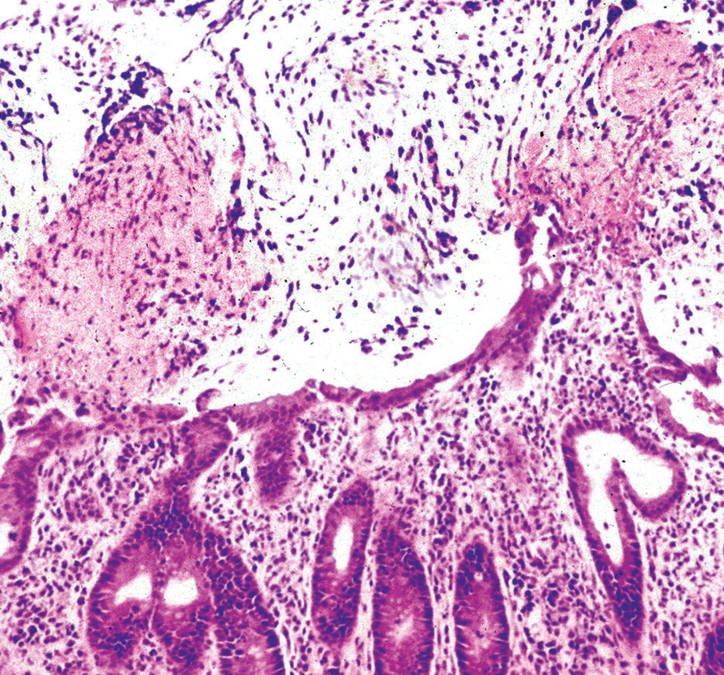
A 75-year-old man is treated by his general practitioner with antibiotics for a urinary tract infection. One week later, he develops severe blood-stained diarrhoea and is admitted via the emergency department. A colonoscopy is performed, showing severe inflammation. The biopsy appearance is shown above. Which ONE of the following statements is correct?

Options:

  • A)

    The organism causing his urinary tract infection is now affecting his colon.

  • B)

    He has amoebic colitis.

  • C)

    He has ulcerative colitis.

  • D)

    Testing for Clostridium difficile toxin should be performed.

  • E)

    He has diverticular colitis.

Chapter 5 Question 2

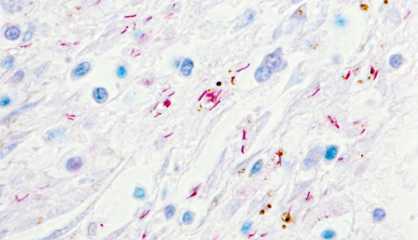
This is a lung biopsy from a 67-year-old patient with a history of chronic cough, night sweats, weight loss and haemoptysis. The biopsy has been stained by a special method to demonstrate the diagnosis. Which ONE of the following statements is correct?

You're Reading a Preview

Become a Clinical Tree membership for Full access and enjoy Unlimited articles

Become membership

If you are a member. Log in here