Physical Address
304 North Cardinal St.
Dorchester Center, MA 02124

Pathologic changes seen in facet joint degeneration include fibrillation, joint space narrowing, articular cartilage thinning, subchondral bone sclerosis, osteophytosis, and development of juxta-facet cysts.
Risk factors for facet joint degeneration include lower spinal level, increasing age, sagittal orientation, and intervertebral disc degeneration.
Facet tropism, gender, increasing body mass index, and ligamentum flavum abnormalities may also be risk factors for facet joint degeneration.
Pathologic and imaging grading systems exist for grading facet joint degeneration, with moderate agreement between the different classifications.
The facet joints, also known as zygapophysial or apophyseal joints, are synovial joints located in the posterior aspect of the vertebral column. The paired facet joints, along with the intervertebral disc (IVD) (see Chapter 1, Chapter 6 ) of the same vertebral level, comprise an entity that is called a spinal motion segment. Functioning in concert with the IVDs, the facet joints participate in the articulation of adjacent vertebrae, spinal load transmission, execution of physiological spinal motions, and stabilization of the vertebral column. The facet joints also play a protective role, in that they provide resistance against deleterious motions such as forward translation, and extreme flexion or extension. Over time, facet joints may undergo degenerative change as seen in other synovial joints, but there is no conclusive evidence of these pathophysiological changes in the facet joints are similar to in other joints. What matters is that these degenerative changes can significantly impact the biomechanical functions of the spine and lead to various spine pathologies such as scoliosis and spondylolisthesis (see Chapter 2, Chapter 3 ).
The lumbar facet joints are mesenchyme-derived, paired, diarthrodial sliding synovial joints that develop from the posterior vertebral arch and form the posterior aspect of the intervertebral foramina along with the ligamentum flavum (see Chapter 1 ) [ , ]. The facet joints are “true” synovial joints in that they are lined with articulating cartilage and are surrounded by a complete, ligamentous capsule. These joints contain two opposing articulating surfaces: a dorsomedially located concave superior facet and a dorsolaterally located convex inferior facet [ ].
Each facet joint is innervated by a medial descending branch from the dorsal ramus of the same spinal level, as well as a medial branch from the dorsal ramus of the vertebral level above [ , ]. The facet joints receive their blood supply from arterial branches of the lumbar segmental artery, which pass through the intervertebral foramina [ ]. Facet joints, along with their capsules, are extensively covered by nociceptors, free and encapsulated nerves, and substance-P carrying nerves [ ]. The rich innervation of the lumbar facet joints makes them a potential source of back pain [ ].
Multiple types of intraarticular inclusions called “meniscoids” or facet folds have been described in facet joints [ ]. Of the most commonly reported types of meniscoids found in facet joints, the first type constitutes fibrous invaginations of the dorsal and ventral portions of the facet joints, and the second type constitutes fat-filled synovial reflections arising from the superior and inferior poles of the facet joints [ ]. These structures, which have been hypothesized to function in covering areas within the facet joint not covered with articular cartilage and protecting the articular processes during flexion and extension, tend to be more fibrous in younger persons, and more of a fatty nature in older patients [ , ].
As stated, the spinal facet joints and the IVDs are part of an entity called the spinal motion segment, the three-joint complex [ ], or the articular triad [ ]. Functioning together, the structures in the spinal motion segments allow for the execution of physiological spinal motions while protecting the spine by preventing activities that can be injurious. While the IVD is more involved in weight transmission and shock absorption, the spinal facet joints' function is to aid in the stabilization of the motion segment through the prevention of forward translation and the restriction of rotation and flexion, spinal motion control, and protection of the neurovascular structures that are conveyed through the intervertebral foramina [ ].
Loads applied to the spine are transferred to the IVD anteriorly and the facet joints posteriorly, which transfer them through the spinal column. Greater loads are applied to the facet joints during spinal extension and axial rotation than during spinal flexion, with reports that facet joints can sustain approximately 33% of dynamic compressive loads [ ]. When the spine is in a normal erect position, the load applied to the lumbar spine is transmitted to, and is shared between the IVDs and facet joints.
As with other synovial joints, the facet joints undergo degenerative change over time. Such change seen in facet joints has been attributed to a variety of causes such as increasing age, repetitive stress, low-grade trauma, IVD degeneration, and extensive motion and loading conditions (see Chapter 2, Chapter 6 ). The degeneration of the lumbar facet joints can lead to spinal instability, which can result in spinal pathologies such as spondylosis, spondylolisthesis, and scoliosis, among others, and thus may be associated with low back pain (LBP).
Pathological changes associated with facet joint degeneration include fibrillation, joint space narrowing, articular cartilage thinning, subchondral bone sclerosis, hypertrophy of articular processes, vacuum jet phenomenon, osteophytosis, juxtafacet cysts, and joint capsule calcifications [ ]. Lewin et al. [ ] found that before the age of 45, minor articular cartilage changes were commonly seen. However, past that time point, more advanced changes in the articular cartilage, subchondral sclerosis, and osteophytosis were prevalent [ , ].
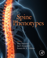
Some of the earliest degenerative changes that can be seen in facet joints are seen within the articular cartilage [ ] initially determined as a loss of facet joint space width. Over time, the organization of the collagen fibril network within the hyaline cartilage is lost, contributing to the early articular changes seen in facet joint degeneration, a phenomenon known as fibrillation [ ]. Fibrillation, which occurs secondary to the loss of matrix proteoglycan, leads to disruption and degradation of the collagen fibril structures [ ]. It starts more superficially and spreads to deeper levels with increasing erosion of the articulating cartilage [ ]. Microscopically, the fibrillated cartilage shows clefts or fissures that begin in the superficial layers and deepen as the cartilage is lost from the surface by tangential flaking, pitting, and grooving ( Fig. 14.1 ) [ , ].
![Figure 14.1, Superior facets showing (A) combined cartilage (asterisk) and bone (arrow) defects or (B/C) sole cartilage defects (asterisks) on the superior pool. Inferior facets displaying (D) large chondral defects (asterisks), leaving cartilage (C) only centrally, (E) osseous defects (arrow), or (F) inferiorly located cartilage defects (asterisks). (G) Totally destroyed cartilage surface (asterisk). (H) Localized circumscribed small defect (arrow) in the cartilage surface (C), covered by meniscoid fold (mF). (I) Corresponding histological section [ 127 ] through a facet joint with superior and inferior facet (sF/iF) (from Ref. [ 127 ] with permission from Thieme Verlag, Stuttgart). Cartilage surface (C) peripherally shows a localized, small circumscribed defect, covered by a meniscoid fold (mF). (K) Superior facet showing osteophyte apposition (asterisk) on the lateral margin. (L) Superior and inferior facets showing corresponding mirror-like defects (asterisks). (M) Inferior facet with large osseous defect inferiorly (arrow) with good preservation of the remaining cartilage surface.](https://storage.googleapis.com/dl.dentistrykey.com/clinical/Facetjoints/0_3s20B9780128227787000158.jpg "Figure 14.1, Superior facets showing (A) combined cartilage (asterisk) and bone (arrow) defects or (B/C) sole cartilage defects (asterisks) on the superior pool. Inferior facets displaying (D) large chondral defects (asterisks), leaving cartilage (C) only centrally, (E) osseous defects (arrow), or (F) inferiorly located cartilage defects (asterisks). (G) Totally destroyed cartilage surface (asterisk). (H) Localized circumscribed small defect (arrow) in the cartilage surface (C), covered by meniscoid fold (mF). (I) Corresponding histological section [ 127 ] through a facet joint with superior and inferior facet (sF/iF) (from Ref. [ 127 ] with permission from Thieme Verlag, Stuttgart). Cartilage surface (C) peripherally shows a localized, small circumscribed defect, covered by a meniscoid fold (mF). (K) Superior facet showing osteophyte apposition (asterisk) on the lateral margin. (L) Superior and inferior facets showing corresponding mirror-like defects (asterisks). (M) Inferior facet with large osseous defect inferiorly (arrow) with good preservation of the remaining cartilage surface.")
Over time, the avascular articulating cartilage decreases, and the subchondral bone is exposed. Changes in the subchondral bone associated with cartilage and facet joint degeneration include hypervascularization, sclerosis, and eburnation [ , , ]. Using a novel validated MR-based technique, Duan et al. [ ] found that subchondral bone thickness increased caudally in the lumbar spine, and that superior facet subchondral bone was consistently thicker than inferior facet subchondral bone. In addition to subchondral bone changes, erosion of articulating cartilage can also lead to narrowing of the facet joint space width, which is a key measure of degradation in other synovial joints. Simon et al. found that in a cohort of healthy subjects ( n = 62) and LBP patients ( n = 34), the joint space width was smaller among the patients, with specific narrowing around the periphery of the facet joints [ ]. This study highlighted the fact that lumbar facet joint space narrowing was dependent on the spinal level and began in the third decade of life. A recent comparison between computed tomography (CT) and ultrasound confirmed that increasing age is associated with decreasing gap width [ ].
Osteophyte formation, bony projections that can grow from the margins of bone, can occur late in the process of facet joint degeneration [ , ], can greatly limit the range of motion, and have been implicated in back pain [ ]. Osteophytes occur commonly in the facet joints of older patients with facet joint degeneration [ , , , , ] but are less common than cartilage defects. Twomey and Taylor [ ] suggested that osteophyte formation may occur as a result of degenerating facet joints' attempt to increase the load-bearing area to deal with the greater loads they experience over time. Otsuka et al. [ ] found that facet joint surface area increased with age and also credited this increase to larger load-bearing in the lower lumbar segments and facet joint degeneration. One study found that osteophyte formation of the lumbar facet joint typically occurred in areas with increased stress secondary to high axial lumbar rotation [ ]. This increased stress may lead to fibrous metaplasia of the dorsal capsule and subsequent osteophyte formation [ ].
The formation of juxtafacet cysts, which are extradural lesions, is also associated with facet joint degeneration. Juxtafacet cysts can occur at any spinal level; however, they are most prevalent at L4/L5 [ ]. Depending on their location, juxtafacet cysts have been identified as possible causes of LBP and radiculopathy [ ]. One study found that juxtafacet cysts were more frequently associated with coronally oriented and arthritic facet joints [ ]. The exact etiology of juxtafacet cyst formation is unknown; however, multiple mechanisms have been proposed. Some proposed mechanisms include synovial fluid extrusion from the joint capsule, latent growth of developmental rest, and myxoid degeneration [ ]. Juxtafacet cysts can arise when the synovium out-pouches through defects in the facet joint capsule or from mucinous degeneration of periarticular connective tissue [ ].
Several factors, such as age, gender, body mass index (BMI), spinal level, tropism, orientation, IVD degeneration, and ligamentum flavum abnormalities have been associated with degenerative change in facet joints.
Become a Clinical Tree membership for Full access and enjoy Unlimited articles
If you are a member. Log in here