Physical Address
304 North Cardinal St.
Dorchester Center, MA 02124


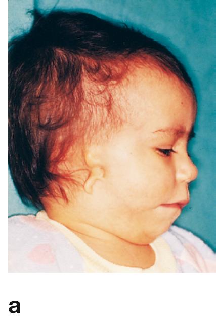
a Inherited external ear defects are common. Severe defects of the external ear and zygomatic bone, receding chin, and cleft palate are typical for the dominantly inherited FRANCESCHETTI's syndrome (mandibulofacial dysostosis). [ E247-09 ]
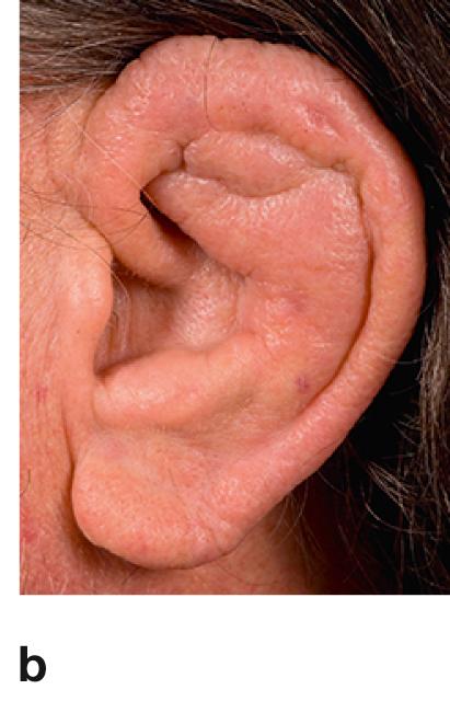
b The cauliflower ear forms as a result of repeated blows to the ear, causing vascular damage and the collection of blood between cartilage and overlying perichondrium providing nutrients to the cartilage. If not drained, the cartilage dies and is replaced by fibrous tissue. [ F217-003 ]
; frontal view.")
, horizontal section (b); schematic drawing.")


Spontaneous perforation and defects in the conductive chain
Spontaneous perforation of the tympanic membrane during putrid middle ear infection (otitis media) tends to occur more frequently in the pars flaccida which is the thinner part of the tympanic membrane.
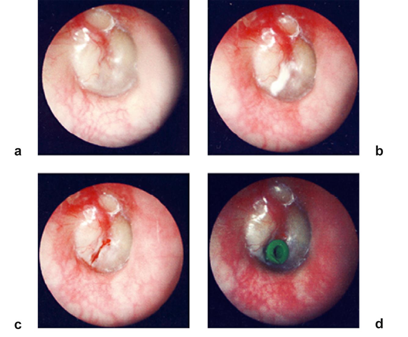
a Serous effusion or pus collecting in the middle ear is visible through the tympanic membrane which bulges outward and looses its light reflex.
b Tympanotomy (tympanostomy, myringotomy) drains liquid from behind the eardrum.
c Incision of the tympanic membrane after tympanotomy and drainage.
d A small tube (grommet) has been inserted into the tympanic membrane to ensure drainage and aeration of the middle ear. Once the middle ear infection is resolved, the grommet is removed and the opening of the tympanic membrane seals.
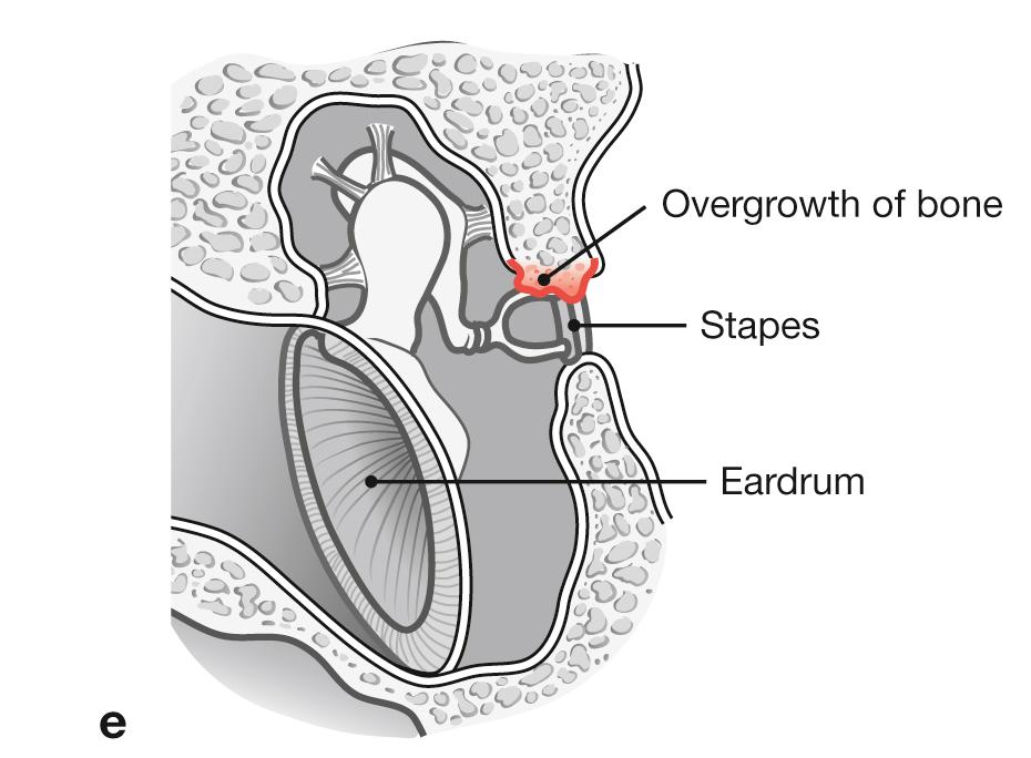
Defects in the conductive chain (tympanic membrane , auditory ossicles, occluded auditory tube) result in conductive hearing loss. e In otosclerosis, the base of stapes progressively fixes to the oval window through ossification of the anular ligament of stapes causing slowly progressing conductive hearing loss. Women at 20–40 years of age are affected two times more frequently than men. In 70 % of cases, otosclerosis affects both ears.
Become a Clinical Tree membership for Full access and enjoy Unlimited articles
If you are a member. Log in here
