Physical Address
304 North Cardinal St.
Dorchester Center, MA 02124
The best morphologic presentation obtained from any cytologic specimen requires an understanding of the factors that went into collecting and preparing the specimen. An accurate cytodiagnosis is based on adequate sampling, proper evaluation of the sample, proper fixation, preparation and staining of the cells. The fundamental principle in cytopreparation is to reduce the specimen to a cellular presentation, which can be interpreted and diagnosed. The final cellular morphology should be free of distortion and artifactual changes, leaving the cells in their most natural state.
Optimal cytologic presentation is directly related to the quality of the specimen, the fixative chosen to preserve the cellular details, and the stain used to render cells viewable. If the specimen is too bloody, too thick, or has been left too long before being taken to the laboratory, the results will be less than optimal. If the specimen is not fixed properly, i.e., the fixative is not correct for the specimen or the fixative is not applied quickly, the results will again be less than optimal. If the chemicals in the stain are old, not properly prepared, or not optimized for time, again the results will not be optimal. The challenge therefore is to develop procedures which will give the best chance at consistently preserving the cells in as close to their natural state as possible.
The processes used in cytopreparation should be continually evaluated especially in light of immunohistochemical and molecular advances in the field. Not only is the goal to produce perfect preparations but thought must go into preserving the residual cellular sample for use in special studies. While these new technologies may require a different approach to sample preparation, the basic principles of fixation, preparation, and staining remain the same, i.e., to obtain an optimal diagnostic cell sample.
The evaluation of the specimen is the initial step in determining the pathway to adequate cellular preparation. A bloody specimen may need additional treatment with a lytic agent such as Cytorich Red (ThermoShandon) or CytoLyt (Hologic) to lyse the red blood cells before it is prepared as a non-gyn ThinPrep. A hypocellular specimen will require techniques that will concentrate the cells into a small area on the slide such as cytocentrifugation using cytospin funnels or a megawell. Urine sent from a distance may be received in Cytospin Collection Fluid, a fixative containing polyethylene glycol (PEG), to preserve cellular integrity (an additional step must be added before staining to remove the PEG). Mucoid specimens may need the addition of a mucolytic agent such as Mucolexx to thin and break up the mucus so it can be processed further. Many options are available and choices must be made to create superior preparations. This chapter explores the different specimen types encountered in the cytoprep laboratory and the methods used to prepare slides for microscopic evaluation.
The purpose of cytologic fixatives is to maintain, as closely as possible, the cytomorphologic characteristics and diagnostically essential cytochemical elements of the cell. An appropriate fixative for cytodiagnostic purposes should perform the following functions:
Penetrate cells rapidly
Minimize cell shrinkage
Maintain morphologic integrity
Deactivate autolytic enzymes
Replace cellular water
Facilitate diffusion of dyes across cell boundaries
Help cells adhere to a glass surface
Provide consistent results over time
Produce a permanent cell record
Stop cellular and microbial growth (antimicrobial).
Historically, Papanicolaou, like Reider before him, used ether and 95% ethanol (1 : 1) as the cytologic fixative of choice. Currently, for safety reasons, 95% ethanol, 80% isopropanol, 100% methanol, 95% denatured alcohol, and proprietary blends such as CytoLyt, PreservCyt and CytoRich Red, along with various commercially available spray fixatives have replaced the ether/ethanol mixture as the fixative of choice. For laboratories using liquid-based techniques, the type of fixative depends on the method selected. Current liquid-based preparations enhance cellular preservation by utilizing fixative solutions that are either ethanol or methanol based. Cell alterations may be minimal among the various fixatives in use. Fixation methods may fall into one of five categories: wet fixation, wet fixation with subsequent air drying, spray fixation, liquid-based fixation, and lysing fixation for bloody samples.
Wet fixation, i.e., submerge the cell sample immediately into the fixative solution. The cell sample remains in the fixative solution until it arrives in the laboratory, where it is accessioned and stained. The cells are not exposed to air at any time during this fixation method. The alcohols – 95% ethanol, isopropanol, and methanol – are all satisfactory fixatives to submit specimens for evaluation.
Wet fixation with subsequent air drying, i.e., submerge the cell sample immediately into the fixative solution, remove the sample from the fixative solution after a specified time, air dry the cell sample, and place it into a container for transport to the laboratory. On arrival in the laboratory, the slide is placed in 95% ethanol or its equivalent prior to staining to rehydrate the cell sample.
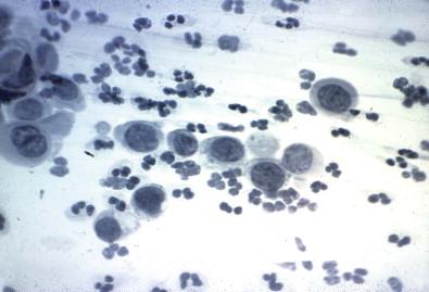
Spray fixation, i.e., immediate fixation of the wet cell sample with a commercially available spray fixative. The spray-fixed cell sample is allowed to air dry and is then placed in a container for transport to the laboratory. Spray fixatives usually consist of an alcohol base blended with a waxy substance (carbowax) that provides a thin protective coating for the cells. When received in the laboratory, the spray-fixed slides must have the waxy substance removed prior to staining. To remove the waxy coating, the slides are placed in two separate rinses of 95% ethanol or two separate rinses of tap water. Slides are placed in the first ethanol rinse for approximately 30 min. One may see opaque, waxy particles float to the surface of the ethanol solution. This first 95% ethanol rinse is discarded. The slides are then placed into the second 95% ethanol rinse for 10–15 min. The use of these two ethanol rinses is usually sufficient to dissolve the waxy substance. In very hot and humid environments, it may be necessary to add a third ethanol rinse. It should be noted that some physicians spray fix more heavily than others, and this factor may add to the difficulty of treating all specimens in the same fashion. Some manufacturers of spray fixatives recommend removal of carbowax with water. We have found 95% ethanol to be more effective and less time-consuming than water rinses. Additionally, after the ethanol rinses, the cells are better prepared for the uptake of dyes than with water rinses alone. However, if availability and cost are factors, water rinses will suffice. It should be noted that if the carbowax is not removed completely, small, waxy particles will be carried into the staining solutions. Nuclei will then appear foggy and lack chromatinic detail and the cytoplasm may exhibit a pale blue color ( Fig. 33-1 ). Alcohol-based, non-aerosol spray fixatives are preferred. One spray container can fix approximately 700–1000 cell samples. It is suggested that one use a commercially prepared fixative designed specifically for cytologic use instead of alcohol-based hairsprays, as has been done in the past. One of the reasons for this recommendation is that companies producing hairsprays may alter their formulas, replacing viable components with substitutes that may alter the quality of cell preservation. When using non-aerosol fixatives, the spray nozzle may sometimes become clogged. It is advisable at the beginning of each day to test the spray emitting from the nozzle to ensure the spray is evenly distributed. Non-aerosol sprays are more likely to produce an uneven spray than aerosol sprays. Non-aerosol spray fixatives may be held closer to the specimen than aerosol sprays. For some products, 3–6 inches is recommended, whereas, for others, 6–10 inches is the recommended distance. One should try to obtain an even spray across the entire slide and avoid spraying the labeled or frosted portion of the slide, where a heavy coating of waxy residue makes it difficult to number or label the slide. If the frosted area is sprayed, it may be necessary to scrape off or otherwise remove the waxy coating on the frosted end of the slide before numbering or applying a label to the slide.

Liquid-based fixation may be methanol or ethanol based, depending on the manufacturer. Two systems are currently in use, while other systems are still undergoing development. Both of the currently used systems collect cell samples into liquid fixative solutions. Human papillomavirus (HPV) DNA testing, immunocytochemistry, tumor classification, and other special studies ( Chlamydia and Gonorrhea ) can also be performed on these preparations directly from the vial. One liquid-based system which requires no special preparative devices was developed in the late 1990s. This technique utilizes an easily made metastable alcoholic gel. It is compatible with many fixation agents, including CytoRich Red, CytoLyt, and PreservCyt. The specimen is received into the laboratory and centrifuged. The supernatant is poured off and an equal volume of the gel is added. The mixture is vortexed into a cell slurry. The resultant sediment can be either placed onto glass slides and evaluated in its entirety or placed onto a slide using the Hettich cytocentrifuge. Because there is no filtering mechanism, cellular debris, mucus, and inflammation will still be part of the presentation. The resulting slides are comparable with the conventional Papanicolaou smear and other ThinPrep preparations.
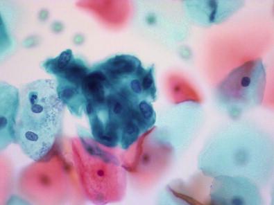
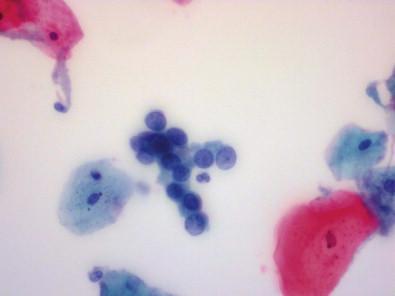
Liquid-based technology further resolves the issue of cells being obscured due to the presence of inflammation and blood. Cellular preservation and sampling problems inherent with the conventional Papanicolaou smear are effectively eliminated with liquid-based Papanicolaou testing. This technology entails direct transfer of cells from the collection device into a fixative solution. Diagnostic confidence is enhanced when the liquid-based method is utilized to obtain a representative sample. Currently, two liquid-based preparation systems are in use that account for approximately 80% of all Papanicolaou collection methods in the USA. ThinPrep and SurePath utilize different processing techniques to achieve the same result: eliminate background milieu and provide a well-fixed concentration of representative epithelial cells ( Figs 33-2 , 33-3 ). ThinPrep technology utilizes a filter process, whereas the SurePath system incorporates a gravity sedimentation process. A ThinPrep specimen may be collected with a “broom,” spatula, or endocervical brush and is then rinsed immediately into a vial containing a methanol-based solution (PreservCyt). This eliminates smearing the sample on a slide and ensures that a greater proportion of cellular material will be captured for microscopic evaluation. The vial containing the specimen is placed in the ThinPrep processor. A plastic cylinder containing a polycarbonate filter membrane is placed onto a holder which is then put into the processor. The filter is immersed into the vial. As processing proceeds, the cylinder is spun gently to produce forces that reduce large cell clusters and break up mucus, blood, and background debris. A vacuum draws the specimen through the filter-trapping epithelial cells and organisms, while mucus, blood, and inflammation pass through the filter. When the pressure across the polycarbonate membrane is high, the processor determines that the filter is full. The vacuum is stopped and the filter is inverted and drained of fluid. Reverse pressure is applied to gently “blow” the cell sample off the filter and on to the glass slide. This produces a thin, monolayered circle of epithelial cells (20 mm) that is fixed immediately and ready for staining.


The SurePath system, as mentioned above, incorporates a gravity sedimentation process. After a specimen is collected, the head of the “broom” can be detached and submitted to the cytology laboratory in the collection vial. The vial contains an ethanol-based solution that fixes the sample immediately. Steps in the preparatory process include layering of the cell sample on to a liquid density gradient, vortexing, and centrifugation. During this process, vortexing breaks up large cell aggregates and mucus. Density gradient centrifugation separates useful cellular elements (filtrate) from obscuring inflammation and debris. The filtrate is placed in a chamber and applied to a glass slide by gravity sedimentation. The end result is an evenly layered circle of cells (13 mm) on the slide that may be stained immediately on the SurePath processor ( Table 33-1 ).
| Conventional Papanicolaou | ThinPrep Papanicolaou Test | SurePath Papanicolaou Test | |
|---|---|---|---|
| Fixation | Ethanol | Methanol | Ethanol |
| Collection | Smear on slide | Sample rinsed in vial | Collection device left in vial |
| Cell sample | Random distribution | Uniform distribution over 20 mm of slide | Uniform distribution over 13 mm of slide |
| Collection device | EC brush, spatula, Cervex-Brush | EC brush, spatula, Cervex-Brush rinsed in vial | Cervex-Brush most effective, tip deposited into vial |
| Preservation artifacts | Air drying, blood, inflammation, irregular distribution of cells | All preservation artifacts greatly reduced | All preservation artifacts greatly reduced |
| Automated processing | Not applicable | Vacuum pressure through TransCyte filter | Gravity sedimentation process |
| Imaging technology | Not applicable | Available | Available |
| Ancillary testing | Not applicable | HPV, Chlamydia , gonorrhea (US FDA-approved) | HPV, Chlamydia , gonorrhea (self-validation required) |
For conventional gynecologic smears, H.W. Boschann (pers. comm. 1962) recommended placing a drop of acetic acid into the 95% ethanol fixative in a Coplin jar both to fix the cells and to lyse the red blood cells. Some laboratories use Carnoy's fixative, others prefer Clarke's fixative or a modification of either fixative. CytoRich Red and CytoLyt (methanol-based) may be used for lysing bloody non-gynecologic specimens for liquid-based preparations. PreservCyt has been granted Food and Drug Administration (FDA) approval for Chlamydia / Gonorrhea and HPV DNA testing. Blood-lysing protocols vary by liquid-based manufacturer. Operating instructions must be followed meticulously for best cytopreparatory results. Various methods for fixation and treatment of bloody cell samples have been suggested. Some recommend fixing the sample first and then placing the slide into a lysing solution, whereas others place the slide directly into the lysing medium. It is important with either approach to rinse the slide thoroughly in 95% ethanol following the lysing solution and before staining in order to stop the action of the lysing solution. Whichever fixation and lysing method is applied to bloody samples, the solutions should be discarded after each use.
The following solutions may be used to lyse red blood cells:
Carnoy's (always prepare fresh): absolute ethanol, chloroform, glacial acetic acid (6 : 3 : 1)
Modified Carnoy's: 95% ethanol, chloroform, glacial acetic acid (7 : 2.5 : 0.5); 95% ethanol, chloroform, glacial acetic acid (6 : 3 : 1); or 95% ethanol, glacial acetic acid (6 : 1). The shelf-life of Carnoy's solution is not known, but it does become hydrochloric acid at some point in time. This is the reason it is suggested that Carnoy's should be mixed fresh each time it is used and discarded after each use. Carnoy's fixative is an excellent nuclear fixative as well as a preservative of glycogen. Because cells may shrink or round up when lysing media are used, it is important to reduce the staining times in hematoxylin and eosin (EA), especially when using Carnoy's fixative
Clarke's solution: absolute ethanol, glacial acetic acid (3 : 1)
One drop of concentrated hydrochloric acid per 500 mL of 95% ethanol
Ten percent glacial acetic acid (this is followed by placing the slide in 95% ethanol)
CytoLyt/Glacial acetic acid (9 : 1) – FDA approved for bloody gyn ThinPrep re-preps. (procedure 4)
CytoLyt is a commercial liquid medium used as a quasi-fixative and an agent to remove red blood cells from specimen samples. It has the ability to hold over specimens so deleterious effects to the cells will not occur. Cell samples prepared from CytoLyt must be fixed for an additional 20 min in PreservCyt before further processing.
Commercially available fixatives such as CytoRich Red (supplied by Thermo-Fisher Scientific) may be used (procedure 5)
CytoRich Red reduces red blood cells and background and fixes cells completely. Removal of the red blood cells allows the diagnostic cells to be visualized, especially in fine-needle aspirates when a rapid interpretation is required.
The solution is said to be a useful fixative for small tissue biopsies and for immunocytochemistry. It works in cytocentrifuged cell samples as well as in liquid-based prepared cell samples.
Fix in 95% ethanol: 15–20 min.
Place in Carnoy's fixative: 10 min or less.
Immerse in 95% ethanol.
Place in Carnoy's fixative for 3–5 min or until material on slide becomes colorless.
Transfer to 95% ethanol or its equivalent.
Place in Clarke's fixative: 10–15 min.
Rinse in 95% ethanol.
Place bloody ThinPrep gyn sample in to a 50 mL tube (saving vial).
Centrifuge sample at 1000 rpm for 10 min.
Pour off supernatant and resuspend in 30 mL CytoLyt/glacial acetic acid.
Centrifuge sample at 1000 rpm for 10 min.
Pour off supernatant and resuspend in 20 mL of PreservCyt.
Pour contents back into original vial.
Prepare new slide on ThinPrep 2000 on setting 4.
Procedure using CytoRich Red (Thomas Jefferson University Hospital cytology laboratory).
Place bloody unfixed fluid or aspirate material in a centrifuge tube with a ratio of one part specimen to two parts CytoRich Red.
Fix for a minimum of 20 min.
Centrifuge specimen at 1000 rpm for 10 min.
Decant supernatant.
Resuspend sediment in non-gyn PreservCyt vial for 15 min.
Prepare a ThinPrep slide on T-2000 on setting 2.
When complete, place slide in 95% alcohol until ready to stain.
Stain in usual manner, using non-gynecologic staining procedure.
Coverslip.
Place on warmer to dry.
The air-dried sample is stained with methods other than the Papanicolaou staining method, such as Wright–Giemsa, Diff-Quik, or May–Grünwald–Giemsa staining procedures. Air-dried cell preparations are well known in the field of hematology and cytology, especially when dealing with rapid evaluation of fine-needle aspirations. The Diff-Quik stain is used to determine the adequacy of a fine-needle aspiration sample. Two slides are labeled with the patient's name, birth date, and specimen source. A drop of specimen is placed on each slide and spread. One slide is placed into 95% alcohol to fix for additional staining. The second slide is allowed to air dry followed by staining.
The stain consists of an alcoholic fixative (12 dips), an eosinophilic (xanthene dye) stain (12 dips), a basophilic (thiazine dye) stain (8 dips), and a water rinse. This stain procedure is performed quickly and easily allowing for a microscopic evaluation in under 5 min. The red blood cells stain red, while the nucleus and cytoplasm stain different shades of blue (metachromasia).
The polychrome Papanicolaou staining method has gained worldwide acceptance for cytologic samples. The staining method and its modifications consist of a nuclear stain and two cytoplasmic counterstains. Hydration prepares the cell sample for uptake of the nuclear dye; dehydration prepares the cell sample for uptake of the counterstains. Dehydration and clearing solutions result in cellular transparency and prepare the cell sample for the final steps: mounting and coverslipping. Papanicolaou described a good staining method as one in which nuclear detail was well defined, transparency of cytoplasm was assured when cells overlapped, and cell types could be differentiated from one another. One might add to this list stability of the stain over time and reproducible results. The stains are commercially available and are tested for dye content and consistency. The performance of the commercial stains is comparable to the performance of the stains made in the laboratory.
Modifications of the technique vary from laboratory to laboratory. Differences are noted visually as well as analytically. Solutions and staining times vary. Of importance to the Papanicolaou staining method is the length of time the slides sit in the hematoxylin and EA dyes. When adjusting staining times, one begins by doubling or halving the time in the dyes to see if adjusting the time in the dyes results in a color change. One then can fine-tune the time changes until one achieves the results desired. The results will yield a purple/blue nucleus, glandular cell cytoplasm and intermediate and parabasal squamous cells will be light blue-green, superficial squamous cells will be pink to orange, and keratinizing squamous cells will be a dense blue or dense orange in color.
Even when the same formulas are selected for use in a particular laboratory, the hydrating and dehydrating solutions may vary. When the regressive method is selected, various formulas are used to obtain the dilute hydrochloric acid rinses to remove the excess hematoxylin. If the progressive method is selected, both the “blueing” solutions selected and the formulas used to make up the solutions often vary. The selection of staining times for each solution and the use of tap water or distilled water (required for the ThinPrep images) differ from laboratory to laboratory. Whichever modification of the Papanicolaou method is used, it is advisable to standardize the staining method as much as possible to achieve reproducible results.
There are several factors that affect the Papanicolaou staining reaction:
Type of fixative used
Type of staining method used (regressive or progressive)
Type of hematoxylin formula selected
Formulas selected for the counterstains
Length of staining times in the dyes
Number of slides stained in each dye
pH and chemical content of tap water (some waters contain an excessive amount of chlorine)
Water temperature
Moisture and humidity in the environment
pH of solutions and dyes
Age of the dyes used
Presence of dye particles in unfiltered solutions
Contamination of dehydrating solutions
Quality of cell sample prepared by the clinician (thick or thin, air-dried or wet-fixed)
pH of the specimen
Presence or absence of inflammatory cell changes.
In the progressive method, the nuclear chromatin is stained with hematoxylin to the intensity desired. The time the cells are exposed to the hematoxylin is not sensitive because the nuclear uptake is complete after a finite time span. The stain usually has a lower hematoxylin concentration and slowly and selectively stains nuclear chromatin. The blueing agent, following the hematoxylin, sets the nuclear dye in place. The cytoplasm is barely tinted. Lithium carbonate, Scott's tap water substitute (STWS), and some commercially available solutions are the most commonly used blueing agents. Tap water may serve as a blueing agent if the pH is higher than 8. Today, ammonium hydroxide is seldom used as a blueing agent because of its variability in formulations and day-to-day instability.
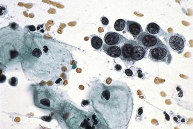
The progressive method is the most commonly used method in laboratories today. It is easier to control and provides more stability on a day-to-day basis. There are differences among laboratories that use the progressive method ( Table 33-2 ). In the University of Chicago section of cytopathology procedure (prior to 1992), the stains were made from scratch according to the formulas described later in this chapter ( Fig. 33-4 ).
| University of Chicago Section of Cytology Laboratory a | University of Rochester Cytopathology Laboratory b | Johns Hopkins Cytopathology Laboratory c | ||||
|---|---|---|---|---|---|---|
| Fixation | Commercial spray fixative | 95% ethanol | 15 min | 95% ethanol | ||
| Removal of carbowax | 95% ethanol | |||||
| 95% ethanol | ||||||
| Hydration | 95% ethanol | 95% ethanol | 15 min minimum | Tap water | 10 dips | |
| Tap water | 70% ethanol | 3 dips | Tap water | 10 dips | ||
| 50% ethanol | 3 dips | |||||
| Running tap water bath | 1–2 min | |||||
| Nuclear stain | Gill's half-oxidized hematoxylin (made by laboratory personnel) | A test slide is used to determine staining times for each batch | Hematoxylin #2 (Richard Allan) | 45 s | Gill's hematoxylin #1 (Harleco) | 1 min (change at 3 weeks) |
| Rinse | Running tap water | Until solution is clear | Running tap water | 1–2 min until clear water is visualized | ||
| Clarifier | Clarifier #2 (Richard Allan) | 2 quick dips – 30 s | ||||
| Rinse | Running tap water bath | 1–2 min | ||||
| Blueing solution | Scott's tap water substitute | 1 min | Blueing reagent (Richard Allan) | 20 s | Tap water | 10 min |
| Rinse | Tap water | 40–50 dips | Running tap water bath | 1–2 min | Tap water | 6 min |
| Dehydration | 95% ethanol | 10 dips | 50% ethanol | 10 dips | 95% ethanol | 10 dips |
| 95% ethanol | 10 dips | 70% ethanol | 10 dips | 95% ethanol | 10 dips | |
| 95% ethanol | 10 dips | |||||
| Cytoplasmic stain | Modified Orange G | 1 min | Orange G (Harleco) | 1 min 15 s | Gill's modified Orange G (Harleco) | 15 s (change at 3 weeks) |
| Rinse | 95% ethanol | 10 dips | 95% ethanol | 3 dips | 95% ethanol | 10 dips |
| 95% ethanol | 10 dips | 95% ethanol | 3 dips | 95% ethanol | 10 dips | |
| 95% ethanol | 10 dips | 95% ethanol | 3 dips | 95% ethanol | 10 dips | |
| Cytoplasmic and nucleolar stain (RNA) specific | Modified EA | 10 min | Eosin (EA) 65 (Harleco) | 3 min 30 s | Gill's modified EA (Harleco) | 10 min (change at 3 weeks) |
| Dehydration | 95% ethanol | 20 dips | 95% ethanol | 3 dips | 95% ethanol | 20 dips |
| 95% ethanol | 20 dips | 95% ethanol | 3 dips | 95% ethanol | 20 dips | |
| 95% ethanol | 20 dips | 95% ethanol | 20 dips | |||
| 95% ethanol | 20 dips | |||||
| Absolute ethanol | 10 dips | 99% absolute ethanol | 3 dips | Absolute ethanol | 10 dips | |
| Absolute ethanol | 10 dips | 99% absolute ethanol | 3 dips | Absolute ethanol | 10 dips | |
| Absolute ethanol | 10 dips | 99% absolute ethanol | 3 dips | Absolute ethanol | 10 dips | |
| Absolute ethanol | 10 dips | Xylene | 3 dips | Absolute ethanol | 10 dips | |
| Clearing | Xylene | 10 dips | Xylene | 3 dips | Xylene | 10 dips |
| Xylene | 10 dips | Xylene | 3 dips | Xylene | 10 dips | |
| Xylene | 10 dips | Xylene | <13 dips | Xylene | 10 dips | |
| Xylene | 10 dips | |||||
| Xylene | 5 min | |||||
| Mounting | Preservaslide | Permount Surgipath #1 coverslip, 1 oz precleaned, 24 × 50 | Permount | |||

The procedure used by the University of Rochester utilizes hematoxylin #2 from Richard Allan and Orange G (OG) and EA from Baxter Harleco. The Richard Allan clarifier #2 is a reagent useful in eliminating excessive background staining by hematoxylin without removing nuclear stain. It enhances cytoplasmic transparency and should be used only with the progressive staining technique.
The Richard Allan blueing reagent is a buffered blueing agent that enhances nuclear detail by changing the nuclear stain from a reddish blue to a crisp blue-purple.
The third procedure ( Table 33-2 ) is a variation of the Papanicolaou progressive staining method as used by the Johns Hopkins cytopathology laboratory (K. Plowden, pers. comm. 2007).
Johns Hopkins uses Gill #1 hematoxylin, Gill EA, and Gill OG from Harleco.
Gill recommends the following steps when staining manually. These steps are part of the staining procedure used by the Johns Hopkins cytopathology laboratory:
Follow times and dips posted on the staining schedule.
A “dip” requires about a second. A dip is gently raising the staining rack until it clears the solution, and without jarring it against the sides or bottom of the staining dish, and lowering the rack until it is totally submerged in the solution. Ten dips effectively replace one solution by another.
Drain solutions well between baths but do not allow the preparations to dry.
Change stains (hematoxylin every 3–4 weeks, OG and EA every other week).
Record changes on the maintenance sheet.
Filter stains through separatory funnels twice daily (see “ Cross-contamination method to avoid floaters ”, below).
Filter xylene through course filter paper to eliminate water, cells, and debris.
A control slide, buccal smear or ThinPrep Papanicolaou, should be run as the first slide through the stain procedure (manual or automated). The slide is evaluated and adjustments are made to the staining times if needed, to achieve the best staining quality.
All the alcohols should be checked daily for stain saturation and the alcohol closest to the stain discarded, the other alcohols moved up and a new replacement alcohol put in the last position. This should be logged on a daily maintenance schedule.
Using the regressive method, one deliberately overstains the nucleus with a non-acidified hematoxylin such as Harris's. The solution has a high concentration of hematoxylin and rapidly stains the entire cell. The excess stain is removed with a diluted hydrochloric acid solution. The decolorizing acid is then removed by a bath of running tap water. Timing in the acid bath is essential for the final appearance of the nuclear pattern. More often than not, hypochromasia rather than hyperchromasia is the result. The cytoplasm as well as the nucleus is decolorized by the diluted hydrochloric acid. If the acid bath is inadequate, the contrast between the chromatin and parachromatin is less and uptake of the counterstains is lessened.
A procedure for the regressive staining method once used by the Papanicolaou laboratory (C.M. Street, pers. comm. 1959) is shown in Table 33-3 . The fixative is 95% ethanol, the same fixative recommended for the progressive staining procedure. The second regressive staining procedure is from the Thomas Jefferson University Hospital cytology laboratory. In place of non-acidic Harris hematoxylin, Gill #3 hematoxylin from Surgipath has been substituted. Please note that, prior to staining, the slides are placed in 95% ethanol for at least 20 min.
| Papanicolaou Laboratory a | Thomas Jefferson University Laboratory b | |||
|---|---|---|---|---|
| This method is used in the Gemini Automated Stainer. | ||||
| Fixation | 95% ethanol | 95% ethanol | 10 min | |
| Hydration | 80% ethanol | 6–8 dips each | 95% ethanol | 10 min |
| 70% ethanol | 6–8 dips each | 70% ethanol | 1 min | |
| 50% ethanol | 6–8 dips each | |||
| Distilled water | 1 min | |||
| Deionized water | 1 min | |||
| Nuclear stain | Harris hematoxylin (without acetic acid) | 6 min (a test slide is used to determine staining times for each batch) | Gill III hematoxylin (Surgipath) (3 : 1); stains are evaluated daily; may be adjusted as needed | 2 min |
| Rinse | Distilled water | Running tap water | 1 min | |
| Distilled water | ||||
| Removal of hematoxylin | 0.25% hydrochloric acid | 6 dips | 0.25% hydrochloric acid | 2 s |
| Rinse | Running tap water (lukewarm) | 6 min | Running tap water | 2 min |
| Dehydration | 50% ethanol | 6–8 dips each | Ammonium hydroxide | 10 s |
| 70% ethanol | 6–8 dips each | Running tap water | 1 min | |
| 80% ethanol | 6–8 dips each | 70% ethanol | 1 min | |
| 95% ethanol | 6–8 dips each | 95% ethanol | 1 min | |
| Cytoplasmic stain | OG-6 | 1.5 min | OG-6 (Surgipath) | 1 min |
| Rinse | 95% ethanol | Gentle rinse | 95% ethanol | 1 min |
| 95% ethanol | Gentle rinse (do not allow slides to stand in alcohol or cells will be discolored) | 95% ethanol | 1 min | |
| Cytoplasmic and nucleolar stain (RNA) specific | EA36 (EA50 or EA65) | 1.5 min | EA50 (Surgipath) | 2 min |
| Rinse | 95% ethanol | Thorough but gentle rinse | 95% ethanol | 1 min |
| 95% ethanol | Thorough but gentle rinse | |||
| 95% ethanol | Thorough but gentle rinse | 95% ethanol | 1 min | |
| Dehydration | Absolute ethanol | 6–8 dips | Absolute ethanol | 1 min |
| Absolute ethanol | 6–8 dips | Absolute ethanol | 1 min | |
| Clearing | Absolute ethanol and xylene (1 : 1) | 6–8 dips | Absolute ethanol and xylene (1 : 1) | 1 min |
| Xylene | 6–8 dips | Xylene | 1 min | |
| Xylene | 6–8 dips | Xylene | 1 min | |
| Xylene | 6–8 dips | Xylene | 1 min | |
| Xylene | 6–8 dips | |||
| Mounting | Permount | Permount | ||
Discovered in 1840 and first used by Bohmer in 1865, hematoxylin is the nuclear dye used in the Papanicolaou procedure. Hematoxylin is, however, one of the most difficult dyes to control and standardize. Luna and Gaffney, in a study of nine commercially available hematoxylins used for hematoxylin and eosin (H&E) staining, concluded, “most H&E staining deficiencies seen on stained slides are not due to the hematoxylin used, but to the quality of fixation and the way the hematoxylin solutions were used.”
Various hematoxylin formulas have been used over the years. Harris hematoxylin is usually used with regressive staining methods, whereas commercially prepared formulas, such as Gill I, Gill II, and Gill III, are usually used with the progressive staining method.
Lillie, in 1974, reported on the variability of hematoxylin samples submitted to the Biological Stain Commission for certification following the reported hematoxylin shortage in 1973. The shortage of hematoxylin resulted in a lower quality product. Some post-1973 samples do not dissolve well in alcohol, and formulas such as Gill's may be more appropriate. Water should be added to the alcohol to help dissolve the hematoxylin; 90% alcohol is suggested. Some batches require two or three times more hematoxylin than was previously required to produce the same results. If the laboratory makes its own dyes, it is advisable to check the batch number and color index (CI) printed on the hematoxylin bottle and include this information in the laboratory procedure manual to ensure the same ingredients are used to make up each batch of hematoxylin. Differences in hematoxylin may also be due to differences in the soil in which its source material is grown.
It is important to date hematoxylin when it is received in the laboratory because the dye may oxidize over time, especially in moist climates. The lighter the crystals, the less oxidized the hematoxylin; the darker the crystals, the more hematein (or oxidized hematoxylin) is present. Lillie stated that overoxidation is one of the main causes of a poor hematoxylin stain.
Information provided by the manufacturer may not indicate the actual shelf life of dyes. It is best to date the bottle when it is received and record the date when it is opened. For stock and working solutions, the date when it was made and the date when it was first used may help the laboratory establish the quality control of these dyes.
Certain components in hematoxylin formulas help transform hematoxylin into a usable nuclear dye. These are: (1) an oxidizing agent; (2) a mordant; (3) a solvent; and (4) a substance used for acidification. The oxidizing agent is used to begin the ripening process that aids in the transformation of hematoxylin to hematein, the active coloring agent. The oxidizing agents most often used are sodium iodate and mercuric oxide. Mercuric oxide is used to oxidize and ripen Harris hematoxylin, and chloral hydrate is added as a preservative, as recommended by Mayer. These are toxic chemicals and should be avoided if possible.
The mordant is a substance that is responsible for the induction of color in the dye. Aluminum sulfate or alum supplies positively charged ions that act as a bridge to chemically unite the negatively charged hematein to the negatively charged phosphoric acid on the DNA chain.
The solvent in hematoxylin is the substance that dissolves hematein into solution. Ethylene glycol is used in certain formulas for this purpose. The solvent acts as a leveling agent and helps reduce the rate of oxidation.
Acidification, the final component, ensures selectivity to nuclear material and aids in preventing oxidation of the dye. It does so by stabilizing the aluminum–hematein complex. Glacial acetic acid or citric acid is used for this purpose.
Soost and coworkers used Harris hematoxylin both progressively and regressively in a controlled study. Their findings showed that with the progressive method the chromatin was a finely distributed structure, hyperchromasia was accentuated, and there was moderate staining of the nuclear envelope. Polychromasia was accentuated and there was one peak of nuclear absorption at 540 nm.
However, with the regressive method, the nuclei were uniformly darkly stained with significantly higher areas of nuclear hyperchromasia. There were textural features as well as contrast between nucleus and cytoplasm. There were two peaks of nuclear absorption at 530 and 590 nm.
These researchers concluded that the progressive method is better for cell measurement and fits the requirements for automated systems. They also found the correlation of visually described chromatin patterns with quantitative data is better. These investigators also reported that the total amount of stain (total extinction values) in the nucleus does increase significantly with the length of time in the staining solution. The total amount of stain bound in the nucleus is not materially different for cells lying singly or in clusters. It was also noted that stained samples stored for 6 months led to optical extinction and readings approximately 6% lower than the original measurements. For the given precision of the technique, this difference is statistically significant.
Wiseman presented findings on a comparative study of six different formulations of hematoxylin. Five formulations were used with the progressive staining method. The sixth was used with a regressive staining method. The half-oxidized formulas were made with varying amounts of hematoxylin, and the oxidizing agent was sodium iodate. Initial studies were planned to determine if such variations could be detected by computerized measurements of the extinction values in cell nuclei of buccal smears prepared with a monolayer technique. A ratio of 2.0 g of hematoxylin dye to 0.2 g of sodium iodate, as recommended by Gill and colleagues, provided the most condensed range of total extinction measurements. These values were then compared with extinction values obtained using the Harris hematoxylin formula with the regressive staining method. The half-oxidized hematoxylin yielded the most condensed range of total extinction values.
The formula for making Gill's half-oxidized hematoxylin is easy to prepare, is stable in solution, and is cost-effective. No scum (the actual coloring agent) forms on the surface of the hematoxylin solution, and it can be used immediately after preparation. Once the staining times have been standardized, little or no alteration is required. In a laboratory with a volume of approximately 40 000 gynecologic slides per year, the hematoxylin solution is changed every 6 weeks, or after every 2000 slides, whichever comes first.
To make 1 L or more of Gill's half-oxidized hematoxylin, the following ingredients are needed. The ingredients should be added in the following order:
Distilled water, 730 mL
Ethylene glycol, 250 mL
Hematoxylin (anhydrous), 2 g
Sodium iodate (a chemical ripening agent), 0.2 g (please note: this step is critical to the formula )
Aluminum ammonium sulfate (alum), 17.6 g
Glacial acetic acid, 20 mL (or 1 g of citric acid).
The ingredients need constant stirring on a magnetic mixer for 1 h at room temperature.
All ingredients must be dissolved. After the staining solution is filtered, it may be used immediately. The shelf-life is over 1 year. It should be noted, however, that in some staining procedures the solution is diluted when in actual use. Some laboratories use the stock solution exclusively, whereas others may use a diluted stock solution for daily work.
As the stain is filtered, a deep purple ring should form at the top of the filter paper. If the ring is brown or brownish-green, discard and use fresh stain.
In a jar of running water, place 5–6 drops of hematoxylin, if the water remains a deep purple, the stain is good. If the water changes color to brown or brownish green, the stain is oxidized and should be replaced.
The Biological Stain Commission certifies most synthetic biologic dyes. In preparing the counterstains OG and EA, it is important to check the dye content on the outside label. OG usually contains 80% dye content; light green, 65%; eosin Y, 80%; and Bismarck brown Y, 45%. Information from the Biological Stain Commission reveals that dye contents for these dyes may be different. In 1997, OG contained 89% dye; light green, 71%; and eosin, 92–94%. To compensate for variations in dye content, one divides the desired amount of total dye content required by the actual percentage of dye content listed on the label. Published staining procedures usually do not contain this information; therefore, in attempting to reproduce a staining procedure, it is necessary to obtain this information directly from its original source.
Become a Clinical Tree membership for Full access and enjoy Unlimited articles
If you are a member. Log in here