Physical Address
304 North Cardinal St.
Dorchester Center, MA 02124
This chapter describes and illustrates systematic dissection sequences and procedures. Although developing a systematic approach to autopsy dissection and organ examination is efficient and generally desirable, the pathologist must be prepared to alter the examination as required for the best demonstration and diagnosis of specific diseases. A given method may be adequate for revealing the pathologic findings of one disease or condition; however, it may be entirely inadequate for identifying or best demonstrating another.
Modern autopsy techniques include modifications of the Virchow, Ghon, or Letulle methods. The method attributed to Rokitansky, characterized by in situ dissection, has not stood the test of time, although many erroneously apply his name to the method of Letulle. Using the Virchow method, the prosector removes organs one by one. In contrast, the pathologist using the Ghon or Letulle method removes the cervical, thoracic, abdominal, and genitourinary organs as separate organ blocks (“en bloc”) or as a single group (“en masse”), respectively.
The Virchow method, removing organs one by one, is excellent for demonstrating pathologic changes in organs but sacrifices interorgan relationships and makes interpretation of regional disease more difficult. The advantages of the Ghon and Letulle techniques include excellent preservation of the interrelationships of the various organs, their regional lymphatic drainage, and their vasculature. The Letulle technique, removing the organs together in toto, allows the most rapid preparation of the body for removal to the mortuary and, because there is less dissection within the confines of the body cavity, probably offers greater safety to the prosector and assistant. However, the examination performed with this method takes a bit longer than that with the Virchow method and is difficult for some to perform without an assistant. The Ghon method, removing the organs in regional and functional groups, is relatively easier for one person to carry out. However, the prosector using the Ghon technique transects the esophagus and aorta at the diaphragm, a disadvantage in cases of aortic dissection or aneurysm and esophageal varices or neoplasm. Skill in the Rokitansky technique (i.e., in situ dissection) is advantageous because it allows one to open and examine organs without removing them from the body, a condition sometimes mandated by autopsy consent restrictions or severe time limitations.
This chapter focuses on postmortem methods based on modifications of the Letulle and Virchow methods. However, the prosector should be prepared to modify any standard autopsy technique to best demonstrate the pathologic changes and important pathologic relationships. Regardless of the method of dissection, well-maintained instruments make the work easier. A list of instruments useful in various standard and specialized postmortem procedures is presented in Box 4-1 . Before starting the prosection, the pathologist should pause to consider whether all administrative preparations have been made and all supplies and equipment are ready ( Box 4-2 ). Useful templates for describing and reporting gross autopsy findings are shown in Appendix A .
Organ knife 10 inches/254 mm or 15 inches/381 mm
Scalpel knife holder and no. 22 disposable blades
Forceps, 1 × 2 teeth, 10 inches/254 mm
Forceps, 1 × 2 teeth, 6 inches/152 mm
Forceps, serrated tips, 6 inches/152 mm
Forceps, Adson 1 × 2 teeth, ![]() inches/121 mm
inches/121 mm
Forceps, Adson, serrated tips, ![]() inches/121 mm
inches/121 mm
Forceps, Rochester Pean, straight and curved, 8 inches/203 mm
Forceps, Halstead mosquito, straight and curved, 5 inches/127 mm
Scissors, Mayo or Doyen abdominal, straight, 9 inches/229 mm
Scissors, Mayo or Doyen abdominal curved, 9 inches/229 mm
Scissors, Metzenbaum, straight round point 5.5 inches/139 mm
Scissors, Metzenbaum, curved round point 5.5 inches/139 mm
Enterotomy (intestinal) scissors, 8 inches/203 mm
Wire cutters
Rib shears
Probe, 1 mm
Postmortem hammer/hook
Virchow skull breaker or chisel
Bone-cutting forceps
Rongeurs, double action
Self-retaining retractors
Oscillating (Stryker) saw
Small rule
Meter stick
Plastic-coated measuring tape
Postmortem needles, serpentine and curved
Administrative preparations:
Review the autopsy consent form to assure proper authorization and identify autopsy restrictions.
Assemble and review relevant clinical records.
Consider medical examiner/coroner notification requirements.
Contact members of the clinical team for additional information as needed, including special points of interest in the autopsy.
Review imaging studies to guide anatomic aspects of the autopsy.
Arrange for special technical assistance , such as cardiology technicians to interrogate implantable defibrillators/pacemakers.
Coordinate with members of the surgical or medical teams who should be additional prosectors at the autopsy.
Notify hospital representatives who are assisting in decedent affairs about the expected timing of the autopsy, as needed.
Preparation of the autopsy room:
Lay out necessary dissection instruments and equipment (see Box 4-1 ).
Prepare materials for ancillary studies, such as microbiologic analysis, electron or immunofluorescence microscopy, and biochemical or molecular analysis (see Chapter 10 ).
Prepare needed equipment for photography during the case (see Chapter 7 ).
Arrange the means to sterilize tissue surfaces for autopsy microbiology cultures.
Label with the correct patient identifiers all materials used to record autopsy information and all specimen containers.
Assure proper protective equipment is worn by all participants in the autopsy.
One of the worst outcomes for an autopsy is to examine the wrong patient, overstep restrictions explicitly written on the autopsy consent form, or lose valuable information or materials from mislabeling. Because of this, many institutions have adopted a simple “time out” method in which all members of the autopsy team, immediately before examining the body, focus on a team statement of the patient name, medical identifiers, and autopsy restrictions for the case they are about to begin. Check the toe tag or other identifier of the body, match it with a copy of the autopsy consent form, and document as needed before continuing. After the autopsy examination, a copy of the consent form, contact information for the autopsy service, and a note that “an autopsy has been performed” may be included with the body. This will facilitate communications with others who may handle the body, including transplant donation networks and those making funeral preparations. Be sure to document and pass along clothes and personal belongings associated with the body.
Remove any bandages, and document any therapeutic devices. Remove superficial, peripheral venous catheters, but leave in place indwelling central lines, endotracheal tubes, feeding tubes, urinary bladder catheters, and so forth until the internal examination confirms their proper location. Measure the body length, and if possible, weigh it. Note the anteroposterior dimension of the chest, and identify any abdominal distention. Determine whether the appearance of the extremities and joint mobility are symmetrical. Document abnormalities by measuring the circumferences of the chest (at the level of the nipples), abdomen (at the umbilicus), or extremities (bilaterally at a specific distance above or below an anatomic landmark such as the superior margin of the patella or the acromioclavicular joint).
Inspect the skin anteriorly and posteriorly. Note its color and elasticity, and characterize any cutaneous lesions. Document surgical and nonsurgical scars. Record any tattoos or other identifying features. While inspecting the dorsum, also inspect the anus. Examine the character and color of the nails. Estimate the degree of rigor mortis by flexing the joints. Note the color, length, and character of the hair. Inspect and palpate the scalp and skull. Examine the eyes, including the condition of the conjunctivae and sclerae and the color of the irides. Measure the diameters of the pupils. Examine the ears and their location. If necessary, use an otoscope to inspect the external ear canal. Inspect the nose, including the nasal mucosa and the integrity of the nasal septum, and note any nasal discharge. Open the mouth and inspect the buccal mucosa and the tongue. Examine the teeth and their state of repair. In the case of unidentified bodies, prepare a detailed dental chart or consult a forensic odontologist.
The illumination provided by an otoscope or penlight may be helpful in examining nasal and oral cavities. Palpate the neck, noting the position of the trachea and the size and consistency of the thyroid gland. Check for cervical, axillary, or inguinal lymphadenopathy. Examine the breasts and palpate for masses. Examine the infraclavicular region for the presence of a pacemaker or implantable defibrillator. Palpate the abdomen. Inspect the genitalia. In the male, palpate the scrotum, determining whether the testes are descended and noting any abnormalities. In the female, separate the legs and inspect the vulva. In some forensic cases, a vaginal speculum is required to complete a detailed inspection of the vagina and cervix that includes appropriate sampling of secretions. Before beginning the internal examination, consider the need to document any significant findings with photographs. Keep a handheld camera ready during the remainder of the dissection for photographs of in situ findings.
After you have completed the external examination, place a block under the shoulders to extend the neck. For fetuses or small infants, a rolled towel may provide adequate elevation of the upper torso. The incision is roughly Y -shaped and most easily made with a sharp scalpel. It begins at the shoulders, anterior to the acromial processes and sparing the top of the shoulders. The upper limbs of the incision penetrate to the ribs and meet at the level of the xiphoid process. Some prefer to extend the upper limb incisions in an arc around the inferior portion of the female breasts. We prefer to direct the upper limb incision medial to the breasts, believing that this results in less chance of fluids inadvertently leaking from the closed body after the autopsy.
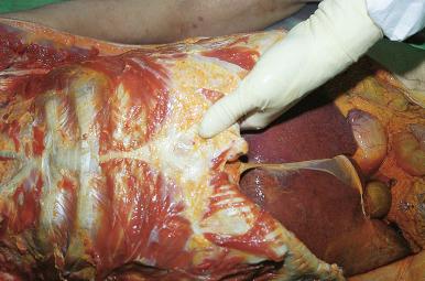
The descending limb of the incision extends along the midline from the xiphoid process to the symphysis pubis, except where it diverts briefly around either side of the umbilicus, through subcutaneous tissue and muscle to the peritoneum. At the level of the umbilicus, measure the thickness of the abdominal fat. At this point, we use scissors to enter the peritoneal cavity to reduce the likelihood of inadvertently piercing the abdominal organs ( Fig. 4-1 ). Expose the abdominal cavity, and prepare for removal of the sternum and anterior portions of the ribs by separating the skin and subcutaneous tissues of the lateral chest and abdominal walls from their bony attachments. Using care not to puncture or “buttonhole” the skin, reflect the skin and subcutaneous tissues of the chest and neck to the level of the hyoid bone. Examine the breasts from their posterior aspects by making parallel incisions through the pectoralis muscles and into the breast tissue, and retain breast tissue as needed.

Inspect the peritoneal lining and omentum. Survey the abdominal viscera and note the location of the organs. Characterize, aspirate, and measure any ascitic fluid. Estimate the height (rib or intercostal space) of the dome of the diaphragm, which should reach to approximately the fourth rib on the right and the fifth on the left, before opening the chest ( Fig. 4-2 ).

Check for pneumothorax (see Chapter 6 ). Open the thorax by cutting the sternoclavicular joint and then the ribs near the lateral margin of the costal cartilage. If greater access of the thoracic inlet is required, use an oscillating bone saw to cut the medial end of the clavicle and first rib. Retract the sternum anteriorly, freeing it from the body. In young individuals, a scalpel penetrates the cartilaginous portions of the ribs quite easily; however, in older patients rib cutters are usually necessary. In patients who have had coronary artery bypass surgery, avoid injury to the bypass grafts during removal of the sternum. Because calcified rib cartilages leave ragged edges, place towels over their cut margins for added safety ( Fig. 4-3 ). Characterize, aspirate, and measure any pleural fluid. Then sweep your hands carefully along the pleural surfaces of the lungs, noting and lysing any thin pleural adhesions. Thicker fibrous adhesions require careful sharp dissection to avoid tearing the visceral pleura. Lung cultures are acquired at this point (see Chapter 10 ); to limit potential microbial contamination, some institutions open the thorax and acquire lung cultures before opening the peritoneum. Examine the thymus. In adults, adipose tissue normally replaces the thymic tissue, although its lobes may still be discernible. In pediatric cases, it is convenient to remove and weigh the thymus at this point. Next, open the pericardium in the midline and examine the pericardial cavity ( Fig. 4-4 ). Characterize, aspirate, and measure any pericardial fluid or blood clot. Unless you plan to fix the heart by perfusion (e.g., in cases of congenital heart disease), open the pulmonary artery above its valve and examine for the presence of saddle or central pulmonary artery emboli by inserting a finger into the main pulmonary artery and its right and left branches. At this time, urine can be collected from the bladder with a syringe and needle.


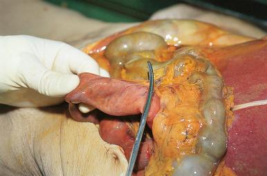
Are there any indications to check blood vessels or surgical anastomoses before removing the intestines? If there are no indications for leaving the intestines attached to the other viscera, remove them now. Displace the omentum and transverse colon superiorly and the small intestine to the right, exposing the ligament of Treitz. Clamp or ligate the small intestine near the ligament, and remove the intestines by cutting (with the rear portion of a large scissors) the mesentery as near as possible to its junction with the serosa ( Fig. 4-5 ). This is facilitated by pulling the intestines toward the scissors and using only the deep hinge portion of its blades ( Fig. 4-6 ). Collect the liberated bowel in a stainless steel basin, noting any obvious serosal lesions during the process. Identify the appendix near the ileocecal junction, and note its location and condition. Examine the external surface of the cecum and ascending colon; then remove it in continuity with the small intestine by lifting it up and cutting the ascending mesocolon and any other fibroadipose tissues securing the bowel. At the hepatic flexure, return the omentum and transverse colon back to their anatomic positions and cut through the transverse mesocolon to the splenic flexure and in turn along the descending colon. Employ double clamps or ligatures at the rectosigmoid junction and transect the bowel. For cases in which the bowel mucosa needs careful microscopic examination, gently instill formalin into the lumen without distention. Otherwise, lay the intestines aside until later.


When the abdominal contents are altered by the presence of numerous adhesions (e.g., with extensive peritoneal metastasis or following peritonitis), it may be necessary to leave the intestines attached to the other abdominal organs. The Letulle autopsy method, described next, coupled with a careful layer-by-layer dissection of the abdominal contents from the posterior aspect, may demonstrate pathologic findings that would otherwise be missed.
After the initial inspection of the organs and body cavities and removal of the gut, prepare for removal of the remaining viscera. Identify and inspect the carotid arteries ( Fig. 4-7 ). If they are flaccid and thin walled, they are best left with the body if later embalming is likely. A long ligature may be placed around each carotid artery where it enters the base of the neck. Using scissors or a scalpel, transect the laryngeal pharynx above the epiglottis through the thyrohyoid membrane or include the hyoid bone by cutting superiorly. Transect the esophagus as well, but avoid injury to the carotid arteries. Reflect the larynx inferiorly, and cut the carotid arteries below their ligatures. It is relatively easy to include the hyoid bone or the tongue and associated tissues as part of the neck dissection, and some pathologists do this routinely because it allows a much better examination of the oropharynx and superior neck. However, the facial artery, a vessel important to the embalmer, is vulnerable to injury during this dissection. Removal of the tongue is facilitated by cutting posterior to the rami of the hyoid bone. Through the neck, reach into the oral cavity, grasp the tongue, flip its tip posteriorly into the neck, and cut the anterior attachments free. More extensive neck dissections are discussed in Chapter 6 .

Reflect the larynx and esophagus downward, and free the neck organs by cutting the vascular and connective tissue attachments at the lateral aspects of the thoracic inlet. Next, cut the right and left hemidiaphragms along their lateral and posterior body walls. On each side, while the posterior aspect of the upper abdominal cavity is exposed, extend the cut through the psoas muscle to the vertebral column. This makes removal of the organ block easier.
Next, turn your attention to the pelvis. Using your hand and fingers, separate the bladder and prostate from the pelvic wall. Extend the plane of dissection posteriorly, separating the rectum along the coccyx ( Fig. 4-8 ). Some physical exertion is necessary here to separate the pelvic organs completely along their entire circumference. Using long-handled scissors, transect at the level of the proximal urethra (distal to the prostate in the male and through the proximal vagina in the female). Continue the cut through the rectum, generally not less than 2 cm above the anorectal junction ( Fig. 4-9 ). Reflect the pelvic organs upward and outward, exposing in turn the iliac vessels bilaterally. Divide these, along with any connective tissue attachments, along the pelvic brim and curve of the sacrum.


At this point, the organ block is ready for removal. Lift the neck and thoracic organs anteriorly and then inferiorly, carefully separating the aorta and other posterior attachments from the vertebral column. Continue applying inferior and upward traction, cutting as needed any remaining diaphragmatic or posterior abdominal wall attachments ( Fig. 4-10 ). An assistant makes this a relatively easy maneuver. If you are working alone, it may be easier to cut posterior attachments by rotating the organ block to one side and then the other before lifting it out of the body.

After removal of the thoracic and abdominal viscera, make a final inspection of the body cavities and their walls. Take samples of sciatic nerve, skeletal muscle (psoas or deltoid), and skin for eventual microscopic examination. Remove the testes by entering the scrotal sac through the inguinal canal from above the pubic ramus. Push and lift the testes and spermatic cords up and out of the inguinal canal, cutting the cords to free the testes ( Fig. 4-11 ).

We place the adult organ block, posterior side up, on a polypropylene or polyethylene cutting board measuring approximately 75 × 50 × 3 cm. Placing this at a workstation that accommodates a chair or elevating the dissection to a height comfortable for a standing examiner reduces stress on one's back. Safety can be maximized—especially if there is more than one dissector—by doing most of the block dissection with both hands occupied by toothed forceps and scissors—with pauses for rinsing, examination, probing, blunt dissection, and use of a large knife. A general strategy for hollow viscera and vascular structures is to open, rinse, and examine as the dissection progresses.
Beginning distally, open the inferior vena cava to the level of the diaphragm, carefully sparing the right renal artery. Note the appearance of the para-aortic lymph nodes, sampling them for microscopy as needed. Next, starting at the distal aortic arch, open the descending aorta and iliac and renal arteries posteriorly, and examine their intimal surfaces ( Fig. 4-12 ). Gently probe the proximal portions of the other major arterial branches of the abdominal aorta for patency. If there are dissections, ruptures, or significant aneurysm of the thoracic aorta, leave the involved portions, or even entire aorta, with the aortic arch. Otherwise, transect the aorta at the distal arch and reflect it downward from the posterior mediastinal tissues. We leave the abdominal aorta attached to the retroperitoneal tissues until dissection of the abdominal organs is completed, but some institutions will remove the aorta along with 1 cm portions of midabdominal tributaries if they are not significantly occluded. In fetuses and infants, the aorta is initially opened only below the level of the diaphragm, at the point where it can subsequently be transected during separation of the thoracic and abdominal organs. Thus the descending thoracic aorta is left in continuity with the aortic arch to aid in evaluation of possible arch malformations.

In cases requiring a more detailed vascular dissection—for example, when indicated by the medical history or by initial autopsy findings such as infarcts, organ atrophy or hypoplasia, primary vascular pathology such as stenosis or occlusion, or the need to demonstrate patency of surgical anastomoses—it may be best to perform the dissection of the pertinent vasculature before removing organs from the autopsy block. Those equipped for postmortem angiography might consider the use of this technique in their dissection plan (see Chapter 8 ).
Open the esophagus along its posterior aspect, and examine it for fistulous communications, mucosal lesions, or abnormalities in the submucosa, such as varices or neoplasms ( Fig. 4-13 ). Reflect it to the level of the diaphragm. Next, remove the adrenal glands by exposing their bed beneath the posterior aspects of the hemidiaphragms. Identify the glands by palpation. Failing that, begin carefully dissecting away the suprarenal fat, starting at the superior pole of the kidneys. The adrenal glands may be difficult to locate in patients with excessive abdominal fat, with hypoplastic glands secondary to corticosteroid therapy, or with altered anatomy related to extensive retroperitoneal fibrosis or tumor metastasis. In such cases, the adrenal glands can be located by dissection of the venous drainage of these organs retrograde from the inferior vena cava.

After locating the adrenal glands, remove them carefully, trim any adherent fat, and weigh them ( Fig. 4-14 ). Sometimes the adrenal glands are so soft and friable from postmortem autolysis that it is better to remove them with their investing adipose tissue and trim them after fixation. Solid organ weights change little with fixation ( Table B-2 , Appendix B ) compared with the error introduced by adherent tissues. In any case, do not slice them until they are adequately fixed because the fragile medulla would become distorted. For identification purposes, it is helpful to know that the right adrenal gland is pyramidal in shape and the left is generally larger with a semilunar shape. Separate the neck and thoracic organs from the abdominal organs by cutting between the inferior aspect of the pericardium and the superior aspect of the diaphragm, thereby transecting the inferior vena cava in the short distance between the hepatic veins and right atrium. Leave the diaphragm with the abdominal block. Take care not to transect the esophagus accidentally.

Remove the lungs, transecting the bronchi at the carina and then the pulmonary arteries and veins near the hilus of the lung unless you plan to perfuse through the artery ( Fig. 4-15 ). Examine the cut ends of the pulmonary arteries for any thromboemboli that could become dislodged with handling of the lungs. Weigh the lungs, inspect their pleural surfaces, and palpate the pulmonary parenchyma. Routinely, we inflate lungs through the bronchi with formalin by simple gravity perfusion with a plastic tube running from a container of fixative positioned approximately 1 m above the specimen. Apparatuses for perfusion at constant, regulated pressure are also easily constructed. Consider perfusing the lungs with formalin through the pulmonary arteries if mucus plugs might have contributed to the patient's death. In either case, clamp the perfused orifice after complete inflation, add fixative to the container, and cover the lungs with a paper towel moistened with fixative to prevent drying of the pleural surfaces. Set aside the perfused lungs for at least 1 hour. Attending to the lungs early in the case allows adequate fixation before slicing and does not result in a delay in the communication of the gross pathologic findings. Many pathologists examine the lungs fresh by cutting sequentially along the arteries, airways, and veins. If you elect to do so, the method of McCulloch and Rutty is best for examination of fresh lungs. Some pathologists compromise, inflating one lung and cutting one fresh. However, we feel that it is difficult to assess subtle parenchymal changes in the fresh lung.

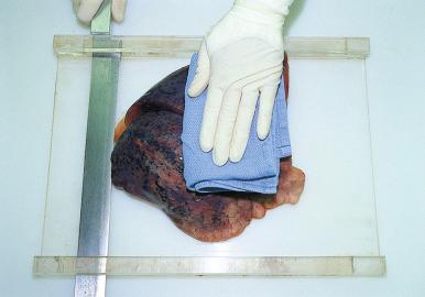
We cut formalin-inflated lungs in 1- to 2-cm parasagittal slices. Coronal slices, including a midcoronal cut through the main stem bronchus, may demonstrate a central carcinoma to advantage. The only advantage of horizontal sections is that they correlate with prior cross-sectional imaging. For slicing lungs, a sharp knife with a long blade is a necessity. Although one can cut the lungs freehand, an acrylic plastic (Plexiglas) cutting board with knife slots on two opposite sides permits finer control ( Fig. 4-16 ). After slicing the lungs, the prosector examines the lung parenchyma with eyes and fingers to identify abnormalities and areas of consolidation or scarring. The larger airways and vessels are opened as needed to complete the examination.

Examine the mediastinum, and sample any abnormal lymph nodes. Open the brachiocephalic veins and superior vena cava to look for mural thrombi in patients who had central catheterization. Open the aortic arch and its tributaries anteriorly. Remove the larynx and trachea. Inspect the still-attached thyroid gland. While removing the thyroid gland from the thyroid cartilage, identify and save any tissue resembling the parathyroid glands. Normally, these small glands are tan or light brown and have a more acutely angled edge than the small lobules of fat, lymph nodes, and extraneous bits of thyroid tissue that masquerade as parathyroid glands. The superior parathyroid glands, frequently found at the level of the middle of the posterior border of each lobe of the thyroid gland, rest in a shallow groove. Unfortunately, the inferior parathyroid glands lie in various positions, including but not limited to the fascial sheath of the thyroid gland near its inferior pole, behind and outside the thyroid gland immediately superior to the inferior thyroid artery, or within the substance of the lobe of the thyroid gland near its inferior posterior border. If there is a question of parathyroid disease, weigh the parathyroid glands, because weight is the best criterion for hyperplasia or hypertrophy. After placing the parathyroid glands in a tissue cassette for safe keeping, continue the examination of the thyroid gland by weighing it, inspecting its surface, and then making nearly complete horizontal sections into its parenchyma. Open the larynx and trachea lengthwise along their softer posterior aspects and view their mucosal surfaces, cracking the thyroid cartilage in the midline as needed in atraumatic cases.
Become a Clinical Tree membership for Full access and enjoy Unlimited articles
If you are a member. Log in here