Physical Address
304 North Cardinal St.
Dorchester Center, MA 02124

The function of the circulation is to serve the needs of the body tissues—to transport nutrients to the tissues, to transport waste products away, transport hormones from one part of the body to another and, in general, to maintain an appropriate environment in all the tissue fluids for survival and optimal function of the cells.
The rate of blood flow through many tissues is controlled mainly in response to their need for nutrients and removal of waste products of metabolism. In some organs, such as the kidneys, the circulation serves additional functions. Blood flow to the kidney, for example, is far in excess of its metabolic requirements and is related to its excretory function, which requires that a large volume of blood be filtered each minute.
The heart and blood vessels, in turn, are controlled to provide the cardiac output and arterial pressure needed to supply adequate tissue blood flow. What are the mechanisms for controlling blood volume and blood flow, and how does this process relate to the other functions of the circulation? These are some of the topics and questions that we discuss in this section on the circulation.
The circulation, shown in Figure 14-1 , is divided into the systemic circulation and the pulmonary circulation. Because the systemic circulation supplies blood flow to all the tissues of the body except the lungs, it is also called the greater circulation or peripheral circulation.
 in the different parts of the circulatory system.")
Before discussing the details of circulatory function, it is important to understand the role of each part of the circulation.
The function of the arteries is to transport blood under high pressure to the tissues. For this reason, the arteries have strong vascular walls, and blood flows at a high velocity in the arteries.
The arterioles are the last small branches of the arterial system; they act as control conduits through which blood is released into the capillaries. Arterioles have strong muscular walls that can close the arterioles completely or, by relaxing, can dilate the vessels severalfold; thus, the arterioles can vastly alter blood flow in each tissue in response to its needs.
The function of the capillaries is to exchange fluid, nutrients, electrolytes, hormones, and other substances between the blood and interstitial fluid. To serve this role, the capillary walls are thin and have numerous minute capillary pores permeable to water and other small molecular substances.
The venules collect blood from the capillaries and gradually coalesce into progressively larger veins.
The veins function as conduits for transport of blood from the venules back to the heart. The veins also serve as a major reservoir of extra blood. Because the pressure in the venous system is low, the venous walls are thin. Even so, they are muscular enough to contract or expand and thereby serve as a controllable reservoir for the extra blood, either a small or a large amount, depending on the needs of the circulation.
Figure 14-1 provides an overview of the circulation and lists the percentages of total blood volume in major segments of the circulation. For example, about 84% of the entire blood volume of the body is in the systemic circulation, and 16% is in the heart and lungs. Of the 84% in the systemic circulation, approximately 64% is in the veins, 13% is in the arteries, and 7% is in the systemic arterioles and capillaries. The heart contains 7% of the blood, and the pulmonary vessels contain 9%.
Most surprising is the low blood volume in the capillaries. It is here, however, that the most important function of the circulation occurs—diffusion of substances back and forth between the blood and tissues, as discussed in Chapter 16 .
If all the systemic vessels of each type were put side by side, their approximate total cross-sectional areas for the average human would be as follows:
| Vessel | Cross-Sectional Area (cm 2 ) |
| Aorta | 2.5 |
| Small arteries | 20 |
| Arterioles | 40 |
| Capillaries | 2500 |
| Venules | 250 |
| Small veins | 80 |
| Venae cavae | 8 |
Note particularly that the cross-sectional areas of the veins are much larger than those of the arteries, averaging about four times those of the corresponding arteries. This difference explains the large blood storage capacity of the venous system in comparison with the arterial system.
Because the same volume of blood flow (F) must pass through each segment of the circulation each minute, the velocity of blood flow (v) is inversely proportional to the vascular cross-sectional area (A):

Thus, under resting conditions, the velocity averages about 33 cm/sec in the aorta but is only 1/1000 as rapid in the capillaries—about 0.3 mm/sec. However, because the capillaries have a typical length of only 0.3 to 1 millimeter, the blood remains in the capillaries for only 1 to 3 seconds, which is surprising because all diffusion of nutrient food substances and electrolytes that occurs through the capillary walls must be performed in this short time.
Because the heart pumps blood continually into the aorta, the mean pressure in the aorta is high, averaging about 100 mm Hg. Also, because heart pumping is pulsatile, the arterial pressure normally alternates between an average systolic pressure level of 120 mm Hg and a diastolic pressure level of 80 mm Hg under resting conditions, as shown on the left side of Figure 14-2 .
 in the different portions of the circulatory system when a person is lying in the horizontal position.")
As the blood flows through the systemic circulation, its mean pressure falls progressively to about 0 mm Hg by the time it reaches the termination of the superior and inferior venae cavae where they empty into the right atrium of the heart.
The pressure in many of the systemic capillaries varies from as high as 35 mm Hg near the arteriolar ends to as low as 10 mm Hg near the venous ends, but their average functional pressure in most vascular beds is about 17 mm Hg, a pressure low enough that little of the plasma leaks through the minute pores of the capillary walls, even though nutrients can diffuse easily through these same pores to the outlying tissue cells. In some capillaries, such as the glomerular capillaries of the kidneys, the pressure is considerably higher, averaging about 60 mm Hg and causing much higher rates of fluid filtration.
At the far-right side of Figure 14-2 , note the respective pressures in the different parts of the pulmonary circulation. In the pulmonary arteries, the pressure is pulsatile, just as in the aorta, but the pressure is far less; pulmonary artery systolic pressure averages about 25 mm Hg and diastolic pressure averages about 8 mm Hg, with a mean pulmonary arterial pressure of only 16 mm Hg. The mean pulmonary capillary pressure averages only 7 mm Hg. Yet, the total blood flow through the lungs each minute is the same as through the systemic circulation. The low pressures of the pulmonary system are in accord with the needs of the lungs because all that is required is to expose the blood in the pulmonary capillaries to oxygen and other gases in the pulmonary alveoli.
Although the details of circulatory function are complex, three basic principles underlie all functions of the system.
Blood flow to most tissues is controlled according to the tissue needs. When tissues are active, they need an increased supply of nutrients and therefore more blood flow than when at rest, occasionally as much as 20 to 30 times the resting level. However, the heart normally cannot increase its cardiac output more than four to seven times higher than resting levels. Therefore, it is not possible simply to increase blood flow everywhere in the body when a particular tissue demands increased flow. Instead, the microvessels of each tissue, especially the arterioles, continuously monitor tissue needs, such as the availability of oxygen and other nutrients and the accumulation of carbon dioxide and other tissue waste products. These microvessels, in turn, dilate or constrict to control local blood flow at the level required for the tissue activity. Also, nervous control of the circulation from the central nervous system and hormones provides additional help in controlling tissue blood flow.
Cardiac output is the sum of all the local tissue flows. When blood flows through a tissue, it immediately returns by way of the veins to the heart. The heart responds automatically to this increased inflow of blood by pumping it immediately back into the arteries. Thus, as long as the heart is functioning normally, it acts as an automaton, responding to the demands of the tissues. The heart, however, often needs help in the form of special nerve signals to make it pump the required amounts of blood flow.
Arterial pressure regulation is generally independent of either local blood flow control or cardiac output control. The circulatory system is provided with an extensive system for controlling the arterial blood pressure. For example, if at any time the pressure falls significantly below the normal level of about 100 mm Hg, a barrage of nervous reflexes elicits a series of circulatory changes to raise the pressure back toward normal within seconds. The nervous signals especially do the following: (a) increase the force of heart pumping; (b) cause contraction of the large venous reservoirs to provide more blood to the heart; and (c) cause generalized constriction of the arterioles in many tissues so that more blood accumulates in the large arteries to increase the arterial pressure. Then, over more prolonged periods—hours and days—the kidneys play an additional major role in pressure control by secreting pressure-controlling hormones and regulating blood volume.
Thus, the needs of the individual tissues are served specifically by the circulation. In the remainder of this chapter, we begin to discuss the basic control of tissue blood flow, cardiac output, and arterial pressure.
Blood flow through a blood vessel is determined by two factors: (1) pressure difference of the blood between the two ends of the vessel, also sometimes called the pressure gradient along the vessel, which pushes the blood through the vessel; and (2) the impediment to blood flow through the vessel, which is called vascular resistance. Figure 14-3 demonstrates these relationships, showing a blood vessel segment located anywhere in the circulatory system.

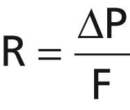
P 1 represents the pressure at the origin of the vessel and P 2 is the pressure at the other end. Resistance occurs as a result of friction between the flowing blood and the intravascular endothelium all along the inside of the vessel. The flow through the vessel can be calculated by the following formula, which is called Ohm’s law :

in which F is blood flow, ΔP is the pressure difference (P 1 − P 2 ) between the two ends of the vessel, and R is the resistance. This formula states that the blood flow is directly proportional to the pressure difference but inversely proportional to the resistance.
Note that it is the difference in pressure between the two ends of the vessel, not the absolute pressure in the vessel, that determines flow rate. For example, if the pressure at both ends of a vessel is 100 mm Hg and no difference exists between the two ends, there will be no flow, despite the presence of 100 mm Hg pressure.
Ohm’s law, illustrated in the preceding formula, expresses one of the most important of all the relationships that the reader needs to understand to comprehend the hemodynamics of the circulation. Because of the extreme importance of this formula, the reader should also become familiar with its other algebraic forms:

Blood flow rate means the quantity of blood that passes a given point in the circulation in a given period of time. Ordinarily, blood flow is expressed in milliliters per minute or liters per minute, but it can be expressed in milliliters per second or in any other units of flow and time.
The overall blood flow in the total circulation of an adult person at rest is about 5000 ml/min. This is called the cardiac output because it is the amount of blood pumped into the aorta by the heart each minute.
Many mechanical and mechanoelectrical flowmeter devices can be inserted in series with a blood vessel or, in some cases, applied to the outside of the vessel to measure blood flow.
An electromagnetic flowmeter, the principles of which are illustrated in Figure 14-4 , can be used to measure blood flow experimentally without opening the blood vessel. Figure 14-4 A shows the generation of electromotive force (electrical voltage) in a wire that is moved rapidly in a cross-wise direction through a magnetic field. This is the well-known principle for production of electricity by the electric generator. Figure 14-4 B shows that the same principle applies for generation of electromotive force in blood that is moving through a magnetic field. In this case, a blood vessel is placed between the poles of a strong magnet, and electrodes are placed on the two sides of the vessel perpendicular to the magnetic lines of force. When blood flows through the vessel, an electrical voltage proportional to the rate of blood flow is generated between the two electrodes, and this voltage is recorded using an appropriate voltmeter or electronic recording apparatus. Figure 14-4 C shows an actual probe that is placed on a large blood vessel to record its blood flow. The probe contains both the strong magnet and the electrodes.

A special advantage of the electromagnetic flowmeter is that it can record changes in flow in less than 1/100 of a second, allowing for the accurate recording of pulsatile changes in flow, as well as steady flow.
Another type of flowmeter that can be applied to the outside of the vessel and that has many of the same advantages as the electromagnetic flowmeter is the ultrasonic Doppler flowmeter, shown in Figure 14-5 . A minute piezoelectric crystal is mounted at one end in the wall of the device. This crystal, when energized with an appropriate electronic apparatus, transmits ultrasound at a frequency of several hundred thousand cycles per second downstream along the flowing blood. A portion of the sound is reflected by the red blood cells in the flowing blood. The reflected ultrasound waves then travel backward from the blood cells toward the crystal. These reflected waves have a lower frequency than the transmitted wave because the red blood cells are moving away from the transmitter crystal. This effect is called the Doppler effect. (It is the same effect that one experiences when a train approaches and passes by while blowing its whistle. Once the whistle has passed by the person, the pitch of the sound from the whistle suddenly becomes much lower than when the train is approaching.)

For the flowmeter shown in Figure 14-5 , the high-frequency ultrasound wave is intermittently cut off, and the reflected wave is received back onto the crystal and greatly amplified by the electronic apparatus. Another portion of the electronic apparatus determines the frequency difference between the transmitted wave and the reflected wave, thus determining the velocity of blood flow. As long as the diameter of a blood vessel does not change, changes in blood flow in the vessel are directly related to changes in flow velocity.
Like the electromagnetic flowmeter, the ultrasonic Doppler flowmeter is capable of recording rapid pulsatile changes in flow, as well as steady flow.
Become a Clinical Tree membership for Full access and enjoy Unlimited articles
If you are a member. Log in here