Physical Address
304 North Cardinal St.
Dorchester Center, MA 02124

Athletic participation frequently places the hand and wrist at risk of injury, which may lead to sprains, fractures, and dislocations. Reports in the literature suggest that 3% to 25% of all athletic injuries involve the hand and wrist. Additionally, overuse in competition and training may lead to tendinopathy and tendon rupture. Certain injuries are more common in particular sports and various playing positions. For instance, baseball players are subject to repeated blunt trauma to the palm from catching and batting; consequently fractures to the hook of the hamate are high on the differential diagnosis. For cyclists, gripping of the handlebar may cause compression of the ulnar nerve at the wrist. Certain sports place exceedingly high demands on the hand and wrist. For instance, gymnasts are reported to have a 49% to 87% incidence of wrist injury over the course of their participation.
Consultants must therefore be familiar with the spectrum of pathology affecting the athlete's hand and wrist and then use a thorough and methodical history and physical examination to narrow the differential diagnosis. With the numerous pathologic conditions that may affect the wrist, we suggest a four-quadrant approach for describing the hand and wrist anatomically. These quadrants are as follows: the radial quadrant , defined by the area from the flexor carpi radialis (FCR) to the extensor carpi radialis longus (ECRL); the dorsal quadrant , defined by the area between the ECRL and the extensor carpi ulnaris (ECU); the ulnar quadrant, defined by the area from the ECU to the flexor carpi ulnaris (FCU); and the volar quadrant , from the FCU to the FCR. This chapter discusses specific conditions encountered within each of these quadrants in an effort to compartmentalize the possible differential diagnosis of an injured athlete ( Table 67.1 ) . Such a differential diagnosis is critical to have in mind in examining the athlete's hand and wrist.
As with any patient encounter, general information should initially be gathered on all patients, including the individual's age, sport, position, hand dominance, any preexisting pain or injury to the involved hand/wrist, worsening or alleviating factors, and associated medical conditions. In obtaining a history, the patient should be asked about the chronicity, mechanism of injury, initial management, subjective weakness, and any associated loss of consciousness. For athletes, it is important to know the player's sport and position as well as the time of season to accurately understand the specific stresses placed on the hand and wrist. This is important not only for diagnosis of the injury but also to guide appropriate return to play (RTP). The identical injury in the throwing hand of a quarterback will likely be managed very differently than the same injury in a defensive lineman.
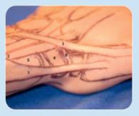
In examining a patient's wrist and forearm, the examiner should be seated across from the patient with the patient's elbow resting on the examination table, the hand toward the ceiling, and the forearm in neutral rotation ( Fig. 67.1 ). General inspection should include any focal areas of swelling, ecchymosis, effusion, open wound or laceration, and any finger or joint misalignment. The patient should be asked to point to the area of maximal tenderness, which should be evaluated last. This is important not only to effectively rule out associated pathology but also to make the patient as comfortable as possible and gain his or her trust.

Next, active and passive range of motion (ROM) should be evaluated with the use of a goniometer. This should be determined for all relevant joints of the digits, thumb, and wrist. Specifically, active and passive motion of the wrist includes flexion and extension, radial and ulnar deviation, as well as pronation and supination. During active or passive motion, any pain or mechanical symptoms of locking or popping should be documented. It is quite common for true carpal pathology to involve some loss of wrist flexion. Normal wrist active ROM is approximately 80 degrees flexion, 70 degrees extension, 30 degrees ulnar deviation, 20 degrees radial deviation, and 90 degrees of both pronation and supination. ROM is always compared with the opposite side.
Next, grip strength should be obtained with the use of a dynamometer and compared with the contralateral, unaffected side. Weakness of grip strength is a very sensitive global assessment tool for wrist and hand pain and can be used to track improvement in subsequent visits and also aid in RTP decisions (many physicians use 85% grip strength as a relative guideline in the RTP algorithm).
The most useful tool in ascertaining the correct diagnosis for hand and wrist pathologies is knowledge of the surface anatomy. The majority of hand and wrist structures are directly palpable once the examiner learns their exact locations. The point of maximal tenderness is the most critical physical examination maneuver in obtaining the correct diagnosis in the hand and wrist.
Lastly no physical examination is complete without a detailed neurovascular examination. Radial and ulnar pulses as well as capillary refill to all fingertips should be documented in all patients. If there is any concern for vascular compromise, an Allen test should be performed. This is described in detail in the physical examination portion of this chapter. Median, ulnar, and radial sensory examination should be carried out preferably with a two-point discriminator, with normal being less than 5 mm. Intrinsic and extrinsic muscle strength should be documented, along with any associated muscle atrophy. Sport-specific neurovascular pathologies exist, such as ulnar nerve entrapment in Guyon's canal in cyclists, ulnar digital nerve injuries in bowlers, and hypothenar hammer syndrome in baseball catchers ; these can help the examiner to focus the exam.
Although discussed in a different chapter, the elbow should always be evaluated as well, since concomitant injuries can occur and be overlooked in the presence of a hand or wrist injury. Finally, the physical examination of the wrist should proceed in a systematic fashion. Each examiner will develop a personal approach, but in general a quadrant-based approach culminating with the region of maximal tenderness is recommended. The remainder of this chapter is devoted to exploring the physical exam of the hand and wrist and a discussion of associated pathology.
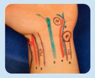
The radial quadrant is defined as the area extending from the FCR to the ECRL ( Fig. 67.2 ). Common pathologies affecting the radial quadrant include FCR tendinopathy, scaphoid or trapezial ridge fractures, De Quervain tenosynovitis, intersection syndrome, thumb carpometacarpal (CMC) synovitis, fracture or dislocation, volar wrist ganglion, superficial radial nerve entrapment, and scaphotrapezotrapezoidal (STT) or thumb CMC arthritis.
 and location of pain in De Quervain tenosynovitis. 2 , ECRL next to ECRB as they cross below APL and EPB and location of pain in intersection syndrome. 3 , EPL. 4 , Scaphoid waist in the anatomic snuffbox. 5 , Radial styloid.")
On examination, the FCR tendon is subcutaneous and can be palpated in its entirety from the musculotendinous junction until it enters the fibro-osseous FCR tunnel at the STT joint on its way to insert on the second metacarpal base. The FCR can be more easily visualized by placing the wrist in flexion and slight radial deviation and having the patient resist wrist flexion. Tenderness along the FCR tendon distally is seen in FCR tendinopathy or FCR tunnel syndrome and may be exacerbated with resisted wrist flexion. Just distal to the wrist flexion crease and in line with the course of the FCR is the bony protuberance of the distal pole of the scaphoid (volar tubercle or scaphoid tuberosity). This is a critical structure to be able to palpate reliably. It will become more prominent with wrist radial deviation and flexion and less palpable with ulnar deviation and extension. This anatomic landmark is critical for wrist examination and for performing a Watson scaphoid shift test, which is described later in this chapter. Distal pole scaphoid fractures and even scaphoid waist fractures and scapholunate (SL) ligament tears will have pain reproduced with a dorsally applied pressure to the volar tubercle.
Trapezial ridge fractures will present with pain in the thenar region after a fall. The trapezial ridge serves as the radial attachment of the transverse carpal ligament and can be avulsed with a sizable bony fragment. A carpal tunnel radiographic view or computed tomography (CT) scan aids in the diagnosis of this easily overlooked fracture.
Moving slightly radial to the FCR, the radial artery is easily palpable. A pulse exam and an Allen test should be documented. The superficial branch of the radial artery can be palpated as it courses obliquely across the volar-radial aspect of the wrist just proximal to the scaphoid tuberosity. The deep branch of the radial artery dives beneath the first dorsal compartment into the proximal aspect of the anatomic snuffbox.
Moving dorsal and radial, the next palpable structure is the radial styloid and first dorsal compartment (abductor pollicis longus [APL] and extensor pollicis brevis [EPB]). The very tip of the radial styloid is easily palpable and represents the distal edge of the first dorsal compartment's tendon sheath. Tenderness at this location may represent radial styloid fracture or radiocarpal ligament sprain in the acute injury setting or, more often, De Quervain tenosynovitis in the subacute or insidious presentation. In the athletic population, De Quervain tendonitis is the most common wrist tendonitis, particularly in racquet sport and rowing athletes. A Finkelstein test has been shown to be reliable and very sensitive (though not always specific) for De Quervain tenosynovitis. The Finkelstein test is performed by the examiner passively flexing the patient's thumb and assessing for pain, whereas the Eichoff test is performed by having the patient place the thumb within a closed fist and ulnarly deviate the wrist ( Fig. 67.3 ). Pain may also be elicited with resisted thumb abduction and extension. Similar pain located about 4 to 5 cm proximal to the radial styloid and slightly more ulnar is typically due to intersection syndrome. Intersection syndrome is caused by inflammation between the first and second extensor compartments, which contain the APL and EPB and the ECRL and extensor carpi radialis brevis (ECRB), respectively. Often, intersection syndrome is accompanied by audible or palpable crepitation at this precise location and may also manifest a positive Finkelstein test. This is frequently seen in rowers, weight lifters, football linemen, and powder skiers due to the repetitive wrist extension and radial deviation that these activities require.

Dorsal to the first extensor compartment is the anatomic snuffbox (bordered by the first extensor compartment volarly, the extensor pollicis longus [EPL] dorsally, and the radial styloid proximally). The location of the scaphoid waist correlates with the anatomic snuffbox. Normally, it has a concave appearance, and fullness with tenderness in this region should always raise suspicion for a scaphoid fracture.
Fractures are common after a collision or fall in sports. These present with immediate pain, swelling, and tenderness. The scaphoid is the most commonly fractured carpal bone, representing roughly two-thirds of all carpal fractures; therefore so a high index of suspicion should always be maintained. The typical mechanism of injury is a fall onto an outstretched hand. These injuries are very common in sports such as, basketball, skiing, snowboarding, and skateboarding. It has been recommended that all athletes with radial-sided wrist pain should carry the diagnosis of scaphoid fracture until proven otherwise and all patients with snuffbox tenderness should have dedicated radiographs to rule out a possible scaphoid fracture. Less common and at times easily missed are fractures of the trapezial ridge. The mechanism of injury is often the same as for a scaphoid fracture but the pain is located in the thenar eminence. If missed, a malunited trapezial ridge fracture has been reported to lead to FCR tendonitis in a professional baseball player. We have seen one case of FCR rupture in a professional football player after this injury ( Fig. 67.4 ).
 Posteroanterior x-ray of the wrist demonstrating a malunited trapezial ridge fracture (blue arrow) . (B) Magnetic resonance imaging demonstrating a flexor carpi radialis rupture from a malunited trapezial ridge fracture.")
Degenerative processes such as STT and CMC arthritis are common in aging athletes and most often present insidiously; however, often an acute event will exacerbate an only mild preexisting pain. Traditionally CMC arthritis is assessed with a CMC axial grind test, but more recently Gelberman and colleagues described thumb adduction and extension provocative testing, which was shown to be more sensitive than the grind test. This is performed by stabilizing the patient's trapezium in one hand (located at the distal edge of the anatomic snuffbox) and loading the thumb axially, all while passively circumducting the thumb ray with the other hand. Pain and crepitation are hallmarks of CMC arthritis with this maneuver. Tenderness over the thumb CMC joint in the setting of trauma should heighten concern for a thumb metacarpal base fracture, including extra-articular metaphyseal base fractures, Bennett, and Rolando fractures. The thumb CMC joint is most easily palpable along the radial side of the thumb metacarpal at the distal edge of the snuffbox, whereas the STT joint is more easily palpable volarly, just distal to the scaphoid distal pole.
Wartenberg syndrome can be a source of radial quadrant pain. This superficial radial nerve entrapment underneath the brachioradialis produces pain, paresthesia, dysesthesia, and/or a positive Tinel sign typically 1 to 2 cm proximal to the radial styloid. This too can produce a falsely positive Finkelstein maneuver.
The radial quadrant is the second most common location for a ganglion cyst (dorsal adjacent to the SL ligament being the most common). Volar wrist ganglions are typically intimately associated with the radial artery adjacent to the FCR tendon and represent the most commonly encountered mass in the radial quadrant. They typically arise from either the FCR tendon sheath or radioscaphoid joint and can be a source of radial quadrant pain. A differential diagnosis of a ganglion cyst in this location is an adventitial cyst, which is a cyst emanating off a branch of the radial artery.
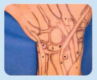
Wrist pathology affecting the dorsal quadrant includes carpal instability (SL ligament tears, midcarpal instability, perilunate injuries); extensor tendinopathy and carpal boss; distal radius, capitate and lunate fractures, Kienbock disease; and dorsal ganglion cysts. Lister's tubercle is the key landmark for identifying dorsal quadrant pathology ( Fig. 67.5 ). This raised dorsal bony prominence of the distal radius separates the second compartment (ECRL and ECRB) from the third compartment (EPL). With wrist ROM, this prominence will remain stationary.
. 2, Second dorsal compartment (ECRL and ECRB) and location of intersection syndrome. 3, Third dorsal compartment (EPL); the fourth and fifth compartments have been omitted for clarity. 4, Sixth dorsal compartment (ECU). 5 , Lunate. 6 , Triquetrum. 7 , Insertion of ECRL and ECRB and location of carpal boss.")
As in the radial quadrant, pain from tendinopathy is most often seen from overuse and repetitive sports injuries. Extensor tendonitis can at times present with rather impressive swelling and pain over the wrist or digital extensor tendons residing just radial and ulnar, respectively, to Lister's tubercle. A bony prominence known as a carpal boss can exist at the insertion of the radial wrist extensors. Although often clinically silent, they can become symptomatic with overuse or after a direct blow to this area is sustained. Grasping the second and third metacarpal heads and shucking one palmar and one dorsal may reproduce pain from a carpal boss. Moving from Lister's tubercle distally about 1 cm toward the long finger, the examiner's finger falls into the concavity of the radiocarpal joint. This point is directly over the dorsal SL ligament and is known as the SL interval. With wrist flexion, the proximal pole of the scaphoid can be palpated just radial to this point and the lunate just ulnar to this point. Tenderness over the proximal pole of the scaphoid may indicate either fracture or scaphoid impaction. Scaphoid impaction, often seen in gymnasts and weight lifters, occurs with repetitive wrist hyperextension resulting in the scaphoid impinging on the dorsal edge of the radius; it presents with pain upon wrist extension with the potential for osteophyte formation.
The SL interval is the most typical location for a dorsal ganglion cyst. In addition to an injury to the SL ligament, an occult dorsal ganglion can produce pain to palpation over this location. Less commonly, anomalous muscles such as the extensor digitorum brevis manus can be located in this area and produce a soft tissue mass and accompanying pain mimicking a ganglion ( Fig. 67.6 ). If uncertainty exists as to whether a mass is a ganglion, simple transillumination or aspiration can easily distinguish a ganglion from anomalous muscles or solid tumors. Along the ulnar edge of the SL interval, the lunate is palpable, particularly with wrist flexion. Tenderness here may represent an SL ligament tear, lunate fracture, lunotriquetral (LT) tear, or Kienbock disease.

Testing the stability of the SL ligament is a critical examination skill for anyone caring for wrist injuries. Carpal instability encompasses a broad spectrum of ligamentous deficiency, including acute and chronic conditions from traumatic and atraumatic causes. In athletes, the most common of these, particularly following an acute traumatic event, is injury of the SL ligament. These are very common injuries in contact sports and the examiner should always have a high index of suspicion, since missed injuries can lead to a chronic debilitating wrist arthritis known as scapholunate advanced collapse (SLAC).
The classic scaphoid shift test was described by Watson and is the most important provocative maneuver in testing SL stability. The maneuver is performed beginning with the patient's wrist in extension and ulnar deviation. The examiner places his or her hand with the thumb over the distal pole of the scaphoid and fingers wrapped around the ulnar side of the patient's hand for stability. The patient's hand is then passively brought into flexion and radial deviation while dorsal pressure is applied to the scaphoid distal pole, resisting its tendency for volar translation ( Fig. 67.7 ). In the setting of a significant SL ligament disruption, the proximal pole of the scaphoid will be temporarily forced onto the dorsal rim of the radius and then clunk back into the scaphoid fossa when pressure is released. The results of the test should always be compared with the opposite side for both stability and pain elicited by the maneuver. It is very common for patients without a history of wrist trauma, especially those with generalized ligamentous laxity, to have painless or minimally painful clunks with a scaphoid shift test. Easterling found that 32% of asymptomatic patients without a history of trauma had bilateral painless “clunking” and another 14% with unilateral painless findings. Asymmetric pain and instability combined with a feasible mechanism of injury are most consistent with an SL ligament tear. When the scaphoid shift test produces only pain, the differential diagnosis includes occult dorsal carpal ganglion, scaphoid impaction, and radioscaphoid or STT chondromalacia. Additional maneuvers to test for SL stability include the scaphoid lift test and scaphoid thrust test, but these are much less comprehensively described and practiced. Both tests seek to understand abnormal carpal motion during dynamic stress testing. Chronic cases of SL instability may lead to pain attributed to a predictable pattern of degenerative changes known as SLAC wrist.
 The examiner places his or her hand with the thumb over the distal pole of the scaphoid and fingers wrapped around the ulnar side of the patient’s hand for stability. (B) The patient’s hand is then passively brought into flexion and radial deviation while dorsal pressure is applied to the scaphoid distal pole, resisting its tendency for volar translation. (C) In a positive test, once the scaphoid is released, it will clunk back into the scaphoid fossa.")
Midcarpal instability may be reproduced with a catch-up clunk test. This maneuver involves the patient beginning with the forearm pronated and the wrist radially deviated. The patient is then asked to move the wrist from radial deviation (during which time the proximal row is flexed and the distal row relatively volar) into ulnar deviation (which extends the proximal row and translates the distal row dorsally). Midway into this maneuver, a visible and audible clunk will occur in the setting of midcarpal instability as the distal row suddenly shifts dorsal and the proximal row shifts into extension. Since an identical clunk can occur in the setting of radiocarpal instability as the proximal row shifts dorsally (rather than into extension as seen in midcarpal instability), this test lacks specificity. An additional test for midcarpal instability reported by Lichtman et al. is the midcarpal shift test. In this test the patient's forearm is placed in pronation with the wrist secured in neutral while a volarly directed pressure is applied to the dorsum of the capitates. The amount of midcarpal translation is noted and compared with the opposite wrist. Next, an axial load is applied, and the wrist is passively deviated ulnarly. The presence of a clunk or report of pain is noted. A grading system of 1 to 4 has been developed and correlated with measured amounts of translation.
Isolated injuries to the capitate are rare; these more commonly represent a spectrum of greater arc injuries including scaphoid waist and capitate neck fractures, termed “scaphocapitate syndrome.” Capitate stress fractures can also be seen in athletes involved in racquet sports. The head of the capitate is palpable just distal to the SL ligament, especially with slight wrist flexion.
Finally, Kienbock disease must be considered in patients with unexplained dorsal wrist pain. Mild insidious complaints of generalized pain, stiffness, or weakness without a history of substantial trauma should prompt further workup for Kienbock. Examination may be largely nonspecific, with lunate tenderness, minor deficits of motion, and reduced grip strength as compared with the contralateral wrist.
Historically the ulnar quadrant has been the least well understood quadrant of wrist pathology and has been termed “the black box” by Kleinman and others. Ulnar-sided wrist pain is arguably the most common athletic wrist complaint and often imparts a diagnostic challenge to even the most experienced specialists. Recent advancements in our understanding of ulnar-sided wrist pain have led to much improved diagnostic and therapeutic care for these injuries. It is useful to distinguish between pathology arising from the radio ulnar, ulnocarpal, and intercarpal articulations and pathology that is extra-articular. This is due to the numerous intimately positioned structures that are prone to traumatic, inflammatory, or degenerative injury. The pathologies include ECU tendinopathy, triangular fibrocartilage complex (TFCC) injuries, distal radioulnar joint (DRUJ) instability, and FCU tendonitis, ulnar impaction, and LT ligament tears.
Most dorsal in the ulnar quadrant of the wrist is the ECU tendon, residing within the sixth dorsal extensor compartment ( Fig. 67.8 ). Physical examination will often show tenderness and sometimes swelling over the course of the ECU tendon ( Fig. 67.9 ). Pain will typically be recreated with resisted wrist extension and ulnar deviation. Tendinopathy of the ECU is extremely common in the athlete, second only to the incidence of De Quervain. There is a predilection for baseball, hockey, golf, and racquet sports, typically with pain generated by backhand volley. When the forearm is pronated and the wrist is in neutral, the tendon has a direct course through its sheath toward its insertion on the base of the fifth metacarpal. This angle increases, however, when the wrist is positioned in supination, ulnar deviation and wrist extension. Excessive extension, as well as rapid flexion, ulnar deviation, and supination can cause tendinopathy or lead to attritional or traumatic ruptures of the ECU subsheath, resulting in painful subluxation or dislocation of the tendon, often associated with a snapping sensation.


Unfortunately tenderness to palpation over the sixth compartment is not specific, as it loads nearby potential sources of pain. To reduce ambiguity, Ruland et al. described the “ECU synergy test”: With the elbow at 90 degrees of flexion and the forearm fully supinated, the examiner grasps the thumb and long finger. When the patient then radially deviates the thumb against resistance, the ECU contracts isometrically and recreates the patient's pain. Instability of the ECU tendon may be elicited with the “FUSS maneuver”—flexion, ulnar deviation, and supination leading to subluxation. This “ice cream scoop” motion will recreate pain with visible or palpable subluxation of the tendon ( Fig. 67.10 ).

Athletic endeavors that involve high-energy or repetitive axial or torsional forces may lead to ulnar impaction syndrome, tearing of the TFCC, and DRUJ instability. Individuals with ulnar positive variance are known to be at higher risk for developing ulnar impaction. Although the incidence of ulnar positive wrists in athletes is not known, it has been noted that football lineman and gymnasts may be at higher risk for developing ulnar positive variance due to chronic distal radial physeal loading and premature arrest.
The TFCC is encountered in the “soft spot” between the ECU and FCU known as the fovea. Foveal tenderness has high sensitivity and specificity (95% and 87%) for TFCC tear. The Nakamura ulnar impaction test loads the TFCC and will cause pain with abutment or tear. While stabilizing the forearm, the examiner takes the patient's ulnarly deviated hand though supination and pronation, all while applying a proximally directed axial load. Ulnar styloid–triquetral impaction (USTI) occurs in supination, as this is the position in which the ulnar styloid is closest to the carpus. A typical history would include pain in the supinated wrist while holding a hockey stick. The USTI test is positive when pain is elicited as the dorsiflexed wrist is taken from pronation to supination. The “press test,” in which the seated patient pushes out of a chair, places the wrist in the same position. This simple test was reported to have 100% sensitivity for TFCC tearing. It should be kept in mind that while they are sensitive, these ulnocarpal loading maneuvers are not specific to TFCC pathology, as they also load the nearby intercarpal joints as well.
Assessment of DRUJ stability completes the evaluation of the TFCC. Injuries that affect the stability of this joint may be isolated or associated with other injuries ranging from frank traumatic dislocation with rotatory mechanical block to more indolent chronic pain and dysfunction with pronosupination activities. On physical examination, the DRUJ is loaded when the ulna is compressed against the radius. Subsequent pain with forearm rotation indicates DRUJ pathology. Increased anterior-to-posterior translation of the radius relative to the ulna indicates instability. Several variations of this test have been described. In the radioulnar ballottement test, the examiner applies palmar and then dorsal pressure to the ulnar head and notes translation relative to the radius in neutral, pronated, and supinated positions. A cadaveric study showed statistically significant examiner accuracy in detecting DRUJ instability with the ballottement test after complete TFCC section, but the performance of these tests in incomplete tears or in the clinical setting is unknown.
Moving distally on the dorsal-ulnar wrist, the intercarpal and CMC articulations are examined. Injury to the LT ligament may occur with a fall, particularly with hyperextension and twisting, although the exact mechanism is controversial. Several maneuvers have been described to specifically elicit instability and pain originating from this joint. First, pain may be elicited with direct palpation of the ligament on the dorsum of the wrist, about a fingerbreadth distal to the DRUJ. Compression of the triquetrum against the lunate with ulnar-to-radial pressure by the examiner may recreate symptoms and has been recommended by some as having the highest sensitivity and specificity. This is similar to the TFCC foveal test, but the anatomic location is immediately dorsal to the pisiform instead of immediately distal to the ulnar styloid. Other tests include the LT “ballottement” as described by Regan or the LT “shear” test as described by Kleinman. Both involve stabilizing the lunate with one hand and the pisotriquetral unit in the other while applying forces in opposite directions across the joint.
CMC fractures at the base of the fifth metacarpal are common following blunt-force injury or after striking with the ulnar side of the hand. Dorsal triquetral fractures are also common after a fall during sports and should be kept in mind when there is focal pain over the dorsal aspect of the triquetrum. A pronated oblique radiograph is sometimes necessary to find this small avulsion fracture. Anatomically, this represents more of an extrinsic ligamentous avulsion than a true carpal fracture.
Due to the proximity of pathology affecting the wrist in the ulnar quadrant, differentiating between one or more diagnoses can be challenging. For example, ECU tendinopathy and TFCC injuries commonly overlap. It can be advantageous to perform a differential lidocaine injection test, which we frequently supplement with corticosteroid once the main area of involvement is discerned. The first location is chosen and a small amount of anesthetic (typically 0.05 mL) is administered. After a few minutes, the amount of pain improvement is noted. It is important to have the patient try to reproduce the previous pain with aggravating wrist positions or offending loading maneuvers. Another selective injection is given at a second (or even third) area and this response of pain improvement is compared to the first response. If performed properly and patiently, this exercise is a powerful diagnostic adjunct with the potential for more accurate therapeutic intervention.
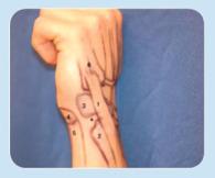
The volar quadrant houses the most critical neurovascular structures of the hand and wrist ( Fig. 67.11 ). The ulnar artery and nerve course deep and radial to the FCU tendon and enter the wrist at Guyon's canal. In this location the ulnar artery is relatively superficial and may be compressed against the underlying hamate. Repetitive blunt trauma may therefore cause local arterial occlusion or aneurysmal dilation. This may further lead to distal embolization or compression of the nearby ulnar nerve. These clinical findings are referred to as the hypothenar hammer syndrome , which is well described in baseball catchers as well as some tennis players and golfers. It usually presents as dysaesthesia or cold intolerance of the ring and small fingers.

The ulnar nerve is more commonly compressed at the elbow, particularly in throwing athletes. Compression or irritation at the wrist, however, may also occur at the Guyon canal. This may be due to an extrinsic mass effect such as ganglion, lipoma, or aneurysmal dilation of the ulnar artery as well as focal prolonged external compression. This last entity is well described in cyclists.
The median nerve, along with the digital flexor tendons, crosses the wrist in a fibro-osseous sheath known as the carpal tunnel. Compression of the median nerve within this sheath is the most common peripheral compression neuropathy but is not considered particularly common in the athletic population. Nevertheless, it has been described in sports that require forceful or prolonged gripping such as cycling and rock climbing or repeated direct volar pressure on the extended wrist as in wheelchair rim propulsion. Additionally, exposure to vibration has been associated with a risk of carpal tunnel syndrome, with implications for motor racing athletes.
From an osseous standpoint, the pisiform and hook of the hamate are important to consider, given their higher predilection for injury during sport. The pisiform is a mobile sesamoid within the FCU tendon, which may then continue to insert on portions of the hamate and base of the fifth metacarpal. The hamate hook protrudes from the hamate body and is a site of muscular and ligamentous attachment. Injuries to these ulnar-sided carpal bones are relatively rare but should be considered in athletes who use bats, racquets, clubs, or sticks, as errant impact is transmitted directly to the hypothenar region of the nondominant palm. A history of a “check swing” in baseball or a “fat” shot in golf should further raise suspicion. A fall onto the outstretched hand may also be responsible for fractures of these structures. Increased participation in sports has led to a recent increase in the prevalence of acute ulnar carpal fractures and chronic stress fractures. Additionally, arthritis may develop between the pisiform and the triquetrum with which it articulates.
Inserting onto the pisiform, the FCU can be a source of volar-ulnar wrist pain. Tendinosis may develop with repetitive microtrauma such as occurs in racquet sports. Occasionally acute calcific tendinitis may develop, which may be mistaken for infection or gout due to the sometimes dramatic erythema and intense pain associated with this condition.
In the volar quadrant, physical examination begins with searching for bony tenderness at prominences of the pisiform and then the hook of the hamate. The pisiform is subcutaneous and easy to locate at the level of the wrist flexion crease, but the hamate hook is deep to the hypothenar musculature. It can be located by placing the examiner's thumb IP flexion crease on the pisiform and aiming the tip of the examiner's thumb radially and distally toward the patient's long finger, but this requires deep palpation. In addition to direct tenderness, a hamate hook fracture will often produce pain with resisted small- and ring-finger flexion, as the hamulus serves as a pulley for these tendons traversing the carpal canal. Delayed flexor tendon injuries may be associated with fractures of the hook of the hamate in up to 17% of cases. It should be kept in mind that fractures of the hook of the hamate may be associated with median or ulnar neuropathy due to the hook's location between the carpal tunnel and Guyon's canal, so concurrent neurologic and vascular assessment is paramount. The pisotriquetral grind test indicates arthritis in this joint and is performed by compressing the pisiform to the triquetrum while flexing and extending the wrist or moving the pisiform medially and laterally. Lastly, radiographic identification of injury to these carpal bones may be difficult because of their three-dimensional shape and overlap with the rest of the carpus. A supinated oblique radiograph of the wrist shows the pisiform in profile well. A carpal tunnel view may identify a fracture of the hook of the hamate, although a CT scan is sometimes necessary for diagnosis in equivocal cases.
More proximally, the FCU tendon is inspected. Physical examination typically reveals tenderness along the tendon proximal to the pisiform as well as pain with wrist flexion and pronation.
Examination of the volar ulnar side of the wrist is not complete without assessing the status of its neurologic and vascular structures. Vascular examination includes examining distal perfusion to the digits and thumb. Altered color, decreased temperature, loss of tissue turgor, sluggish capillary refill, and even ulcerated or gangrenous tips all indicate critical ischemia. Next, the presence of an ulnar artery pulse should be established. Examination with a handheld Doppler is a reliable method for evaluating areas of occlusion, pathways of perfusion, and even the quality of blood flow. The Allen test establishes the patency of the radial and ulnar arteries and the state of the superficial palmar arch connecting them. This test is performed by manually compressing the radial and ulnar arteries and then having the patient open and close his or her fist several times to exsanguinate the hand. Pressure is then released from one of the arteries, which in a normal state (complete superficial palmar arch) should reperfuse the entire hand in less than 3 seconds. The test is then repeated with release of the other artery, and resulting flow is documented ( Fig. 67.12 ).
 This test is performed by manually compressing the radial and ulnar arteries, then having the patient open and close his or her fist several times to exsanguinate the hand. (B) Pressure is then released from one of the arteries, which in a normal state (complete superficial palmar arch), should reperfuse the entire hand in less than 3 seconds. (C) The test is then repeated with the release of the other artery, and the resulting flow is documented.")
The function of the ulnar nerve should be assessed by testing two-point sensibility along the small finger. Intrinsic muscle bulk, tone, and function should likewise be examined. The Froment sign assesses key grip and the function of the adductor pollicis, which is innervated by the terminal deep motor branch of the ulnar nerve. The patient is asked to forcefully key pinch a piece of paper, which the examiner attempts to pull away. If the adductor is weak, the flexor pollicis longus (FPL) is recruited as a compensatory mechanism and the IP joint assumes a flexed posture ( Fig. 67.13 ).

Concluding the examination of the volar surface of the wrist is an assessment of the median nerve. History will include numbness in the radial digits during offending activities. Physical examination of the carpal tunnel is well described. Durkan's compression test, which is performed by applying pressure over the carpal tunnel for 30 seconds, is reported to be the provocative test with the highest sensitivity and specificity. Two-point discrimination should be tested for in the volar thumb and index. The thenar musculature should be inspected for atrophy. Asking the patient to keep their thumb out of their palm against resistance tests the motor function of the abductor pollicis brevis.
The hand and digits are especially vulnerable to injury during athletic participation and in fact are the most commonly fractured body sites, making up 32% of all high school sports–related fractures. Both phalangeal and metacarpal fractures are extremely common in sports, with phalangeal fractures representing over half of all sports-related hand fractures. Additionally, injuries to certain ligaments and tendons occur with such frequency that they carry the name of the offending sport, such as skier's thumb or boxer's knuckle.
History should center on the activity and mechanism of injury and acuity or chronicity of the complaint. A relatively violent mechanism with sudden onset of pain—along with swelling, ecchymosis, and crepitus—typically creates a high level of suspicion for fracture. Following any hand injury, radiographs must be obtained to rule out fracture or joint incongruity. Angulation and rotational deformity are noted. Evaluation of neurologic and vascular status as well as that of the overlying skin is emphasized.
Fingertip crush injuries are extremely common in sport and may result in distal phalangeal fractures and nail bed injuries. Distal phalangeal fractures are the most common of all hand fractures. In the skeletally immature athlete, a unique dorsally displaced transphyseal distal phalanx fracture known as a Seymour fracture can present with associated nail bed incarceration in the fracture site ( Fig. 67.14 ). Axial loads to the end of the finger can rupture the terminal tendon producing the inability to extend the distal interphalangeal joint (DIP) joint, which is termed “mallet finger” or “baseball finger.” This injury is particularly common in baseball, football, and basketball and is the most common closed tendon injury in athletes. On the opposite side of the digit, an avulsion of the flexor digitorum profundus (FDP) from its insertion may occur with swift pulling away of a forcefully flexed finger. This frequently occurs while grabbing the jersey of an opponent in football or rugby and therefore is commonly called “jersey finger.” The ring finger is involved in over 75% of cases, although this injury has been reported in all digits. It is well established that timely repair of the flexor tendon, especially when it is retracted into the palm, improves results.
 , known as a Seymour fracture, which can present with associated nail bed incarceration in the fracture site.")
Physical examination starts with noting the resting posture of the affected digit during a passive wrist tenodesis test. The DIP joint will be abnormally extended in the setting of FDP avulsion, particularly when the wrist is passively extended. In this setting, the patient will be unable to flex the DIP joint but should be able to independently flex the proximal interphalangeal joint (PIP) joint, since the FDS is typically not involved simultaneously. Active DIP joint flexion should be tested while blocking PIP and MCP joint motion, as should the adjacent digits. PIP joint flexion may be diminished, however, due to the avulsed FDP tendon stump lodged in the flexor sheath, which impedes the gliding of the FDS tendon of the involved digit.
Impact to the extended finger may rupture the terminal tendon, which is the insertion of the extensor mechanism on the dorsal epiphyseal portion of the distal phalanx. Examination of these “mallet fingers” reveals a flexed DIP joint posture and inability to fully extend it actively. Typically full passive extension is possible, and tenderness is present dorsally at the DIP joint. There may be an associated hyperextension posture of the PIP joint or a swan-neck deformity, particularly in a chronic mallet finger or individual with PIP volar plate laxity. Radiographic evaluation differentiates between bony or purely tendinous injury and assesses congruity of the joint.
The PIP joint is next considered. According to Rettig, the PIP joint is the most commonly injured structure of the hand in athletes. Disruption of the extensor mechanism, collateral ligaments, and/or volar plate may occur in this highly constrained hinge joint. These are usually partial injuries, often referred to as the “jammed finger,” but they can range from minor sprains to open fracture/dislocations. A 10-year review of the National Football League database found dorsal PIP dislocations to be the most common hand injury. Dislocations present with gross malalignment and are often reduced “on the field” by the player or trainer with manual traction. Following closed reduction, the joint should be assessed clinically by applying gentle angular force and ensuring that a stable, concentric ROM is present. It should also be evaluated radiographically to rule out associated fracture and confirm congruent reduction is present. Radiographic stress views may be helpful in detecting subtle instability.
At the level of the PIP joint, the insertion of the extensor mechanism dorsally is known as the central slip . Disruption of the central slip can lead to a boutonnière deformity, and prompt recognition of this injury is paramount.
The extensor mechanism at this joint may be injured in a manner similar to mallet finger but with force concentrated at the middle phalanx instead of the distal phalanx. This may lead to rupture of the central slip, which is the insertion of the extensor mechanism on the dorsal middle phalanx base. The patient may be able to extend the PIP joint in the acute setting, but over time an extension lag and boutonnière deformity may develop. This refers to a posture of flexion at the PIP joint and hyperextension at the DIP joint as well as inability to initiate PIP joint flexion due to volar subluxation of the lateral bands. In the acute setting, an Elson test can aid in the diagnosis of a central slip injury before a boutonnière deformity develops. This test is performed by having the patient flex the involved PIP joint 90 degrees over the end of a table. The examiner then blocks active PIP joint extension with counterpressure over the dorsum of the middle phalanx. In a normal state, there is supple passive flexion of the DIP joint by the examiner, but in the setting of an acute central slip injury, force transmission to the terminal tendon places the DIP joint in rigid extension ( Fig. 67.15 ).
 joint 90 degrees over the end of a table. The examiner then blocks active PIP joint extension with counterpressure over the dorsum of the middle phalanx. In a normal state, there is supple passive flexion of the distal interphalangeal (DIP) joint by the examiner, but in the setting of an acute central slip injury, force transmission to the terminal tendon places the DIP joint in rigid extension.")
A pseudoboutonnière deformity refers to an isolated flexion contracture at the PIP joint without concurrent DIP joint hyperextension. This deformity is secondary to scar contraction of the volar plate after sprain or rupture. It often develops when a small bony avulsion of the volar plate insertion at the middle phalanx base is present and the joint is mistakenly immobilized for a prolonged period. The examiner must distinguish greater tenderness at the volar plate than at the dorsal central slip and also confirm a negative Elson test.
Become a Clinical Tree membership for Full access and enjoy Unlimited articles
If you are a member. Log in here