Physical Address
304 North Cardinal St.
Dorchester Center, MA 02124
The first report on the use of “needle puncture” is referred to in The Kitab al-Tarif ( The Method of Medicine ), the most influential book of Arab medieval medicine, authored by Albucasis or Abu al-Qasim Khalaf ibn al-Abbas Al-Zahrawi (936–1013). Albucasis was the court physician to the caliph of Andalucia and described the first account of fine-needle aspiration (FNA) of the thyroid gland, employing instruments resembling the modern aspiration needle. In 1847, Kun described the use of a needle as a “new instrument for the diagnosis of tumors”. Various sporadic reports follow this initial description, including Leydon (1883), who described the use of needle aspiration to isolate microorganisms from pneumonic effusions, Menetrier (1887), who diagnosed pulmonary carcinoma utilizing needle aspiration, and Griegg and Gray (1904), who diagnosed trypanosomiasis from lymph node aspirates in patients with sleeping sickness. In 1912, a German hematologist, Hans Hirschfield (1873–1944), reported the use of FNA for the diagnosis of cutaneous lymphomas. In his manuscript entitled Über isolierte aleukämische Lymphadenose der Haut , he eloquently described the FNA procedure and making air-dried smears which were stained with May–Giemsa. This method was further improved on by Gordon Ward, a British hematologist, in 1914, in the diagnosis of lymphoblastoma. Similar accounts of obtaining cellular material by needle puncture for diagnosis have been furnished by Anastasios Aravandinos in 1916, a professor of internal medicine in Athens University, and Guthrie from Johns Hopkins Hospital in 1921.
Dudgeon and Patrick (1927) in the UK and Martin and Ellis (1930) at New York's Memorial Hospital for Cancer and Allied Diseases are generally credited with the first description of sampling of tumors by means of a narrow-gauge needle. Fred W. Stewart was the pathologist responsible for the microscopic evaluation of the smears in the New York group, and, in 1933, he described his experience with 2500 tumors analyzed by the aspiration method. In this communication, he discussed several points to be considered for a successful needle aspiration of tumors. Despite this emerging publicity of FNA, there was still distrust in the FNA procedure, particularly by surgeons, who feared the possibility of tumor seeding the needle tract and tumor spread, limiting its widespread application in the USA.
Increasing interest in the FNA technique occurred in the 1950s and 1960s in Northern Europe and Scandinavia, particularly by hematopathologists such as Soderstrom and Franzen in Sweden and Lopes-Cardozo in Holland. They introduced the use of higher gauge needles for performing the aspiration (22 gauge or higher), as opposed to the standard 18-gauge needle that had been used previously in the USA, and defined precise diagnostic criteria to be applied to aspiration biopsies, increasing sensitivity and specificity of the procedure. The Swedish experience with FNA of tumor lesions of the breast, salivary gland, and thyroid in the 1960s and 1970s served as a model for the development of FNA clinics throughout the world. In fact, several clinicians and pathologists from the USA studied with the group in Sweden in the early 1970s, aiding the resurgence of FNA as a useful biopsy method in the USA.
Since the 1970s, improvements in imaging, including computed tomography (CT) scan and ultrasound, in combination with the development of new specialties such as interventional radiology has fueled the use of FNA among clinicians and radiologists. Small and deep-seated lesions, most found incidentally during the workup of a patient with rather vague symptoms, are now easily identifiable and precisely located, making them amenable to FNA. Even more recently, the widespread use of endoscopic ultrasound has allowed gastroenterologists, radiologists, and pulmonologists to target lesions in the chest and abdomen, which previously required open biopsy for their evaluation.
The dominant body sites for FNA procedures remain the thyroid, salivary glands, and superficial lymph nodes, where it has been shown to be efficient, safe, and cost-effective. Advances in high-resolution mammography and stereotactic needle placement have allowed a shift in recent years to core needle biopsies when dealing with breast lesions. In cases of deep-seated lesions, there has been a shift in recent years from FNA only to a combination with micro-core biopsies. In these cases, both the pathologists and the radiologists are involved in the procedure, and a considerable amount of time may be required to localize the lesion and obtain a suitable sample. A combination of FNA and micro-core biopsy assures that the lesion in question is being biopsied and enough tissue is available for evaluation and potential ancillary studies that might be needed for further workup of the case.
The dramatic popularity and demand for FNA cytology is evidenced by the increasing number of journals and meetings devoted primarily to the cytopathology specialty in the USA and European countries. Essentially, every pathology practice in large hospitals has a devoted FNA service. Small tissue samples obtained and interpreted by FNA have become essential in providing a precise diagnosis in the current era of personalized medicine.
The FNA biopsy can be performed on superficially located/palpable and easily accessible lesions, as well as on deep-seated lesions in close proximity to important and vital body organs. Whether the FNA procedure is performed by a clinician, interventional radiologist, or a cytopathologist, an understanding of the anatomy at the site of the lesion is of utmost importance. This is crucial to avoid injury of vital structures that might be in close proximity to the target lesion. This knowledge of basic anatomy is important even in a case of a superficially located lesion. Aspirating a superficially located lesion in the neck might appear of no consequence until one takes into account the presence of major blood vessels traversing the superficial and deep neck structures. Likewise, an aspiration of a chest wall lesion can easily be complicated by a pneumothorax if the performer does not take into consideration the close anatomic relationship of the lesion with the pleural space. This basic knowledge of anatomy is equally important during evaluation of the on-site cytologic preparations. It is important to recognize “normal” contaminants that might be present in a FNA sample to avoid over- or underdiagnosis; for example, the occasional presence of hepatocytes in a smear of a right lower lobe lung lesion or the presence of normal intestinal-type epithelium in a sample of a pancreatic lesion obtained via endoscopic ultrasound-guided FNA.
An appropriate bedside manner is essential for performing an FNA biopsy procedure. This is particularly important when a cytopathologist, whose daily activities do not usually include direct patient contact, is performing the procedure. It allows for a certain rapport to be established with the patient, reducing any anxiety related to the FNA procedure. A cytopathologist might need to brush up on basic clinical skills, including obtaining a basic medical history and performing a physical examination. The physician performing the FNA procedure should discuss the pertinent medical information with the patient, and not always rely on the information provided by the clinicians, which, at times, might be suboptimal. Significant prior medical history, the evolution and duration of the lesion, potential allergies and exposures are all important information that might help in the procedure, diagnosis of the lesion in question, and preventing complications.
The success of an FNA biopsy depends greatly on the technique employed in performing the procedure and making the on-site smears. It is of no use to locate and accurately target a lesion if an incorrect aspiration technique yields only blood and precludes the retrieval of diagnostic material. Likewise, a successful aspiration biopsy can lead to suboptimal results if the cytomorphology is hampered by on-site smears prepared by untrained hands. If the aspiration or the smears are performed by a clinician, it is of utmost importance for them to understand the proper FNA techniques and on-site smear preparation. The clinicians and interventional radiologists performing FNA biopsies should also be aware that, unlike in surgical pathology, in aspiration biopsies an increased quantity of material assessed visually is not necessarily better because a small amount of diagnostic material is more adequate than a large amount of blood obscuring the lesional cells.
In order to triage a specimen adequately, the FNA operator must be aware of the various stains, preparations, and ancillary tests that may be required to arrive at an accurate diagnosis. For example, making a smear and rinsing the syringe in a fixative solution may be suboptimal in cases where flow cytometric analysis may be needed to exclude a lymphoproliferative disorder. At a minimum, a cytotechnologist should be present during most aspiration biopsies to assess the adequacy and guide the clinician as to what additional steps might be necessary to obtain an adequate and representative FNA specimen. Evaluation of adequacy should not be attempted by clinicians or radiologists who lack the basic histologic knowledge. This evaluation requires pattern recognition and high-power evaluation of the material on the slides; experience is required in order to recognize normal tissue from diagnostic lesional material. It is the authors' opinion that a cytopathologist should be involved in the initial on-site evaluation and in the final evaluation of all aspiration biopsies, in order to maximize the success of the procedure. However, in certain instances (especially for thyroid FNA) where the on-site adequacy evaluation by a pathologist is not possible, a clinician can be trained to make on-site smears and differentiate between epithelial cells, inflammatory cells, and non-cellular contaminants.
The FNA biopsy is the procedure by which a needle is inserted into a mass; cells and material from the mass are aspirated and a cytologic diagnosis is rendered based on the cytomorphologic findings. Generally, 25–27-guage needles are utilized, varying in length from 1 to 1.5 inches. However, the specifications of the needle employed may vary according to the site and characteristics of the lesion. As such, for a very small cutaneous lesion such as cutaneous metastasis of a breast carcinoma, a 0.5–1 inch, 25–27-gauge needle is useful, whereas for densely fibrotic and highly vascularized lesions a 25-gauge needle performs better. Significantly longer needles are required when performing an aspiration of a deep-seated lesion, such as transthoracic aspiration of a lung nodule or an aspiration of a metastatic liver lesion.
In an attempt to increase the sensitivity of FNAs, various needle designs have been implemented. The Chiba needle is characteristically used by most radiologists for transthoracic and transabdominal procedures. These needles vary from 18 to 22 gauge and can reach up to 20 cm in length. Re-puncture of the lesion is necessary if additional material is required with the Chiba needle. For this reason, the coaxial needle is preferred by some radiologists. With the coaxial needle, an outer bore serves as a guide to an inner bore. Once the outer bore is in place, the lesion can be re-aspirated multiple times by use of the inner needle. This setting is also of use when aspirating lesions within bone, in which the cortex of the bone must be breached in order to access the lesion.
The Franseen needle, with a notched crown shape, allows for better tissue cutting and retrieval. It is most useful when aspirating fibrous lesions which might not shed enough material with a Chiba needle. The Franseen needle also allows for tissue micro-core retrieval. Therefore, this needle is commonly employed for targeting soft-tissue lesions, which are often fibrotic, and on which additional material for surgical pathology is required for interpretation.
The use of FNA for diagnosis of prostate lesions is no longer standard practice in the USA. The advances in transrectal ultrasound-guided biopsies of the prostate have allowed for an easy, safe, systematic, and highly sensitive method of pathologic evaluation of the prostate gland, which has entirely replaced FNA. Similarly, the use of FNA of breast lesions has declined steadily during the last two decades. This is largely due to the increase in the sensitivity of radiologic imaging and the advances in core needle biopsy techniques, which have made it fast, safe, and easy to obtain tissue for diagnosis and hormone status testing. Moreover, there are certain significant limitations of breast FNAs, such as obtaining sufficient tissue for diagnosis in sclerotic lesions or clearly distinguishing between invasive or in situ tumors.
Complications associated with FNA of superficial lesions are minimal, when utilizing 22–77-gauge needles. They are usually limited to small hematomas, even in patients with hemostatic defects. Pneumothorax can be a rare complication of FNA of breast, supraclavicular lymph nodes, or axillary lesions. The complication rates can be higher in cases of deep-seated lesions. Pneumothorax and intrapulmonary hemorrhage have been reported as a complication of transthoracic FNA in 9.7% and 9.1% cases, respectively. These are considerably lower than the described rates of pneumothorax and intrapulmonary hemorrhage when a transthoracic core needle biopsy is performed (31.5% and 14.7% of cases, respectively). There were no deaths described in one review of 5300 transthoracic FNAs. The rate of complications of percutaneous abdominal FNAs remains much lower than that of transthoracic FNAs, and includes peritonitis, pancreatitis, infection, carcinoid crisis, and tumor seeding in the tract of the needle; the mortality rate from these complications ranged from 0.006% to 0.018%. Although seeding of the needle tract with tumor was a major concern during the early days of FNA, it has been shown that the frequency of this occurrence is minimal and overall limited to rare cases.
The individual performing an FNA should be competent in history-taking and performing a basic physical examination. The prior medical and surgical history of the patient, together with the presenting symptoms, location, growth pattern, and size of the lesion are crucial pieces of information in the interpretation of an aspiration specimen. Similarly, a sound knowledge of anatomy is essential in order to avoid potential complications, especially performing aspirations of deep-seated lesions. Any cytopathologist who is considering performing FNA should re-train and gain competency in these tasks.
The clinicians performing FNA procedures should be educated about the merits, realistic expectations, and potential pitfalls of FNA interpretations by means of personal discussion, timely feedback of results, and discussion of cases during tumor boards and clinicopathologic conferences. A clear discussion about each particular case should occur prior to any procedure, during which the cytopathologists can be informed of all relevant clinical information and particular concerns/questions the clinician might have. If available, a review of the radiology reports and the radiologist's impressions can complement this discussion. The clinical information obtained from the clinician, radiologist, and directly from the patient will allow the cytology team to adequately triage and evaluate the specimen. It is of utmost importance to plan ahead and have all necessary resources available at the time of the procedure in order to assure adequate handling of the sample, including media for flow cytometric studies and fixative solutions for potential molecular studies.
Evidently, technical experience in smear preparation and solid knowledge of smear interpretation is necessary on the part of any member of the cytology team involved in on-site slide preparation and evaluation. Furthermore, a clear understanding of ancillary studies is necessary to successfully triage the specimen. The interpreting cytopathologist must have a sound knowledge of surgical pathology and cytopathology, and render diagnostic reports in accordance with the most recent literature.
Informed consent should be obtained from the patient or their designee undergoing FNA, most commonly as a written consent depending on institutional policies and state regulations, a copy of the form should be retained in the patient's medical records. As a general guideline, the informed consent should include a clear and understandable description of the FNA procedure and any potential risks and complications that could arise. It is also important to mention the limitations of the procedure. All information should be presented in a manner that is clear to the patient. Enough time should be spent in answering questions and addressing any doubts that the patient might have.
A list of the necessary equipment to perform successful FNA is given in Box 20-1 . It is essential to have a supply of an assortment of needles, ranging from 22 to 27 gauge and of varying lengths. The 25-guage needles that are 1–1.5 inches in length are commonly used for aspirating superficial lesions as well as fibrotic lesions. A 27-gauge needle is typically used when aspirating vascular organs, such as the thyroid, and small cutaneous lesions. Larger, 22-gauge needles can be employed to sample soft-tissue tumors or when more cell volume is required for additional ancillary studies. It has been found that needles with a beveled tip generally work better than flat cutting tip needles. The needles should have a clear hub. It is necessary to see when the aspirated material has filled the needle hub, before it reaches the syringe, where it is likely to clot.
Needles
Disposable plastic syringes, 10 mL and 20 mL
Syringe handle
Sterile gauze pads and Band-Aids
Microscopic glass slides
Fixative alcohol solution
Ingredients for rapid stains
Balanced salt solution and RPMI medium
Culture media
Anesthetic
Gloves
Sharps container
Specimen transport container and slide storage box
A syringe handle is typically used when aspirating a palpable lesion. The preferred syringe size is 10 mL because it is easier to control the device when a smaller syringe is in place. Larger (20 mL) syringes can be used when evacuating cystic fluid. The syringe holder, syringe, and needle apparatus should be assembled prior to the start of the procedure.
The use of local anesthesia is optional, except when aspirating deep-seated lesions. The FNA procedure is not usually associated with significant patient pain or discomfort. However, the use of anesthesia can help to relax the apprehensive patient and provide a more comfortable experience. If local anesthesia is to be used, the local anesthetic of choice is 1% lidocaine or lidocaine 2% with epinephrine 1 : 100 000. Preparing the smears on slides with frosted ends is useful for labeling with patient identification information.
To increase the cell yield, the needle should be rinsed in a balanced salt solution or Roswell Park Memorial Institute (RPMI) medium. This rinse is then held in reserve, and can be used for cytospin, cell block preparation, or flow cytometry, depending on the necessity of the case.
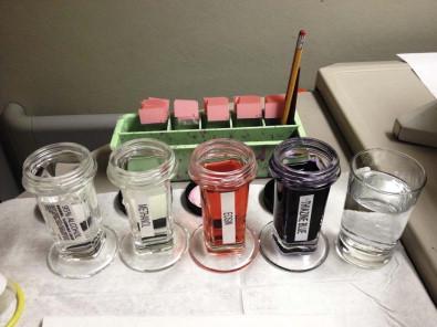
Although the exclusive use of either air-dried or wet-fixed smears is satisfactory, it is recommended to use both of these methods simultaneously. The stains used on the air-dried and wet-fixed smears complement each other and facilitate interpretation of the cytologic features present in the biopsy material. Air-dried smears are Romanowsky-stained, with May–Grünwald–Giemsa stain, Wright–Giemsa stain, or a modified Wright–Giemsa stain, such as the Diff-Quik ® stain. An ultrafast Papanicolaou staining technique has been developed and can also be used to rapidly stain air-dried smears. Slides are wet-fixed by immediately immersing the slides in an alcohol solution, either 95% ethanol or 70–90% methanol. Slides can also be sprayed with fixative solution and then immersed in the alcohol solution. Alcohol-fixed slides are then stained by either the Papanicolaou or hematoxylin–eosin method. An example of an on-site staining set-up is shown in Figure 20-1 .

Virtually all of the special techniques that have been developed for use on tissue samples are applicable on cytology specimens, including FNA biopsy specimens, and include immunohistochemistry, flow cytometry, molecular and cytogenetic techniques, electron microscopy, and microbiology culture.
Cytology preparations are amenable to immunohistochemical and special stains. The cell block sections are preferred for the detection of antigens by immunocytochemistry to help delineate differentiation and features of the cells of interest. Cytospin, Thin Prep ® , and Surepath ® slides can also be used for immunohistochemical stains to help characterize lesions.
Flow cytometry performed on FNA specimens has been shown to be an accurate method of classifying non-Hodgkin lymphoma. Flow cytometry coupled with immunohistochemistry has also been shown to improve the characterization of tumor subtypes, and more often leads to a diagnostic result for tumors sampled by endoscopic ultrasound FNA.
FNA specimens can be used to acquire target DNA for molecular testing. FNA biopsy has been reported to be advantageous over core needle biopsy for several reasons: rapid on-site evaluation of adequacy, sampling of a wider surface area of the lesion, and the ability to acquire more tumor cells with less stromal cell contamination. The reported literature shows the excellent potential of using material obtained from FNA for almost any type of molecular test and the list of molecular tests that can be applied to non-gynecologic cytology specimens is quickly expanding.
Examples of molecular and cytogenetic techniques and applications used on FNA biopsy material include: polymerase chain reaction-based assays to determine the presence of gene rearrangements and mutations in lymphoproliferative disorders, soft-tissue neoplasms, thyroid neoplasms, lung carcinoma, and melanoma. Other ancillary tests performed on FNA specimens include proteomic profiles of tumor protein expression by the surface-enhanced laser desorption–ionization time of flight (SELDI-TOF) methodology to characterize breast lesions, fluorescent in situ hybridization (FISH) to evaluate human epidermal growth factor receptor 2 (HER2/neu) gene amplification in breast cancer, methylation-specific polymerase chain reaction (PCR) to identify methylation profiles, which may have therapeutic implications in breast cancer as well as adjunctive information for diagnosis, and comparative genomic hybridization (CGH) analysis for the differential diagnosis of renal tumors. FNA material can also be stored to maintain RNA integrity for the use of future gene expression analysis.
FNA specimens can also be used for electron microscopic analysis. Applications of electron microscopy include: assessing abdominal fat pad aspirates for amyloid in early disease and subclassifying tumors as epithelial or mesenchymal.
Culture of FNA material can be a useful tool if performed when there is a clinical suspicion of infection or when there is cytologic evidence of infection.
Ultrasound-guidance of FNA (US-FNA) biopsy allows for better characterization of the lesion and for a more thorough sampling of the lesion. At present, US-FNA is commonly employed in sampling thyroid nodules, head and neck lesions, hepatic nodules, musculoskeletal lesions, and superficial abdominal and omental lesions.
Ultrasound along with FNA has proven to be an effective tool in the management of thyroid nodules. The practice guidelines of the American Association of Clinical Endocrinologists and American Thyroid Association recommend using ultrasound guidance to select a suspicious nodule(s) within a multinodular gland, obtain an adequate and representative sample from a suspicious thyroid nodule by sampling multiple sites within the nodule, and sample solid foci within cystic nodules to minimize false-negative thyroid FNA results. It is also recommended that initial non-diagnostic thyroid nodules should undergo repeat FNA under ultrasound guidance to obtain adequate cellular material for diagnosis. The US-FNA combined with on-site evaluation significantly improves the diagnostic accuracy of thyroid FNA specimens. Ultrasound guidance has also been shown to improve the sensitivity and specificity of thyroid FNA significantly and decreases the non-diagnostic rate in comparison with manual thyroid FNA. Ultrasonography is particularly useful in thyroid lesions that are small and difficult to palpate, arise in the posterior portion of the gland, or nodules associated with diffuse pathologic processes. To perform US-FNA, high-ultrasound imaging equipment with high-frequency linear array transducers, appropriate diagnostic skills, and documented experience in performing US-FNA are needed.
In the early 1980s, ultrasound probes were attached to endoscopes and allowed improved visualization of the gastrointestinal wall and abdominal organs. The resolution of these images has improved due to the proximity of the ultrasound transducer to the lesion and the use of a high-frequency ultrasound probe. Endoscopic US-FNA has emerged as a successful technique for tissue diagnosis in patients with lesions of various organs and is commonly incorporated into the management algorithms of patients with suspected mediastinal or abdominal neoplasms. This method has been used to visualize, recognize, and differentiate lesions not only of the luminal gastrointestinal tract, but also of the gastrointestinal tract wall, intrathoracic and intra-abdominal periluminal lymph nodes, pancreas, liver, left kidney, spleen, adrenal glands, central lung, and peribronchial/hilar lymph nodes. The procedure uses a linear-array echoendoscope and a needle-aspiration device. Lesions as small as 5 mm can be aspirated using endoscopic ultrasound guidance. The number of needle passes required for diagnosis is variable, and depends on the type and size of the lesion, and the difficulty of the needle passage. Rapid on-site evaluation to assess the adequacy of the material aspirated under endoscopic ultrasound guidance has proven to be helpful in obtaining an adequate diagnostic specimen and effective triage for ancillary studies.
The initial step in performing a successful FNA is a thorough review of the patient's medical history to establish and confirm the indication for this procedure. Relevant clinical data should be recorded on a requisition form, including the patient's name, identification number, sex, age, tumor size and location, physical examination findings, presenting symptoms and duration, imaging characteristics of the lesion (solid or cystic, single or multiple), and the clinical impression. In addition, history of relevant past or present infectious disease, malignancy, and use of chemotherapy or radiotherapy should be obtained.
The person performing the FNA should discuss the steps of the procedure with the patient, including the possible risks and complications, and answer any questions. An informed consent should be obtained. Depending on local or institutional policies, a written consent may be required. However, there should always be documentation of informed consent from every patient in the medical record. Educating the patient about the procedure is an important part of informed consent and it is necessary to inform and advise the patient that FNA is a sampling test (a biopsy) and that there is a possibility that the specimen may not be representative of the entire lesion.
It is imperative to examine the patient and palpate the lesion. This is the time to plan the procedure, locate the lesion, and estimate its depth. Decide the best angle and entry point to access the lesion while minimizing the amount of soft tissue the needle will traverse before entering the lesion. Most often, needle entry should be perpendicular to the skin surface and lesion. It is also helpful to assist the patient to a comfortable position in which the lesion is easily palpated. For head and neck lesions, it is helpful to place a small pillow under the upper back to extend the neck. Every effort should be made to avoid the sternocleidomastoid muscle, as it is painful for the patient if it is pierced, and, furthermore, the needle can become plugged with skeletal muscle. The last preliminary step is to pause and confirm the patient's identification and lesion site.
Prior to performing the actual FNA procedure, it is important to set up an area with fixative (alcohol solution or proprietary solution supplied by the manufacturer), ingredients for rapid stains, a solution (either Normosol or proprietary solution supplied by the manufacturer) for the needle wash, and slides labeled with patient identification information (usually initials and site and side of lesion) ( Fig. 20-1 ). If a syringe handle is to be used, the syringe holder, syringe, and needle apparatus should be assembled prior to approaching the patient for FNA.
Universal infection precautions should be used. The first step in performing an FNA procedure is to palpate and stabilize the lesion with one hand, typically with two fingers, or push the lesion into a fixed position. Local anesthesia is not necessary due to the small bore of the FNA needle; however, it can be used to relieve patient anxiety. The skin surface at the site of needle puncture is prepped with an alcohol sponge, the needle is placed against the skin at a predetermined angle and puncture site and is then inserted through the skin and into the subcutaneous tissue in a swift smooth motion.
The needle is advanced into the lesion; feel for resistance when penetrating a capsule or note that the lesion moves with the needle if you gently swing it side to side. After the needle is within the lesion, apply suction with the syringe. With suction, move the needle forward and backward in short rapid strokes in a swift cutting motion. Move the needle in the same direction of the original needle tract while increasing the angle. The area biopsied should be reminiscent of a triangle with the puncture site representing the tip of the triangle. This method increases the cellular yield as well as the area sampled. It has been shown that there is a significant difference between a single-planar multipass technique and multiplanar multipass technique, with the latter giving a greater mass yield. The cellular sample is acquired by the cutting action of the needle and is maintained in the needle core by forward motion and capillary tension. The inherent suction provided by surface tension of smaller diameter needles can make devices for additional suction unnecessary. This phenomenon is valuable when aspirating vascular lesions, such as thyroid lesions. If the lesion is visualized with ultrasound, various areas of large masses should be sampled. An approximate dwell time of 2–5 s within the nodule with two or three forward and back needle strokes per second usually maximizes cellular yield and minimizes bloody artifact.
During the aspiration procedure, watch the needle hub for the presence of sample. It is critical to keep the aspirated sample within the needle. The cellular material will start to dry almost immediately if it reaches the syringe. When there is sample present at the hub, release suction and allow the vacuum to normalize. There are times when material is not visualized in the needle hub. After 2–5 s within the nodule with two or three needle strokes per second, suction should be released. If a cyst is encountered during aspiration, evacuate as completely as possible, palpate for a residual mass, and re-aspirate any residual lesion. It is also important to communicate with the patient and inform them that the procedure will take longer while the cyst is being drained.
Without suction, the needle is withdrawn from the lesion in a straight line along the needle tract. If suction is still applied, a portion of the sample will be pulled from the needle into the syringe. The needle should not be removed at an angle because it could cause a laceration of the patient's skin. Pressure is then applied to the puncture site with a sterile gauze pad. This should be done by an assistant, so that the person performing the FNA can quickly start to prepare the slides.
When using FNA for deep lesions, anesthesia should be used. Typically, a guide needle of 18 gauge is placed within the lesion using imaging to verify its position. Then a 21- or 22-gauge needle is inserted through the guide needle, and the position within the lesion is confirmed. The sample is obtained while the patient is holding his/her breath to reduce trauma, and, in the case of the lungs, reduce the risk of pneumothorax. It is imperative that the radiologist and pathologist collaborate and communicate through rapid on-site evaluation to obtain a high-quality sample and sufficient material for ancillary studies.
In cases of endoscopic US-FNA, endoscopic ultrasound is used to target the lesion and position the endoscope at the site of the lesion. Color Doppler imaging is used to identify the vascular anatomy and to prevent insertion of the needle into vessels within the projected needle path. Then, the needle–catheter assembly is passed through the biopsy channel of the echoendoscope. A sheath is used so that the needle does not damage the biopsy channel. The sheath is slowly advanced forward in the biopsy channel until it is visible through imaging. The distance from the middle of the lesion to the tip of the sheath is measured. The needle stop on the system is then locked at this distance and the stylet is removed. Next, the needle is advanced under endoscopic ultrasound guidance to the middle of the lesion by using a quick firm thrust of the handle. When the needle has been clearly placed into the target lesion, the stylet is pushed to the tip of the needle and then completely withdrawn. A syringe is attached to the head of the needle device and the needle is oscillated back and forth approximately 10–20 times within the lesion with suction. Suction is then released, the needle is withdrawn into the catheter, and the whole system is removed from the echoendoscope.
Immediately after the needle is withdrawn from the patient, remove the needle from the syringe, fill the syringe with air, and reattach the needle. Place the needle tip near the center toward the frosted end of a glass slide, touching the bevel against the pre-labeled slide. Delicately advance the syringe plunger to express a small droplet of the sample (2–3 mm diameter) onto the slide. There are several methods described to smear the aspirated material.
Three commonly used techniques are described below.
Once a drop of the sample material is placed on a slide, invert another slide over the droplet. The specimen drop will spread by the weight of the slide and capillary action. As the droplet is spreading, pull the slides away from each other in a horizontal direction using a gentle single motion. Then, place one slide in fixative and allow the other to air dry.
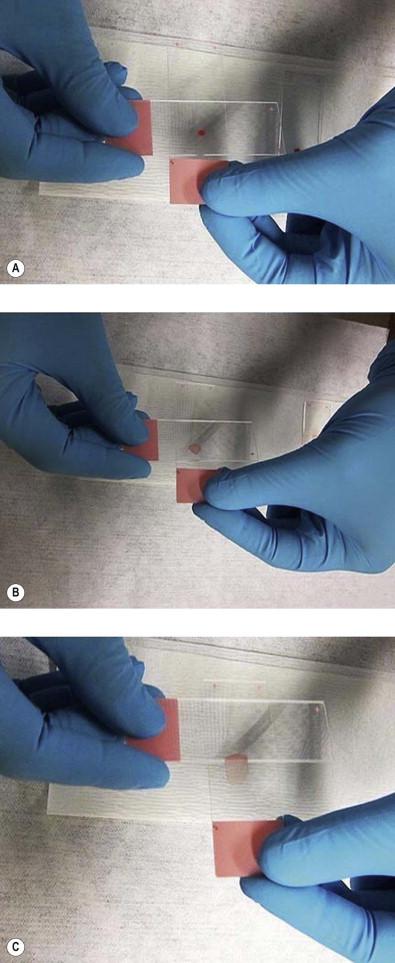
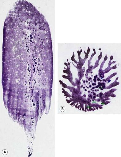
Another method uses a “spreader” slide. The “spreader” slide is gently lowered onto the sample droplet on the initial slide. The droplet starts to spread via capillary action and the droplet is divided between the two slides. A portion of the droplet becomes adherent to the “spreader” slide and a portion remains on the initial slide; the two slides do not come into contact. The “spreader” slide is then gently lowered onto a third slide, causing the droplet to spread, and is pulled in a single smooth motion down length of the slide ( Fig. 20-2 ). This step is then repeated on the initial slide. This method produces two slides with smeared sample and a “spreader” slide that should retain little or no material. One slide is then placed in fixative and the other is air dried ( Fig. 20-3A ). The “spreader” slide can be discarded.


A pop/butterfly/bookend smear can also be made by inverting a second slide on the droplet of the original slide using very little pressure and pulling the slides apart vertically. This method is typically most useful when working with fluid specimens as opposed to semisolid specimens. This technique produces two slides with a smeared sample ( Fig. 20-3B ). One slide is placed in fixative for wet fixation and the other is allowed to air dry.
A cell suspension is then created by rinsing the needle in rinse solution. Occasionally, additional FNA passes are added to the needle rinse for the purpose of creating a cell block or for ancillary studies, such as flow cytometry.
The smearing procedure is repeated for each pass. It is important to keep the material from reaching the edges of the slides. It is also important for the smear to be limited to a segment rather than whole surface area of the slide; this minimizes the area to be evaluated. The slides that are put in fixative for wet fixation should be immersed in the fixative quickly and not slowly. Slow immersion can create alternating bands of cellular and non-cellular areas on the slide, a so-called “ribbing effect”.
The idea that more is better does not necessarily apply to cytology and smear preparation. Large amounts of blood or other bodily fluids not only dilute the diagnostic material, but also have a tendency to cause clotting of the specimen. Diagnostic cells are trapped and obscured in the blood clots, making their cytologic interpretation a challenge. Furthermore, excess blood on the slides causes uneven drying of the sample and distorts the architectural patterns necessary to provide a diagnosis.
Certain organs are more prone to yield a bloody specimen, and this situation is most commonly encountered when dealing with thyroid aspiration biopsies. Independent of the site of the procedure, it is important to realize that certain maneuvers during the aspiration itself might reduce the amount of blood drawn. A vigorous motion of the needle with excessive negative pressure applied to the syringe will cause bleeding in the target lesion. Once this occurs, additional passes are unlikely to retrieve anything but more blood. In addition, the longer the needle stays in the target lesion allows more time for blood to clot in the lumen of the needle, making interpretation of the specimen very difficult.
Once a needle with a bloody specimen is received, the goal is to identify small tissue fragments present in the material and attempt to separate them from the excess blood to allow for adequate smear and interpretation. Various techniques have been used for this purpose. In general, the first step is similar for all techniques. A small sample of the material is expelled onto a slide, towards the frosted end. Due to the liquid nature of the specimen, the fluid will spread to form a 5–10 mm drop on the slide. The next step involves isolating the small tissue particles in the sample, which can be done in a variety of ways:
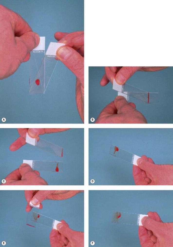
The slide with the material is held by the frosted end in the dominant hand. A “spreader” slide is taken with the other hand, and the lateral edge of this “spreader” slide is placed in contact with the specimen, allowing the fluid to spread along the edge of the “spreader” slide. The “spreader” slide should be maintained at a 30–45° angle. The “spreader” slide is gently moved away from the frosted end of the slide with the sample, until half way down the slide. At this point, the “spreader” slide is lifted and the slide with the specimen is tilted in such a way that the fluid runs back towards the frosted end of the slide. Small tissue fragments will remain in the leading edge of the smear, towards the middle of the slide, and will be separated from the blood. At this point, a new “spreader” slide is taken and the lateral edge is placed over the tissue fragments. The fragments can then be smeared towards the non-frosted end of the slide ( Fig. 20-4 ).

The slide with the material is tilted in such a way that the excess fluid drains towards one of the long edges of the slide. Small tissue fragments, which are heavier than the fluid, will remain in the middle of the slide and be separated from the excess fluid. The short edge of a second slide is taken and the small tissue fragments are scraped off the original slide, in such a way that they become attached to the short edge of the second slide and are not smeared across the original slide. The short edge of the second slide with the small tissue fragments attached to it is now placed over a third slide, and the tissue fragments are smeared across the third slide.
Occasionally, large amounts of bloody fluid might be retrieved from certain procedures, such as paracentesis or pleurocentesis. However, the large quantity of fluid obtained precludes using the techniques for single slide preparation described above. In these cases, initial centrifugation of the specimen allows for separation of the solid material in the sample from the liquid. The supernatant can be discarded and the remaining button can be used for slide preparation. Evidently, red blood cells are still present in the button. To reduce obscuring red blood cells in the smear, hemolysis can be performed, either prior to the slide elaboration by addition of commercially available combined fixative and hemolytic agents, or after slide preparation, by placing the slide into a hemolytic agent (such as 2M urea) for 30 s prior to placing the slide in the fixative solutions.
The presence of a cytopathologist during image-guided aspirations is greatly beneficial by giving the radiologist or clinician performing the aspiration immediate feedback in relation to the quality of the specimen obtained. The goal of on-site evaluation is not to provide an instant diagnosis, but rather to make sure that the best possible specimen is obtained for morphologic interpretation and for potential special studies.
In this sense, the mere fact of informing the performing clinician that diagnostic material is seen in the smears performed confirms that the target lesion has been successfully localized and that viable material is available. If only normal tissue or necrotic tissue is seen, additional passes are requested to increase the yield of the procedure. Once diagnostic material is obtained, a preliminary evaluation of the smears can give the radiologist and the clinical team a general sense regarding the characteristics of the lesion. On-site evaluation is also helpful in providing triage of the specimen, as additional passes may be required to obtain material for special studies, including elaboration of a cell block for immunohistochemistry, flow cytometry, and molecular studies.
Whether it is a cytopathologist or a cytotechnologist who is present to assist during image-guided aspirations, a solid knowledge and practical experience in smear preparation and interpretation is necessary. Likewise, understanding all available ancillary studies, and, most importantly, how to preserve the specimen for these studies in order to adequately triage the cases, is necessary.
In certain instances, particularly when dealing with aspirations of deep-seated lesions performed utilizing a Franseen-style needle, a micro-core tissue biopsy can be retrieved by inserting a stylus into the needle and dislodging small tissue fragments present inside the needle. Smearing of these tissue fragments between two slides, as performed with regular smears, should be avoided, as these are generally too big for complete smearing and excess pressure applied might cause artifacts (usually crushing artifact) that preclude morphologic evaluation. Instead, performing touch preparations, by gently tapping or rolling the tissue fragments onto a slide, is preferred. The slides can then be air-dried or fixed as performed routinely with other smears for routine cytologic evaluation. The tissue fragments can be processed as a routine cell block or placed in 10% buffered formalin for routine surgical pathology specimen processing. If needed, a portion of the micro-core can also be placed in appropriate solutions to perform flow cytometry, electron microscopy, or molecular studies. The interval between obtaining the micro-core and placing it in the appropriate fixative solution should be as minimal as possible, to avoid drying of the tissue leading to compromise of the morphology.
Become a Clinical Tree membership for Full access and enjoy Unlimited articles
If you are a member. Log in here