Physical Address
304 North Cardinal St.
Dorchester Center, MA 02124

Complete transposition of the great arteries (TGA) is a congenital cardiac anomaly in which the aorta arises entirely or largely from the right ventricle (RV) and in which the pulmonary trunk arises entirely or largely from the left ventricle (LV), known as ventriculoarterial discordant connection . 1 Although the phrase complete transposition of the great arteries may properly be applied whenever this situation exists, this chapter uses TGA to denote a cardiac anomaly with atrioventricular concordant connection as well as ventriculoarterial discordant connection. Thus, the term TGA is not applicable to patients with transposed great arteries and tricuspid or mitral atresia or double inlet left or right ventricle (see Chapter 41, Chapter 56 ) or atrioventricular discordant connection (congenitally corrected transposition of the great arteries; see Chapter 55 ).
1 The adjectives left and right used to modify atrium or ventricle always mean morphologically left or right. The position of a chamber or valve is referred to as right-sided or left-sided .
The first morphologic description of TGA is attributed to Baillie in 1797. The term transposition of the aorta and pulmonary artery was coined by Farre when he described the third known case of this anomaly in 1814 using the word transposition ( trans , “across”; ponere , “to place”), meaning that the aorta and pulmonary trunk were displaced across the ventricular septum. In subsequent pathologic descriptions that included attempts to explain its embryologic basis, the word transposition was used to describe an anterior position of the aorta relative to the pulmonary trunk, and by the early 1900s, it had become accepted practice to include any abnormal position of the aorta, regardless of its ventricular origin, under this heading. This broad confusing definition was clarified by Van Praagh and colleagues in 1971, when they strongly advocated return to Farre's original definition of transposition, and introduced the useful term malposition to describe those abnormal positions of the aorta in which both great arteries fail to be displaced across the ventricular septum. This literal meaning of transposition is now accepted by most pathologists and surgeons.
Recognition of TGA during life resulted from observations of Fanconi in 1932 and Taussig in 1938. Importance of the early appearance of pulmonary vascular disease , even when the ventricular septum was intact, was described by Ferguson and colleagues in 1960 and Ferencz in 1966.
Surgery for TGA commenced in 1950 when Blalock and Hanlon at Johns Hopkins Hospital described a closed method of atrial septectomy designed to provide mixing of pulmonary and systemic venous return at the atrial level. Edwards, Bargeron, and Lyons modified the Blalock-Hanlon procedure in 1964 by resuturing the septum so as to connect the right pulmonary veins to the right atrium.
In 1953, Lillehei and Varco described a “partial physiologic correction” (or atrial switch) consisting of anastomosis of right pulmonary veins to right atrium, and inferior vena cava (IVC) to left atrium, a technique that became known as the “Baffes operation.” Baffes incorporated use of an allograft aortic tube to connect the IVC to the left atrium.
Palliation of TGA was revolutionized when Rashkind and Miller in Philadelphia introduced balloon atrial septostomy (BAS) in 1966. However, in 1971 at Great Ormond Street Hospital in London, Tynan showed that BAS did not allow all babies with TGA to survive until repair. A modification of this procedure was introduced in 1975 by Park and colleagues with their substitution of a blade rather than a balloon at the end of the catheter.
Throughout the 1950s there were attempts to correct TGA surgically either at the atrial or the great artery levels. The concept of a physiologic correction at the atrial level by switching the atrial septum so that systemic venous return is directed to the LV and pulmonary venous return to the RV was first proposed by Albert at a meeting of the American College of Surgeons in 1954. This concept was amplified by Merendino and colleagues in 1957. The first successful operation of this type was accomplished by Senning in 1959, who refashioned the walls of the right atrium and the atrial septum to accomplish atrial-level transposition of venous return. Modifications were suggested by many, including Schumaker in 1961 and Bernard and colleagues in 1962. At the Mayo Clinic the Senning procedure was used between 1960 and 1964 with some successes (a few of these patients were still alive and well 30 years later) but with many disappointing results, related in part to the fact that most of the infants and children had a large ventricular septal defect (VSD) and varying degrees of pulmonary vascular disease.
The Mustard procedure , in which the atrial septum is excised and a pericardial baffle used to redirect systemic and pulmonary venous flow, was devised in an attempt to create larger atria than were produced by the Senning procedure and was successfully introduced at the Toronto Sick Children's Hospital in 1963 and reported in 1964. (Actually, Wilson and colleagues described essentially the same operation in 1962. ) Mustard's initial results were better than had been achieved with the Senning procedure, at least in part because he had access to a reservoir of young children with TGA and intact ventricular septum who had been palliated by a Blalock-Hanlon operation.
The Mustard technique soon was adopted in almost all cardiac surgical centers. However, a slightly modified Senning repair was reintroduced by Quaegebeur, Rohmer, and Brom in 1977, mainly because of persisting problems with baffle obstruction and arrhythmia after the Mustard procedure.
It became conventional to delay this atrial switch definitive procedure for 12 to 24 months after BAS. In occasional patients the Mustard procedure was extended to smaller infants by Dillard and colleagues in 1969, Bonchek and Starr in 1972, and Subramanian and Wagner in 1975. The first substantiated proposal that repair was necessary and possible in the first 3 months to avoid considerable pre-repair mortality was by Barratt-Boyes and colleagues.
TGA with large VSD remained a difficult problem throughout this early era because of high hospital mortality after repair and rapid development of pulmonary vascular disease in many patients. However, enough successes were obtained with the atrial switch procedures to demonstrate the value of continuing to treat patients in this subset surgically. In 1972, Lindesmith and colleagues introduced the use of a palliative Mustard procedure in which the VSD was left unclosed for patients with high pulmonary vascular resistance. The modification in which a large VSD was created in TGA with intact ventricular septum was used by Stark and colleagues in 1976.
Successes were few in patients with TGA, VSD, and important left ventricular outflow tract obstruction (LVOTO) in this early period of intracardiac surgery for TGA. Daicoff and colleagues in 1969 reported a few successful repairs by direct relief of the LVOTO associated with an atrial switch by Mustard's technique. Later, in 1969, Rastelli and colleagues combined intraventricular tunnel repair (LV to aorta) of the double outlet RV operation (see “Intraventricular Tunnel Repair of Simple Double Outlet Right Ventricle” under Technique of Operation in Chapter 53 ) with a rerouting valved extracardiac conduit (RV to pulmonary trunk) and closure of the origin of the pulmonary trunk from the LV to produce an anatomic repair of TGA, VSD, and LVOTO.
Somewhat disappointing results of the atrial switch operation for TGA and large VSD continued to be a stimulus for developing an arterial switch operation, particularly because the right (systemic) ventricle sometimes failed late postoperatively in these patients. Much earlier, in 1954, Mustard and colleagues had described unsuccessful attempts to perform an arterial switch operation in seven patients, with transfer of the left coronary ostium to the pulmonary trunk and use of a monkey lung as the oxygenator. Other reports of unsuccessful operations of this general type were those of Bailey and colleagues in 1954 and Kay and Cross in 1955. Idriss and colleagues attempted such a procedure in two patients with an intact ventricular septum in 1961 using cardiopulmonary bypass (CPB), transferring the great arteries and a ring of aorta carrying the coronary arteries. Interest then lagged in many centers, but a few groups persisted with efforts to perfect this approach. Jatene and colleagues in Brazil achieved a major breakthrough in 1975 with the first successful use of an arterial switch procedure (Jatene procedure), applying it in infants with TGA and VSD. Soon after, Yacoub and colleagues reported successful cases. An important technical modification of the original Jatene procedure was the demonstration by Lecompte and colleagues that direct anastomosis of both great arteries without interposition of a tube graft is possible when the pulmonary bifurcation is transferred in front of the distal ascending aortic arch. Aubert and colleagues successfully used intraarterial baffling and creation of an aortopulmonary tunnel to correct simple TGA by an arterial switch in 1978.
Yacoub's attempts in London to perform an arterial switch procedure in three infants with TGA and intact ventricular septum were unsuccessful in 1972, but reports by Mauck in 1977 and Abe in 1978 with their colleagues indicated that such a repair was possible in infancy. However, most infants with TGA and intact ventricular septum did not survive arterial switching. Yacoub approached this problem of the low-pressure LV not being prepared for sustaining systemic pressure by performing pulmonary artery banding as a first stage. The matter was resolved when Radley-Smith and Yacoub in London, Quaegebeur in Holland, and Castaneda in Boston with their colleagues demonstrated feasibility and safety of repair of simple TGA in the first few days of life by an arterial switch operation.
The RV is normally positioned, hypertrophied, and large in TGA. Its inflow and sinus portions are essentially normal in architecture. In about 90% of cases, there is a subaortic conus, and the aorta is rightward and anterior and ascends parallel to the posterior and leftward pulmonary trunk ( Fig. 52-1 ). Such hearts also have an infundibular septum, which in the absence of a VSD, joins normally with the ventricular septum between the limbs of the trabecula septomarginalis (septal band; TSM). The infundibulum does not deviate to the left as in the normal heart, but projects directly superiorly from the sinus portion of the ventricle ( Fig. 52-2 ).

 septum inserts in a normal position between the two divisions of trabecula septomarginalis (septal band). These structures and right ventricular free wall are hypertrophied. Infundibulum projects directly superiorly from sinus portion of ventricle rather than superiorly, anteriorly, and leftward as in normal heart, and gives origin to aorta and aortic valve. Key: AoV, Aortic valve; IS, infundibular septum; RV, right ventricle; TSM, trabecula septomarginalis; TV, tricuspid valve (one chorda has been cut).")
There is less wedging of the pulmonary trunk between the mitral and tricuspid valves in TGA than of the aorta in normal hearts. As a result, a larger area of contiguity exists between the mitral and tricuspid valves than normally. These atrioventricular (AV) valves may be at virtually the same level, and the AV septum and membranous interventricular septum are then smaller than usual or (rarely) absent. The right fibrous trigone of the central fibrous body is abnormally shaped and attenuated.
In about 10% of hearts with TGA and intact ventricular septum, the subaortic conus in the RV is absent or very hypoplastic. Then the aorta is either directly anterior or anterior and to the left of the pulmonary trunk origin or (rarely) posterior. In a few cases, however, a posteriorly placed aorta is associated with a subaortic conus.
The LV infrequently contains a conus; typically pulmonary-mitral fibrous continuity exists, comparable with aortic-mitral continuity in the normal heart ( Fig. 52-3 ). In about 8% of hearts with TGA, and most often in those with a VSD, a subpulmonary conus is present in the LV. The subpulmonary conus is frequently stenotic. In most of these cases, the aorta still lies anteriorly and to the right, but it may be leftward or posterior.
. Approximate course of left bundle branch is shown by cross-hatched area. Key: LV, Left ventricle; MV, mitral valve; PV, pulmonary valve.")
In the normal heart, the LV wall is thicker than the RV wall in utero. After birth, LV wall thickness increases progressively, whereas the RV wall becomes relatively thinner.
In TGA, the RV wall is considerably thicker than normal at birth and increases in thickness with age. When the ventricular septum is intact and no important pulmonary stenosis is present, the LV wall is of normal thickness at birth. Wall thickness remains static, however, leading to less-than-normal thickness within a few weeks of birth and a relatively thin wall by age 2 to 4 months. When a VSD is present, LV wall thickness increases slightly less than in the normal heart, but remains well within the normal range during the first year of life. With LVOTO (pulmonary stenosis) the evolution is similar, although when obstruction is severe and the ventricular septum is intact, LV wall thickness eventually exceeds RV wall thickness. Although not equivalent to LV work potential, LV wall thickness reflects the ventricle's functional capacity.
In infants with TGA, the LV cavity is the usual ellipsoid in shape at birth but soon becomes banana shaped. Alteration in LV function accompanies this geometric change.
RV function is usually normal in TGA in the perinatal period. Thereafter, when the ventricular septum is intact, RV end-diastolic volume is increased and RV ejection fraction decreased. Depressed RV ejection fraction is unlikely to be caused by increased afterload or decreased preload and probably results from depressed RV function from relative myocardial hypoxia or the geometry of the chamber.
LV end-diastolic volume is increased in TGA, and LV ejection fraction is normal. RV/LV end-diastolic volume ratio, normally 1.0, is increased to 1.5 ± 0.33.
The atria are normally formed in TGA. Right atrial size is usually larger than normal, particularly when the ventricular septum is intact.
The AV node and bundle of His lie in a normal position, although the AV node is abnormally shaped and may be partly engulfed in the right trigone. The left bundle branch originates more distally from the bundle of His than usual and arises as a single cord rather than a sheath. Therefore, damage to the bifurcation of the bundle at VSD closure is more likely to produce complete heart block than in the normally structured heart.
The aorta is most often directly anterior or slightly to the right ( Table 52-1 ). In the Taussig-Bing heart, great arteries may be side by side, with the aorta to the right (see “Taussig-Bing Heart” under Morphology in Chapter 53 ). Rarely the aorta is directly posterior.
| Position | Number | Percent of 330 |
|---|---|---|
| Ao anterior 0° | 203 | 62 |
| Ao anterior 30°R | 54 | 16 |
| Ao anterior 60°R | 41 | 12 |
| Ao-PT side-by-side 90°R | 24 | 7 |
| Ao anterior 30°L | 6 | 2 |
| Ao anterior 60°L | 2 | 0.6 |
| Subtotal | 330 | 100 |
| Unknown | 183 | — |
| T otal | 513 |
a Data based on 513 neonates with simple transposition of the great arteries (TGA) or TGA with ventricular septal defect undergoing arterial switch operation, 1985 to March 1, 1989, Congenital Heart Surgeons Society multiinstitutional study.
Some refer to the aortic sinuses of Valsalva as “left posterior–facing” or “right posterior–facing” sinuses and “nonfacing” sinus. This becomes awkward, however, in the 25% of cases in which positions of the great arteries are different from the usual anteroposterior locations. A more universally applicable scheme is the Leiden convention , in which sinus 1 is on the right of an imagined observer standing in the nonfacing noncoronary aortic sinus of Valsalva looking toward the pulmonary trunk. Proceeding counterclockwise, the next sinus is sinus 2 .
In 13% to 30% of patients with TGA, aortic and pulmonary commissures are not precisely aligned because of malalignment of either the aortic or mitral valve. In one study, commissural malalignment was found in nearly 40% of patients undergoing an arterial switch procedure. Recognition of commissural malalignment is important in planning the coronary transfer as well as preventing neoaortic valve regurgitation.
Coronary arteries in TGA usually arise from the aortic sinuses that face the pulmonary trunk, regardless of the interrelationships of the great arteries. Thus, the noncoronary sinus is usually the anterior one. Most often the left anterior descending (LAD) and circumflex (Cx) coronary arteries arise as a single trunk ( left main coronary artery [LCA]) from aortic sinus 1 and distribute in a normal manner, although the Cx system is often small ( Table 52-2 ). The right coronary artery (RCA) arises from sinus 2 and follows this artery's usual course.
| Origin of Coronary Arteries | No. | Percent of 513 | |
|---|---|---|---|
| Sinus 1 | Sinus 2 | ||
| LCx | R b | 367 | 72 |
| L | R | 1 | 0.2 |
| LCxR | — | 5 | 1 |
| — | LCxR | 36 | 7 |
| Single ostium or two very closely placed ostia c | 26 | 5 | |
| Two ostia, one far to left and near commissure d | 10 | 2 | |
| LR | Cx | 13 | 3 |
| LR | LCx | 2 | 0.4 |
| R LCx | — | 8 | 2 |
| L | CxR | 75 | 15 |
| RV LCx | CxR | 1 | 0.2 |
| Malaligned aortic commissures e | 2 | 0.4 | |
| Unknown | 3 | 1 | |
| T otal | 513 | 100 | |
a Data based on 513 neonates with simple TGA or TGA with ventricular septal defect undergoing arterial switch operation (see Table 52-1 ).
b Three of the 367 had malaligned pulmonary commissures.
c LAD or left main coronary artery passed between aorta and pulmonary artery (intramural) in two (one death) of the 26.
d All passed between aorta and pulmonary artery (intramural).
e In one, all coronaries arose from a posterior-facing sinus. In one, LCx arose from a posterior-facing sinus, R from a right-sided sinus.
An almost infinite number of deviations from this usual pattern exist. Typical patterns are observed when both sinus 1 and sinus 2 give rise to a major coronary artery ( Fig. 52-4 ). Patterns in which the Cx or LCA passes behind the pulmonary trunk deserve special attention by the surgeon (see “ Arterial Switch Operation ” under Technique of Operation later in this chapter).
 gives origin to a major coronary artery in hearts with transposition of great arteries. Key: Cx, Circumflex; L, left anterior descending coronary artery; R, right coronary artery.")
All three main coronary arteries may arise from a single sinus (single coronary artery), most frequently, and of utmost concern to the surgeon, from sinus 2. Usually the arteries all arise from a single ostium in the center of the sinus (see Table 52-2 ). Alternatively, they may arise from a double-barreled ostium consisting of two ostia immediately adjacent to each other and constituting essentially a single ostium. Regardless, often these patients have an infundibular branch arising from sinus 1, making true single RCA uncommon. Coronary artery distribution in this situation has a typical pattern ( Fig. 52-5 ). At times, however (in a pattern not shown in Fig. 52-5 ), the LCA or LAD passes forward between aorta and pulmonary trunk in an intramural course to emerge anteriorly. In this situation, instead of all three main coronary arteries arising from an essentially single, more or less centrally positioned, ostium, the LCA or LAD alone nearly always arises from an entirely separate ostium far to the left of the RCA ostium, adjacent to or just above the valvar commissure between sinus 2 and sinus 1 ( Fig. 52-6 ). An LCA or LAD arising in this location passes forward in an intramural course (see Fig. 52-6 ) and entirely within the aortic wall ( Fig. 52-7 ). It emerges from the aorta anteriorly and has the same appearance externally as when the artery originates from sinus 1.
 in sinus 2 or sinus 1 in hearts with transposition of great arteries. Key: Cx, Circumflex; L, left anterior descending coronary artery; R, right coronary artery.")
. A, Origin of left coronary artery and right coronary artery from posterior sinus. Aortic and pulmonary orifices have a side-to-side relationship, and there is alignment of interostial commissures. B, Origin of coronary arteries from posterior sinus. In this case, left circumflex originates separately, whereas left anterior descending and right coronary arteries initially have a common origin and intramural course. Aortic and pulmonary orifices have a side-to-side relationship. Interostial commissures are not aligned, and sinuses do not completely face one other. Key: Ao, Aorta; Cx, circumflex; L, left anterior descending; LCA, left coronary artery; P, posterior sinus; Pu, pulmonary orifice; R, right coronary artery.")
. Key: AD, Adventitia; Ao, aorta; C, commissure; L, left sinus; LCA, left coronary artery M, media of aorta; P, posterior sinus; Pu, pulmonary orifice.")
In patients with situs inversus, the coronary arteries are a mirror image of situs solitus but seem to have a predilection for all coronary arteries to arise from a single sinus.
Rarely the RCA may be intramural as it passes to the right and forward from its usual origin from sinus 2 in an otherwise typical pattern of “sinus 1: LAD, Cx; sinus 2: RCA” (or 1LCx-2R).
In 88 autopsy specimens, origins of coronary arteries were at or above the level of the sinutubular junction in 20%, paracommissural origin occurred in 3%, and angle of exit from the aortic wall was not orthogonal but tangential in 7%. Those with high takeoff were all intramural.
A conus artery frequently arises separately and from its own ostium in sinus 1. It may supply at least a considerable part of the anterior wall of the infundibulum of the RV.
The course of the sinus node artery may be important in the atrial switch (Mustard or Senning) operation. This artery usually arises from the RCA close to its origin and passes superiorly and rightward, usually partly embedded in the most superior portion of the limbus of the atrial septum, where it can be damaged if this portion of the atrial septum is widely excised. Then the sinus node artery usually passes behind or branches to form an arterial circle around the cavoatrial junction.
Now that repair of simple TGA is usually performed in the first week or two of life, and repair of TGA with VSD is usually performed in the first month or two of life, pulmonary vascular disease has almost disappeared (see Natural History and Results later in this chapter), just as it has in many other types of congenital heart disease. However, it becomes important in many patients with TGA when early surgical treatment is not performed ( Table 52-3 ).
| Intact Ventricular Septum | Large VSD | |||||||||||
|---|---|---|---|---|---|---|---|---|---|---|---|---|
| 3-12 Months a | >12 Months a | 3-12 Months a | >12 Months a | |||||||||
| Study | n | No. | % | n | No. | % | n | No. | % | n | No. | % |
| Ferencz | 13 b | 3 | 23 | 12 | 7 | 58 | 14 | 1 | 7 | 18 | 10 | 56 |
| Vilesetal | 4 | 1 | 25 | 3 | 2 | 67 | 6 | 3 | 50 | 9 | 9 | 100 |
| Newfeld et al. | 12 | 0 | 0 | 26 | 4 | 15 | 17 | 5 | 29 | 28 | 26 | 93 |
| Clarkson et al. | 6 b | 2 | 33 | 9 | 4 | 44 | 7 | 2 | 29 | 5 | 2 | 40 |
| T otal | 35 | 6 | 17 | 50 | 17 | 34 | 44 | 11 | 25 | 60 | 47 | 78 |
When pulmonary vascular disease develops in TGA, histologic changes in the pulmonary arteries are comparable with those found in isolated large VSD and can be similarly graded by the Heath-Edwards or Reid criteria (see “Pulmonary Vascular Disease” under Morphology in Section I of Chapter 35 ). In addition, however, pulmonary microthrombi are present in about 25% of lungs examined at autopsy or on lung biopsy. Pulmonary microthrombi produce a variety of intimal lesions, including eccentric cushion lesions and occlusion with recanalization of nonlaminar intimal fibrosis that can result in irregular fibrous septa within vessel lumen. These changes occur with and without laminar and circumferential changes secondary to hypertensive pulmonary vascular disease and are of uncertain etiology and importance. The changes are seldom severe enough to cause an increase in pulmonary vascular resistance (Rp) and occur with equal frequency in TGA with intact ventricular septum, large VSD, and large VSD and LVOTO. Using lung biopsy specimens, Wagenvoort and colleagues have also described wall thinning and dilatation of pulmonary arteries and, to a lesser extent, pulmonary veins in TGA with intact ventricular septum, particularly when the hematocrit is high.
About 75% of neonates presenting with TGA have no important coexisting cardiac anomaly other than a patent foramen ovale or an atrial septal defect. About 25% to 40% have a large or small VSD. Only about 5% have associated LVOTO. Some VSDs close spontaneously in the first few weeks or months of life, and some patients without LVOTO during the first few weeks of life develop obstruction later. Also, some with TGA, VSD, and LVOTO are asymptomatic as neonates and present later in life.
The same types of VSD occur with TGA, and with the same definitions, as occur in hearts with a primary VSD, and they occur in about the same proportions ( Table 52-4 ) (see “Location in Septum and Relationship to Conduction System” in Morphology in Section I of Chapter 35 ). Conoventricular defects of the several different varieties are most common and may not necessarily be juxtapulmonary (on LV side) ( Fig. 52-8 ). In some hearts with conoventricular VSDs, the infundibular septum is malaligned and fails to insert within the Y of the TSM. The septum may be displaced leftward, resulting in a variable degree of LVOTO ( Figs. 52-9 and 52-10 ), or rightward, tending to result in RV (subaortic) obstruction ( Fig. 52-11 ). VSD with malalignment may not be juxtatricuspid, as in tetralogy of Fallot, but the malaligned infundibular septum may be hypoplastic, varying from the usual tetralogy of Fallot (see Morphology in Section I of Chapter 38 ).
| Type of VSD | No. | % of 81 |
|---|---|---|
| Conoventricular: | 47 | 58 |
| Without outlet septal malalignment | 27 | |
| With outlet septal malalignment: | 20 | |
| Displaced to left | 10 | |
| Displaced to right | 10 | |
| Juxta-aortic | 4 | 5 |
| Juxta-arterial | 4 | 5 |
| Inlet septal (AV canal type) | 4 b | 5 |
| Muscular: | 22 c | 27 |
| Basal (posterior, inflow) | 7 | |
| Midseptal | 12 | |
| Apical | 1 | |
| Anterior | 3 | |
| T otal | 81 | 100 |
a Data are based on GLH autopsy study.
. A, From right ventricular side, VSD is seen to be adjacent to tricuspid valve anulus and extends inferiorly beneath it. Infundibular septum is normally aligned with trabecula septomarginalis. B, From left ventricular side, VSD is separated from pulmonary valve in part by an anomalous bulky fibrous pouch (arrow) that originates from left side of septal tricuspid leaflet and is a cause of left ventricular outflow tract obstruction. There is mitral-tricuspid continuity across floor of defect. Key: AoV, Aortic valve; IS, infundibular septum; LV, left ventricle; MV, mitral valve; PV, pulmonary valve; RV, right ventricle; TSM, trabecula septomarginalis; TV, tricuspid valve.")

 and leftward displacement of relatively small conal septum. A, From right ventricular side, fibrous tag (arrow), which also contributes to left ventricular outflow tract obstruction, is seen through VSD. B, From left ventricular side. Key: AoV, Aortic valve; IS, infundibular septum; LV, left ventricle; MV, mitral valve; PV, pulmonary valve; RV, right ventricle; TV, tricuspid valve; VSD, ventricular septal defect.")
 and rightward deviation of infundibular septum that tends to produce subaortic obstruction. A, From right ventricular side, VSD is best appreciated by noting deep position of tricuspid valve relative to infundibular septum (compare with Fig. 52-8, A ). B, From left ventricular side, gap between infundibular septum and ventricular septum is obvious. Anomalous tricuspid chordae are attached to edge of VSD. Key: IS, Infundibular septum; LV, left ventricle; MV, mitral valve; PV, pulmonary valve; TSM, trabecula septomarginalis; TV, tricuspid valve.")
When the infundibular septum is displaced to the right, the pulmonary trunk may be biventricular in origin and over a juxtapulmonary VSD. Hearts with this arrangement are similar to those with double outlet right ventricle and juxtapulmonary VSD (see “Taussig-Bing Heart” under Morphology in Chapter 53 ) and may be associated with subaortic stenosis or aortic arch obstruction (arch hypoplasia, coarctation, or interruption).
Occasionally the VSD is juxta-aortic and associated with a malaligned but nondisplaced infundibular septum. The infundibular septum may be absent or almost gone, and the VSD is then juxta-arterial (doubly committed) ( Fig. 52-12 ).
. Infundibular septum is absent, and confluent aortic and pulmonary valves form upper margin of VSD. Defect is thus doubly committed. There is mild overriding of pulmonary artery and valve into right ventricle. A, From right ventricular side. B, From left ventricular side, there is pulmonary-mitral continuity. Key: Ao, Aorta; AoV, aortic valve; LV, left ventricle; MV, mitral valve; PT, pulmonary trunk; PV, pulmonary valve; TV, tricuspid valve.")
Inlet septal defects that are also juxtatricuspid are slightly more common in hearts with TGA than in those with a concordant ventriculoarterial connection, in which the bundle of His passes from the AV node along the posteroinferior margin of the VSD. The juxtacrucial type of inlet septal defect, with its characteristic tricuspid straddling and abnormal AV node position, probably also occurs more often in hearts with TGA than other defects (see “Inlet Septal Ventricular Septal Defect” in Morphology in Section I of Chapter 35 ).
Most muscular VSDs are in the midseptum but may occur in other areas ( Fig. 52-13 ).
. B, From left ventricular side. Key: AoV, Aortic valve; D, ventricular septal defect; LV, left ventricle; MV, mitral valve; PV, pulmonary valve; RV, right ventricle; TV, tricuspid valve.")
Development of LVOTO, which produces subpulmonary obstruction, is part of the natural history of many patients with TGA. The obstruction may be dynamic or anatomic. LVOTO occurs in an important way at birth or within a few days in only 0.7% of patients with TGA and intact ventricular septum. Obstruction is present in about 20% of patients born with TGA and VSD. LVOTO may become apparent or develop after birth in other patients, thus reaching an overall prevalence of 30% to 35%.
Dynamic type of LVOTO, developing in patients with TGA and intact ventricular septum, is the result of leftward bulging of the muscular ventricular septum secondary to higher RV than LV pressure. Dynamic LVOTO is particularly likely to occur if the aorta lies anterior and more to the left than usual, with increased wedging of the subpulmonary area. The septum impinges against the anterior mitral leaflet in combination with abnormal systolic anterior leaflet motion (SAM). Thus, the mechanism is similar to that present in hypertrophic obstructive cardiomyopathy (HOCM), but there is no asymmetric septal hypertrophy (see “Dynamic Morphology of Septum and Mitral Valve” under Morphology in Chapter 19 ). The gradient may be contributed to by the high velocity of blood flow produced by the usually large pulmonary-to-systemic blood flow ratio and the deformation of the LV outflow tract. When dynamic obstruction is severe, a ridge of endocardial thickening is produced on the septum at its point of contact with the mitral leaflet ( Fig. 52-14 ). In patients with TGA and intact ventricular septum, rarely a subvalvar fibrous ridge may produce LVOTO. The ridge extends onto the anterior mitral leaflet near its hinge. This lesion is analogous to discrete subvalvar aortic stenosis occurring in otherwise normal hearts with ventriculoarterial concordant connection; it is usually localized but may be the tunnel type (see “Tunnel Subvalvar Aortic Stenosis” under Morphology in Section II of Chapter 47 ). LVOTO in these patients rarely may be caused by fibrous tags arising from the mitral apparatus or membranous septum. Valvar stenosis occurs infrequently in this situation, and anular hypoplasia is even less common.

In patients with TGA and VSD, stenosis is usually subvalvar and valvar. Subvalvar stenosis is in the form of a localized fibrous ring, long tunnel-type fibromuscular narrowing, or muscular obstruction related to protrusion of the infundibular septum into the medial or anterior aspect of the LV outflow tract ( Figs. 52-15 and 52-16 ). An important but fortunately rare form of subvalvar stenosis is attachment of the anterior mitral leaflet to the muscular outflow septum by anomalous fibrous or chordal tissue ( Fig. 52-17 ). This stenosis can occur in combination with a cleft anterior mitral leaflet with or without overriding or straddling. Other rare causes of subvalvar pulmonary stenosis are parachute mitral valve, accessory mitral leaflet tissue, and aneurysm of the membranous ventricular septum. An aneurysm may bulge as a windsock into the LV outflow tract. Its walls are thick, and the VSD is either below the aneurysm or within its sac. However, most of these “aneurysms” are examples of redundant fibrous tissue prolapsing through the VSD from the tricuspid valve (see Fig. 52-8 ) or accessory fibrous tags (see Fig. 52-10 ) in association with the anterior mitral valve leaflet.
, and left ventricular outflow tract obstruction. VSD is associated with infundibular septal malalignment and leftward displacement into left ventricular outflow tract. A, From right ventricular side. B, From left ventricular side, infundibular septum has fused with base of anterior mitral valve leaflet, which is cleft (arrow). Pulmonary valve ostium is displaced posteriorly and is severely stenotic. Key: AoV, Aortic valve; IS, infundibular septum; MV, mitral valve; PV, pulmonary valve ostium; RV, right ventricle; TV, tricuspid valve.")

, and left ventricular outflow tract obstruction, viewed from left ventricular side. Left ventricular outflow tract is a stenotic fibrous tunnel formed by bulky fibrous tissue (arrow) extending from mitral leaflet to septal surface superior to VSD. Pulmonary valve is in its normal position. Key: LV, Left ventricle; MV, mitral valve leaflet; PT, pulmonary trunk; PV, pulmonary valve.")
Valvar stenosis is caused by anular hypoplasia and when present is typically associated with subvalvar lesions. The pulmonary valve may be bicuspid. Rarely, there is a stenotic muscular subpulmonary infundibulum.
Patent ductus arteriosus (PDA) is more common in hearts with TGA than in hearts with ventriculoarterial concordant connection. At initial cardiac catheterization at an average age of 2 weeks, Waldman and colleagues found a PDA present in almost half the cases, but it was functionally (although not necessarily anatomically) closed at 1 month. Persistence of a large PDA for more than a few months is associated with an increased prevalence of pulmonary vascular disease.
The tricuspid to mitral anulus circumference ratio, normally greater than 1, is less than 1 in 46% of patients (Calder L: personal communication; 1984). This reduced ratio is most marked in hearts with associated coarctation ( Fig. 52-18 ). Functionally important tricuspid valve anomalies are present in only about 4% of surgical patients ( Table 52-5 ). In autopsy studies, however, a considerably higher proportion are found, particularly when there is a VSD.
 compared with 17 normal hearts. “Control” TGA specimens were those with a completely intact ventricular septum, with or without a small patent ductus arteriosus, atrial septal defect, or patent foramen ovale. Only unoperated specimens and those obtained within 30 days of an intracardiac repair are included. Vertical bars indicate one standard deviation. Individual P values are noted. Key: Coarct, Coarctation; PS, pulmonary stenosis; TV/MV, tricuspid to mitral valve; VSD, ventricular septal defect.")
| Anomaly | No. | % of 260 |
|---|---|---|
| No associated anomaly | 93 | 36 |
| Ventricular septal defect: | 126 | 49 |
| Small | 31 | 12 |
| Moderate | 30 | 12 |
| Large | 65 | 25 |
| Multiple | 19 | 7 |
| Patent ductus arteriosus b : | 37 | 14 |
| Small c | 21 | 8 |
| Moderate | 7 | 3 |
| Large | 9 | 4 |
| Left ventricular outflow tract obstruction d : | 67 | 26 |
| Essentially intact ventricular septum | 27 | 10 |
| Ventricular septal defect | 40 | 15 |
| Tricuspid valve anomalies | 10 | 4 |
| Mitral valve anomalies | 11 | 4 |
| Coarctation (or interrupted arch) | 11 | 4 |
| Right ventricular hypoplasia: | 8 | 3 |
| Mild | 7 | 3 |
| Moderate | 1 | 0.4 |
| Large aortopulmonary collateral arteries e | 1 | 0.4 |
| Atrial situs inversus | 1 | 0.4 |
| Miscellaneous | 4 | 1.5 |
a Data from series of 260 patients undergoing operation at GLH, 1964-1984. Totals are not cumulative.
b Status at time of intracardiac repair.
c Excludes 29 patients in whom small patent ductus arteriosus was only possibly present.
Rarely, in hearts with intact ventricular septum, minor tricuspid valve anomalies may lead to severe regurgitation early in life. In hearts with TGA and VSD, anomalous chordal attachments around the edges of conoventricular VSDs are even more common than in isolated VSD. These may complicate transatrial VSD closure and the construction of an intraventricular tunnel in the Rastelli operation.
The tricuspid leaflets can be redundant and dysplastic in TGA. Accessory tricuspid tissue may prolapse through the VSD and produce LVOTO (see “ Left Ventricular Outflow Tract Obstruction ” in previous text).
The tricuspid anulus may be dilated, resulting in some regurgitation, or in other cases the valve may be hypoplastic in association with underdevelopment of the RV sinus. Anular overriding or tensor straddling or both can occur, the latter being more common.
Important structural anomalies of the mitral valve are present in 20% to 30% of hearts with TGA, mostly in combination with a VSD, but the majority are not functionally important. There may be slight hypoplasia of the valve ring, often with clockwise rotation (viewed from LV apex). Mitral valve anomalies can be categorized into four groups as those affecting the:
Leaflets
Commissures
Chordae tendineae
Papillary muscles
The most important from a surgical standpoint are those of mitral valve overriding or straddling, in which the mitral valve leaflet is frequently also cleft.
Coexisting aortic obstruction can be discrete (coarctation or less often, interrupted aortic arch) or caused by distal arch hypoplasia. Rarely, it occurs when the ventricular septum is essentially intact, but it occurs in 7% to 10% of patients with TGA and VSD. This coexistence is more frequent when the VSD is juxtapulmonary and the pulmonary trunk is partly over the RV in association with rightward and anterior displacement of the infundibular septum and with some subaortic narrowing. (Coarctation is also common in the Taussig-Bing type of double outlet right ventricle; see “Taussig-Bing Heart” under Morphology in Chapter 53 .) The ductus usually also remains patent to the aorta below the coarctation (preductal coarctation).
When there is associated coarctation, underdevelopment of the RV sinus is more common and, as noted earlier, tricuspid-to-mitral anulus circumference is less than in other TGA subsets.
Right aortic arch occurs in about 5% of patients with TGA. It is more common when there is an associated VSD than when the ventricular septum is intact and when there is associated leftward juxtaposition of the atrial appendages.
Leftward juxtaposition of the atrial appendages occurs in about 2.5% of patients with TGA coming to repair. It is associated with a higher than usual prevalence of important underdevelopment of the RV sinus. Bilateral conus and dextrocardia seem more common in TGA associated with leftward juxtaposition than in TGA generally.
RV hypoplasia was found to some degree in 17% of the autopsy series of TGA reported by Riemenschneider and colleagues.
Rarely, TGA coexists with congenital valvar aortic stenosis, and very rarely with total anomalous pulmonary venous connection. TGA can also coexist with complete AV septal defect (see “Complete Atrioventricular Septal Defect” under Morphology in Chapter 34 ).
When the great arteries are transposed in hearts with AV concordant connection, systemic and pulmonary circulations are in parallel. Unless there is shunting between the two, this defect is incompatible with life for more than a short time. With this arrangement, pulmonary ( ![]() ) and systemic (
) and systemic ( ![]() ) blood flow can vary independently, and shunting between the two circulations over more than very short periods must be equal in both directions, or eventually all the blood will be in one or the other circulation. Magnitude of bidirectional shunting is highly variable and is referred to as degree of mixing.
) blood flow can vary independently, and shunting between the two circulations over more than very short periods must be equal in both directions, or eventually all the blood will be in one or the other circulation. Magnitude of bidirectional shunting is highly variable and is referred to as degree of mixing.
Symptoms and clinical presentation in patients with TGA depend in large part on degree of mixing between the two parallel circulatory circuits. When there is a high degree of mixing and large ![]() , arterial oxygen saturation (Sa o 2 ) may be near normal, and unless there is pulmonary venous hypertension, symptoms are minimal. When mixing is minimal, Sa o 2 is low and symptoms of hypoxia are severe. Adequate mixing can occur only when there are communications of reasonable size at atrial, ventricular, or great artery levels. With adequate-sized communications, mixing tends to be directly related to
, arterial oxygen saturation (Sa o 2 ) may be near normal, and unless there is pulmonary venous hypertension, symptoms are minimal. When mixing is minimal, Sa o 2 is low and symptoms of hypoxia are severe. Adequate mixing can occur only when there are communications of reasonable size at atrial, ventricular, or great artery levels. With adequate-sized communications, mixing tends to be directly related to ![]() . Factors that reduce
. Factors that reduce ![]() , such as LVOTO and increased Rp, reduce mixing and increase cyanosis.
, such as LVOTO and increased Rp, reduce mixing and increase cyanosis.
Symptoms and clinical presentation also depend in part on left atrial and pulmonary venous pressure. When ![]() is even moderately elevated, these pressures tend to become elevated and produce symptoms. Both LV and RV failure usually result.
is even moderately elevated, these pressures tend to become elevated and produce symptoms. Both LV and RV failure usually result.
Clinical features and diagnostic criteria of patients with TGA fall into three groups based on these criteria, as discussed in the text that follows.
TGA with essentially intact ventricular septum includes infants without a VSD or with a VSD 3 mm or less in diameter. A patent foramen ovale or naturally occurring atrial septal defect (ASD) is usually present. Cyanosis is apparent in half these infants within the first hour of life and in 90% within the first day and is rapidly progressive. ![]() is usually increased to a pulmonary-systemic blood flow ratio (
is usually increased to a pulmonary-systemic blood flow ratio ( ![]() ) of about 2, but because of poor mixing across the small communication, this does not alleviate hypoxia. The baby becomes critically ill with tachypnea and tachycardia and dies from hypoxia and acidosis without appearance of frank heart failure. This rapid downhill course is usually obviated with a naturally occurring ASD of adequate size, because cyanosis is less severe. In surviving infants, appearance of moderate or severe dynamic LVOTO is associated with increasing cyanosis and hypoxic spells even after an adequate atrial septostomy.
) of about 2, but because of poor mixing across the small communication, this does not alleviate hypoxia. The baby becomes critically ill with tachypnea and tachycardia and dies from hypoxia and acidosis without appearance of frank heart failure. This rapid downhill course is usually obviated with a naturally occurring ASD of adequate size, because cyanosis is less severe. In surviving infants, appearance of moderate or severe dynamic LVOTO is associated with increasing cyanosis and hypoxic spells even after an adequate atrial septostomy.
Clinical signs in most newborns are unimpressive. Generally, patients are of average birth weight and in good general condition, although with severe cyanosis. Clubbing of fingers and toes is absent and generally does not appear unless the infant survives to about age 6 months. There is mild increase in heart and respiratory rates. The heart is not hyperactive, and the liver is barely palpable. A faint mid-systolic ejection-type murmur is present along the midleft sternal edge in less than half these infants. This murmur is more prominent with organic or dynamic LVOTO, first appearing at age 1 or 2 months with the dynamic form and then gradually increasing in intensity. The second heart sound is unremarkable (often apparently single or narrowly split), and the third heart sound and apical mid-diastolic flow murmur are both rare.
Chest radiography ( Fig. 52-19 ) has three characteristic features:
An oval- or egg-shaped cardiac silhouette with a narrow superior mediastinum
Mild cardiac enlargement
Moderate pulmonary plethora

In the first week of life, however, the chest radiograph may be normal; occasionally cardiac enlargement may be more marked. The narrow mediastinum is caused in part by the great artery positions and by shrinkage of the thymus, usually associated with stress, and the plethora is caused by the increase in ![]() . Plethora is less marked when there is important LVOTO.
. Plethora is less marked when there is important LVOTO.
The electrocardiogram (ECG) is often normal at birth, with the usual neonatal RV pattern. By the end of the first week, persistence of an upright T wave in the right precordial leads indicates abnormal RV hypertrophy, and right-axis deviation predominates. The vectorcardiogram shows a clockwise horizontal plane loop indicative of a near-normal LV systolic pressure and a dominant RV mass. When important LVOTO is present or Rp is elevated, ECG evidence indicates biventricular hypertrophy.
Presentation in this TGA group generally occurs in the latter half of the first month, with mild cyanosis and signs of heart failure resulting from pulmonary venous hypertension and myocardial failure. Tachycardia, tachypnea, important liver enlargement, and moist lung bases are present. The heart is more active and usually larger than in the poor-mixing group.
A large VSD is associated with a moderate-intensity pansystolic murmur along the lower left sternal edge that may not be present initially. There is usually an apical mid-diastolic murmur or gallop rhythm and narrow splitting of the second heart sound with accentuation of the pulmonary component. With a large PDA, a continuous murmur, bounding pulses, and an apical mid-diastolic murmur are present in less than half the patients, even when the ventricular septum is intact. Sudden spontaneous closure of a large PDA when there is no VSD results in an increase in cyanosis (see Natural History later in this chapter).
Chest radiography may show more cardiomegaly, more plethora, and a wider superior mediastinum than in the poor-mixing group. Development of pulmonary vascular disease is associated with reduction in ![]() and less plethora, particularly in the peripheral lung fields, as well as reduced heart size, but these features generally appear after the neonatal period.
and less plethora, particularly in the peripheral lung fields, as well as reduced heart size, but these features generally appear after the neonatal period.
The ECG shows biventricular hypertrophy and, when there is a persistent large VSD, a Q wave in V 6 . Isolated LV hypertrophy is rare and suggests RV hypoplasia with tricuspid valve overriding.
When coarctation of the aorta coexists with VSD and PDA, femoral pulses are usually normal because the coarctation is preductal and ductus arteriosus large. Rarely, differential cyanosis can occur, with cyanosis confined to the upper torso. All patients with this combination present early in life in heart failure and respond poorly to decongestive treatment. Isolated LV hypertrophy may be present on ECG because of frequent association of coarctation with RV hypoplasia.
Large VSD with LVOTO is the least common of the three TGA groups. LVOTO is associated with a decreased ![]() and poor mixing, but pulmonary venous hypertension and associated symptoms and signs do not develop because of lack of increase in
and poor mixing, but pulmonary venous hypertension and associated symptoms and signs do not develop because of lack of increase in ![]() . Heart failure is therefore not present. Clinical findings are similar to those of tetralogy of Fallot with severe pulmonary stenosis or pulmonary atresia (see Clinical Features and Diagnostic Criteria in Section I of Chapter 38 ), and cyanosis is severe from birth. The heart is not overactive, and there is a pulmonary ejection murmur and often a single heart sound without an apical gallop or mid-diastolic murmur. Chest radiography shows a near normal–sized heart with normal or ischemic lung fields, and ECG shows biventricular hypertrophy.
. Heart failure is therefore not present. Clinical findings are similar to those of tetralogy of Fallot with severe pulmonary stenosis or pulmonary atresia (see Clinical Features and Diagnostic Criteria in Section I of Chapter 38 ), and cyanosis is severe from birth. The heart is not overactive, and there is a pulmonary ejection murmur and often a single heart sound without an apical gallop or mid-diastolic murmur. Chest radiography shows a near normal–sized heart with normal or ischemic lung fields, and ECG shows biventricular hypertrophy.
Definitive diagnosis of TGA can be made using two-dimensional (2D) echocardiography. Two-dimensional echocardiography is also particularly valuable in detecting tricuspid valve abnormalities, including overriding and straddling, and the varieties of subpulmonary stenosis, including dynamic obstruction. Echocardiographic features of dynamic LVOTO include leftward deviation of the ventricular septum, abnormal fluttering and premature closure of the pulmonary valve, SAM of the mitral leaflet (about 50% of cases), and prolonged diastolic apposition of the anterior mitral valve leaflet to the septum. Echocardiography can also define with reasonable accuracy morphology of the coronary arteries, including number, origin, major branching pattern, and other features such as intramural course ( Fig. 52-20 ). With two-reader methodology, the sensitivity of echocardiography to detect coronary variants is 86%, with a negative predictive value of 91% ( Fig. 52-21 ).

: right coronary artery (RCA) is first branch that passes anterior to aortic valve and to right, with left main coursing leftward and anterior to pulmonary valve trunk (PT) . B, Single RCA: LCA is seen passing behind pulmonary trunk and bifurcating into left anterior descending artery (LAD) and left circumflex artery (LCx) . C, Intramural LCA: there is significant displacement of orifice of LCA arising from right-facing sinus. Proximal LCA runs in wall of aortic root where there is double-border appearance of intramural segment. Key: Ao , Aorta; L Main, left main coronary artery.")
Fetal echocardiography may be helpful in identifying abnormalities of the foramen ovale or ductus arteriosus, which is associated with neonatal hypoxia and death, and of the ventricular septum. Fetal diagnosis may improve perinatal care and reduce perinatal mortality and postoperative morbidity.
Cardiac catheterization and cineangiography are not performed routinely, particularly in neonates, with major reliance for diagnosis placed on echocardiography. Nonetheless, knowledge of the information from these studies remains important.
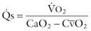
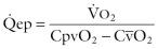
A full study includes calculation of ![]() and
and ![]() and pressures, including those across the LV outflow tract. Because of intracardiac communications in patients with TGA, the Fick method is usually the only practical way of measuring
and pressures, including those across the LV outflow tract. Because of intracardiac communications in patients with TGA, the Fick method is usually the only practical way of measuring ![]() and
and ![]() . Despite complexity of the circulation, standard calculations apply. Meticulous care is required in measuring oxygen consumption using a closed-box technique in infants. Equations are as follows:
. Despite complexity of the circulation, standard calculations apply. Meticulous care is required in measuring oxygen consumption using a closed-box technique in infants. Equations are as follows:
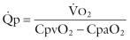
where
![]() = Pulmonary blood flow
= Pulmonary blood flow
![]() = Oxygen consumption, mL · min −1
= Oxygen consumption, mL · min −1
Cpv o 2 = Pulmonary venous oxygen content, mL · L −1
Cpa o 2 = Pulmonary arterial oxygen content, mL · L −1
![]() = Systemic blood flow
= Systemic blood flow
Ca o 2 = Systemic arterial oxygen content, mL · L −1
![]() = Mixed venous oxygen content, mL · L −1
= Mixed venous oxygen content, mL · L −1
![]() = Effective pulmonary blood flow
= Effective pulmonary blood flow
![]() represents flow of blood from the systemic to the pulmonary circuit at atrial, ventricular, and great arterial levels. Flow must be equal in the opposite direction (anatomic left-to-right shunt or effective systemic blood flow), or over time one circuit would be deprived of blood.
represents flow of blood from the systemic to the pulmonary circuit at atrial, ventricular, and great arterial levels. Flow must be equal in the opposite direction (anatomic left-to-right shunt or effective systemic blood flow), or over time one circuit would be deprived of blood.
Inherent errors occur in measuring these flows. When ![]() is high and therefore pulmonary arterial oxygen saturation (Spa o 2 ) is high, the Fick calculation tends to be inaccurate. This error may be compounded by difficulties in recovering a truly mixed Spv o 2 . Fortunately, these errors are greatest in patients with a very high
is high and therefore pulmonary arterial oxygen saturation (Spa o 2 ) is high, the Fick calculation tends to be inaccurate. This error may be compounded by difficulties in recovering a truly mixed Spv o 2 . Fortunately, these errors are greatest in patients with a very high ![]() , when concern is minimal about a high Rp. Calculations are more accurate when the
, when concern is minimal about a high Rp. Calculations are more accurate when the ![]() is low and Rp correspondingly high. Potential for error exists if pulmonary arterial sampling is made proximal to site of entry of sizable systemic (bronchial) collaterals. Truly mixed Spa o 2 would then be lower than that measured, and
is low and Rp correspondingly high. Potential for error exists if pulmonary arterial sampling is made proximal to site of entry of sizable systemic (bronchial) collaterals. Truly mixed Spa o 2 would then be lower than that measured, and ![]() correspondingly lower, but in practice this situation is uncommon.
correspondingly lower, but in practice this situation is uncommon.
Thus, with careful technique, Rp in patients with TGA can be calculated with reasonable accuracy. A specific problem arises, however, if hematocrit is particularly high; viscosity of the blood increases sharply when hematocrit is greater than 60%. The effect of viscosity on ![]() may then become important, and calculated Rp may be higher than that dictated by the pulmonary vascular bed alone. The only solution to this is to repeat the measurements after lowering the hematocrit by venisection.
may then become important, and calculated Rp may be higher than that dictated by the pulmonary vascular bed alone. The only solution to this is to repeat the measurements after lowering the hematocrit by venisection.
![]() is the flow upon which life depends. This flow is relatively fixed, typically only about 1.0 to 1.5 L · min −1 · m −2 . This places a major constraint on oxygen supply to the patient. These relationships become evident in rewriting the Fick equation as follows:
is the flow upon which life depends. This flow is relatively fixed, typically only about 1.0 to 1.5 L · min −1 · m −2 . This places a major constraint on oxygen supply to the patient. These relationships become evident in rewriting the Fick equation as follows:
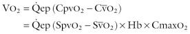
where
Spv o 2 = Pulmonary venous oxygen saturation
![]() = Mixed-venous oxygen saturation
= Mixed-venous oxygen saturation
Cmax o 2 = Oxygen capacity per gram of Hb
Hb = Hemoglobin concentration, g · L −1
On this basis, any reduction in hemoglobin will reduce oxygen uptake, and compensation for it is not possible in patients with TGA. If stress or exercise increases oxygen requirement, the difference in Cpv o 2 and ![]() must widen, and because Cpv o 2 cannot increase,
must widen, and because Cpv o 2 cannot increase, ![]() (and thus tissue P o 2 ) must fall.
(and thus tissue P o 2 ) must fall.
Using appropriate views, cineangiography demonstrates the cardiac connections and great artery positions ( Fig. 52-22 ), position and number of VSDs ( Fig. 52-23 ), site of any LVOTO ( Fig. 52-24 ), size and function of AV valves, size and function of both ventricles, pattern of the coronary arteries, and presence of other cardiac anomalies.

. A, Small midmuscular VSD is demonstrated by right ventricular injection in long axial view. B, Large VSD in inflow portion of septum is demonstrated by right ventricular injection in four-chamber position. C, Large conoventricular VSD is shown with left ventricular ejection in long axial view. D, Multiple muscular VSDs are demonstrated with a right ventricular injection in long axial view. Key: Ao, Aorta; LV, left ventricle; PT, pulmonary trunk; RV, right ventricle.")
, and left ventricular outflow tract obstruction (LVOTO). A, Subvalvar LVOTO is associated with large conoventricular VSD, as shown by left ventricular injection and four-chamber view. B, Long subvalvar LVOTO is associated with large conoventricular VSD, as shown by left ventricular injection and four-chamber view. C, Discrete subvalvar LVOTO with large VSD and mild overriding of aorta onto left ventricle, as shown by left ventricular injection and four-chamber view. Key: Ao, Aorta; IS, infundibular septum; LV, left ventricle; PT, pulmonary trunk; PV, pulmonary valve; RV, right ventricle.")
Although these newer modalities are more accurate than echocardiography in evaluating anatomy, particularly coronary anatomy, they are not routinely used in the neonate. Cardiac computed tomographic angiography (CTA) and image postprocessing with volume rendering can give an accurate diagnosis of the coronary pattern, even in neonates ( Fig. 52-25 ). These modalities are used more frequently in postoperative patients in whom coronary imaging is indicated.

TGA is a common form of congenital heart disease, occurring in 1 : 2100 to 1 : 4500 births and accounting for 7% to 8% of all congenital heart disease. Prevalence might be reduced more than 50% by maternal preconceptional multivitamin use or may be reduced by avoiding pesticides during the first trimester. In the Auckland area of New Zealand, prevalence over a 10-year period was 1 : 2400, whereas in New England (U.S.), it was 1:4000 (P < .005). Before the advent of effective treatment, at least 16% of deaths from congenital heart disease during childhood were caused by TGA.
Male-to-female ratio is 2 : 1. Male predominance increases to 3.3 : 1 when the ventricular septum is essentially intact and disappears in complex forms.
When patients with all varieties of TGA are considered, 55% survive 1 month, 15% survive 6 months, and only 10% survive 1 year ( Fig. 52-26 ). Mean life expectancy is 0.65 year, rising to 4 years for those who survive to 12 months and to 6 years for the few who survive for 10 years. Thereafter, life expectancy declines rapidly (see Fig. 52-26 ).
 of all types, all of whom died between 1957 and 1964; 73 living children and 14 miscellaneous deaths are excluded. Group is impure in that about 15% of the total had either single ventricle, hypoplasia of left ventricle with mitral stenosis or atresia, or hypoplasia of right ventricle with tricuspid stenosis or atresia. However, trends are representative of patients with TGA.")
Survival without treatment is different among subsets. It is particularly poor in untreated patients with TGA and essentially intact ventricular septum : 80% at 1 week but only 17% at 2 months and 4% at 1 year. Survival in this group is better when there is a true ASD ( Fig. 52-27 ).

In patients with TGA and important VSD , early survival is higher: 91% at 1 month, 43% at 5 months, and 32% at 1 year. It is lower when the patient has a very large ![]() (see Fig. 52-27 ). The combination of large VSD and aortic obstruction (coarctation, interrupted arch) is particularly lethal; all patients die within a few months of birth with severe heart failure. Paradoxically, obstructive pulmonary vascular disease in patients with TGA and VSD improves early survival to 40% at 1 year, but with rapid decline thereafter and none alive by age 5 years.
(see Fig. 52-27 ). The combination of large VSD and aortic obstruction (coarctation, interrupted arch) is particularly lethal; all patients die within a few months of birth with severe heart failure. Paradoxically, obstructive pulmonary vascular disease in patients with TGA and VSD improves early survival to 40% at 1 year, but with rapid decline thereafter and none alive by age 5 years.
In patients with TGA , VSD , and LVOTO , early survival is still better, reaching 70% at 1 year and 29% at 5 years, because in many patients LVOTO is only moderate initially.
Leibman and colleagues found that PDA increased risk of early death in all subsets of patients. This is particularly the case when the ductus is large.
Poor survival in patients with TGA and essentially intact ventricular septum is related primarily to hypoxia. Intercurrent pulmonary infections may develop and are particularly lethal because they reduce ![]() and lead rapidly to increasing hypoxia, acidemia, and death. Death in this group may also result from cerebrovascular events, usually caused by the polycythemia and increased blood viscosity secondary to severe cyanosis, particularly in association with dehydration. However, hypoxia plus hypochromic microcytic anemia has also been implicated in the etiology of these events. Nonfatal cerebrovascular events occur in about 6% of patients treated by BAS and include cerebral abscess.
and lead rapidly to increasing hypoxia, acidemia, and death. Death in this group may also result from cerebrovascular events, usually caused by the polycythemia and increased blood viscosity secondary to severe cyanosis, particularly in association with dehydration. However, hypoxia plus hypochromic microcytic anemia has also been implicated in the etiology of these events. Nonfatal cerebrovascular events occur in about 6% of patients treated by BAS and include cerebral abscess.
Patients with TGA and important VSD usually die with heart failure. Modes of death described for patients with simple transposition sometimes pertain to this group as well and include frequent intercurrent pulmonary infections.
Hypoxia is the primary cause of morbidity and mortality in patients with TGA, VSD, and LVOTO.
PDA is present at age 1 week in about half the patients with TGA, but thereafter the prevalence falls rapidly. When patent, the ductus is small (<3 mm in diameter) in about two thirds of patients and seems to have little influence on natural history. When it is large, LV output is increased and hypoxia lessens, but heart failure becomes more severe. Under these circumstances, acute and often early closure of the ductus results in sudden increase in hypoxia and clinical deterioration. This is related not only to decreased mixing at the ductus level but also at the atrial level because of the fall in left atrial pressure that results from decreased pulmonary venous return.
In patients with TGA, the patent foramen ovale tends to close at the usual rate. This is the major cause of the time-related increase in hypoxia and death in patients with TGA and essentially intact ventricular septum without an important PDA. A true ASD, on the other hand, remains unchanged in size and palliates the patient longer. The same is true for those rare examples of coexisting partial anomalous pulmonary venous connections.
Large VSDs close or narrow in probably a smaller proportion (≈20%) of patients with TGA than in patients with isolated VSD (see “Spontaneous Closure” under Natural History in Section I of Chapter 35 ). In most cases, however, the closing VSD is initially small and often muscular, and spontaneous closure has been documented to occur as late as the last part of the first decade of life. This process was rarely documented before the era of BAS, because so few patients survived beyond the first few months of life.
Dynamic LVOTO is not present at birth but can appear within several weeks. It gradually progresses in severity. Awareness of this tendency has increased since the era of BAS, after which LVOTO frequently develops. When dynamic LVOTO becomes important, hypoxia returns and life expectancy is shortened. LVOTO develops infrequently in patients with TGA and important VSD.
When TGA occurs as an isolated lesion (simple TGA) , pulmonary vascular disease rarely develops in the first few months of life. After about 6 to 24 months, however, its prevalence increases to 10% to 30%. Its development reduces ![]() and increases hypoxia.
and increases hypoxia.
In patients with TGA and moderate or large VSD, pulmonary vascular disease develops more rapidly than in patients with simple TGA, as it does in those with persistently large PDA. Among those dying at about age 6 months, 25% have developed severe pulmonary vascular disease (≥grade 3), and 50% of infants dying by age 12 months have developed it. These prevalences are much higher than in patients with primary VSD, and mechanisms may include hypoxemia and a prominent bronchopulmonary collateral circulation.
At birth in TGA, as in normal patients, slightly more blood flows to the right lung than to the left. In contrast to normal flow, however, flow to the right lung in TGA increases as age increases. In addition to age, magnitude of the increase is affected by the angle between takeoff of the right pulmonary artery and pulmonary trunk; the wider this angle (and thus the more the pulmonary trunk faces directly into the right pulmonary artery), the greater the blood flow to the right lung. The tendency of infants with intact ventricular septum to develop dynamic LVOTO after the first few months increases the velocity of flow, which increases the momentum effect toward the more directly aligned vessel.
Once right lung flow increases, the right vascular bed grows more and there is a relative increase in Rp and reduced compliance in the left lung, which further reduces left lung flow. It is unlikely that this phenomenon importantly affects the natural history of untreated TGA.
Currently, the arterial switch operation is advised for most patients with TGA except those with important fixed LVOTO. An atrial switch operation (Mustard or Senning type) may be appropriate rarely, and in highly selected patients. Patients with poor mixing, typically those with intact ventricular septum and a small ASD, come to the operating room receiving an infusion of prostaglandin E 1 and usually having had BAS. In current cardiology practice, septostomy is performed through transvenous access using echocardiographic guidance. These preoperative maneuvers usually result in adequate mixing and a stable patient.
Preparation of the patient for operation, anesthesia, placement of monitoring devices, and details of the median sternotomy and initial dissection are the same as in other operations in neonates and young infants (see “Preparation for Cardiopulmonary Bypass” in Section III of Chapter 2 ). Positioning of the baby with extension of the neck is particularly important for exposure of the great arteries. Three general types of support systems are in use for arterial switch operation:
Continuous CPB , usually at 18° to 25°C, with reduced flow rate after reaching the target temperature. In some centers, mild hypothermia or normothermia is used. The IVC and superior venae cavae (SVC) are cannulated directly for venous return.
Near-continuous CPB at 18° to 20°C and with reduced flow rates (0.5 to 10 L · min −1 · m −2 ), but with a single venous cannula inserted through the right atrial appendage (see Sections III and IV in Chapter 2 ). Hypothermic circulatory arrest is established only for closure of the ASD, which is done through the opening in the tip of the right atrium or a small right atriotomy after removing the venous cannula. After this closure, the venous cannula is reinserted, CPB reinstituted, and full flow restored for rewarming of the patient.
Operation primarily is performed during hypothermic circulatory arrest after the patient has been cooled to 18°C by CPB, with rewarming also accomplished by CPB.
Preference for these methods is in the order presented.
Myocardial management is also variable among institutions achieving good results. A prevalent method is infusion into the aortic root through a large-bore needle of a cold, hyperkalemic, sanguineous solution just after clamping the ascending aorta, and no more. Another method is use of the same protocol but with an asanguineous cardioplegic solution.
The aorta and pulmonary trunk must be dissected apart and the ductus arteriosus dissected. The right and left pulmonary arteries are extensively mobilized to their lobar branches and beyond if needed. As much of this as convenient is performed before CPB, but it may be necessary to complete these steps after CPB is established. The aortic purse-string stitch is placed as far downstream as possible to facilitate work on the aortic root and ascending aorta ( Fig. 52-28, A ). When using two venous cannulae, purse-string sutures are placed in the superior and inferior venae cavae as they enter the right atrium. A suture ligature is placed around the aortic end of the ductus (see Fig. 52-28, A ) Another purse-string stitch is placed in the right superior pulmonary vein as it enters the left atrium (not shown in Fig. 52-28, A ).
![Figure 52-28, Arterial switch operation for transposition of great arteries, with aorta anterior and rightward, and usual coronary artery pattern (1LCx-2R). A, Placement of cardiopulmonary bypass (CPB) purse strings is shown in a patient who will undergo operation using continuous bypass and bicaval cannulation. Note that aortic purse string is placed as high on ascending aorta as possible to provide room for great artery manipulation. Venous purse strings are placed directly into superior and inferior venae cavae. A suture ligature has been placed around aortic end of ductus arteriosus. Tissue between great arteries is dissected prior to establishing CPB. A helpful maneuver is to place marking sutures on neoaorta for identifying sites of coronary implantation. Left and right branch pulmonary arteries are mobilized into first-order branching vessels. Cannulation proceeds in standard fashion and ductus arteriosus is immediately ligated. A separate suture ligature is placed on pulmonary artery end of ductus arteriosus and tied, and ductus is transected. A separate purse-string suture is placed in right upper pulmonary vein as it enters left atrium, and a vent catheter is introduced through purse string into left atrium, across mitral valve, and positioned into left ventricle (vent is not shown in this figure). B, After target core temperature is achieved, aorta is clamped and cardioplegia introduced into aortic root by one of standard methods. The aorta is transected just above sinutubular junction, and coronary arteries are carefully examined to confirm their positions and to rule out possibility of any unusual variations, such as eccentric coronary ostia or intramural coronary arteries. Using sharp dissection with fine scissors, coronary arteries are removed from their sinuses with at least a 1- to 2-mm cuff of sinus tissue surrounding ostia. Ligated and divided ductus arteriosus is also shown. C, Coronary buttons have been completely mobilized. Pulmonary trunk is transected at its midportion and sites of coronary implantation (with help of marking sutures, if present) are identified on proximal neoaorta (dashed lines) . Various techniques can be used to prepare implantation sites. Most common variation is shown here, in which implantation site is prepared by removing a horseshoe-shaped segment of pulmonary trunk wall. Implantation sites can also be prepared with a simple incision (slit) without resection of any proximal neoaortic tissue. D, Coronary implantation is performed sequentially using a running 8-0 or 7-0 monofilament suture. Following implantation, it is important to visualize course of coronary artery and, if any doubt remains as to its patency, a 1- to 1.5-mm probe is passed into its proximal portion to demonstrate patency. E, Coronary arteries are fully implanted. Lecompte maneuver has been performed, as indicated by branch pulmonary arteries now located anterior to aorta. Anastomosis between proximal neoaorta and ascending aorta is performed end to end using a running 7-0 monofilament suture technique. After completing aortic anastomosis, coronary explantation sites on proximal neopulmonary trunk are reconstructed with individual patches of glutaraldehyde-treated autologous pericardium. Individual pericardial patches are tailored to be slightly larger than defects that resulted from coronary explantations. Proximal neopulmonary trunk is then connected to distal pulmonary trunk, end to end, using a running monofilament 7-0 suture technique. F, Completed great artery reconstruction is shown, along with closed CPB cannulation sites. Careful examination of proximal coronary arteries and their relation to anteriorly positioned pulmonary trunk and pericardial patches is routinely performed to ensure coronary arteries are not distorted or compressed. (Atrial septal defect [ASD] is closed using standard technique as described in text. ASD can be closed at any point in procedure. The common technique used with continuous CPB is to close ASD after aortic reconstruction is complete, but before embarking on pulmonary trunk reconstruction. In this way, ASD is closed with the aortic clamp still in place, aiding intracardiac visualization. Aortic clamp is then removed prior to performing pulmonary trunk reconstruction, minimizing myocardial ischemia time.)](https://storage.googleapis.com/dl.dentistrykey.com/clinical/CompleteTranspositionoftheGreatArteries/31_3s20B9781416063919000528.jpg "Figure 52-28, Arterial switch operation for transposition of great arteries, with aorta anterior and rightward, and usual coronary artery pattern (1LCx-2R). A, Placement of cardiopulmonary bypass (CPB) purse strings is shown in a patient who will undergo operation using continuous bypass and bicaval cannulation. Note that aortic purse string is placed as high on ascending aorta as possible to provide room for great artery manipulation. Venous purse strings are placed directly into superior and inferior venae cavae. A suture ligature has been placed around aortic end of ductus arteriosus. Tissue between great arteries is dissected prior to establishing CPB. A helpful maneuver is to place marking sutures on neoaorta for identifying sites of coronary implantation. Left and right branch pulmonary arteries are mobilized into first-order branching vessels. Cannulation proceeds in standard fashion and ductus arteriosus is immediately ligated. A separate suture ligature is placed on pulmonary artery end of ductus arteriosus and tied, and ductus is transected. A separate purse-string suture is placed in right upper pulmonary vein as it enters left atrium, and a vent catheter is introduced through purse string into left atrium, across mitral valve, and positioned into left ventricle (vent is not shown in this figure). B, After target core temperature is achieved, aorta is clamped and cardioplegia introduced into aortic root by one of standard methods. The aorta is transected just above sinutubular junction, and coronary arteries are carefully examined to confirm their positions and to rule out possibility of any unusual variations, such as eccentric coronary ostia or intramural coronary arteries. Using sharp dissection with fine scissors, coronary arteries are removed from their sinuses with at least a 1- to 2-mm cuff of sinus tissue surrounding ostia. Ligated and divided ductus arteriosus is also shown. C, Coronary buttons have been completely mobilized. Pulmonary trunk is transected at its midportion and sites of coronary implantation (with help of marking sutures, if present) are identified on proximal neoaorta (dashed lines) . Various techniques can be used to prepare implantation sites. Most common variation is shown here, in which implantation site is prepared by removing a horseshoe-shaped segment of pulmonary trunk wall. Implantation sites can also be prepared with a simple incision (slit) without resection of any proximal neoaortic tissue. D, Coronary implantation is performed sequentially using a running 8-0 or 7-0 monofilament suture. Following implantation, it is important to visualize course of coronary artery and, if any doubt remains as to its patency, a 1- to 1.5-mm probe is passed into its proximal portion to demonstrate patency. E, Coronary arteries are fully implanted. Lecompte maneuver has been performed, as indicated by branch pulmonary arteries now located anterior to aorta. Anastomosis between proximal neoaorta and ascending aorta is performed end to end using a running 7-0 monofilament suture technique. After completing aortic anastomosis, coronary explantation sites on proximal neopulmonary trunk are reconstructed with individual patches of glutaraldehyde-treated autologous pericardium. Individual pericardial patches are tailored to be slightly larger than defects that resulted from coronary explantations. Proximal neopulmonary trunk is then connected to distal pulmonary trunk, end to end, using a running monofilament 7-0 suture technique. F, Completed great artery reconstruction is shown, along with closed CPB cannulation sites. Careful examination of proximal coronary arteries and their relation to anteriorly positioned pulmonary trunk and pericardial patches is routinely performed to ensure coronary arteries are not distorted or compressed. (Atrial septal defect [ASD] is closed using standard technique as described in text. ASD can be closed at any point in procedure. The common technique used with continuous CPB is to close ASD after aortic reconstruction is complete, but before embarking on pulmonary trunk reconstruction. In this way, ASD is closed with the aortic clamp still in place, aiding intracardiac visualization. Aortic clamp is then removed prior to performing pulmonary trunk reconstruction, minimizing myocardial ischemia time.)")
After cannulation is completed, CPB is established and cooling begun. Another suture ligature is placed around the pulmonary end of the ductus and the ductus divided. If the two venae cavae have been directly cannulated, adjustable snares are placed around them and tightened, and a small (13F) angled vent catheter is placed through the purse string in the right superior pulmonary vein, positioning its tip across the mitral valve into the LV. The cardioplegic infusion needle is inserted, the aorta clamped, and infusion given.
The aorta is transected and better exposure is obtained by turning back the proximal segment to facilitate further dissecting apart the great arteries ( Fig. 52-28, B ) . The pulmonary trunk is transected just proximal to its bifurcation ( Fig. 52-28, C ). The aortic button around the orifice of the LCA is excised from its sinus, and this is inserted into the left-facing sinus of the neoaorta (originally, pulmonary trunk). The aortic button around the orifice of the right coronary artery is excised and inserted into the right-facing sinus of the neoaorta ( Fig. 52-28, B-D ).
After the Lecompte maneuver ( Fig. 52-28, E ), the neoaorta is constructed by anastomosing the proximal segment of the original pulmonary trunk to the distal aortic segment.
The stretched or torn foramen ovale (or ASD) is closed through an incision in the right atrium, usually with a running stitch. A patch may be used if the ASD is large. The right atrium is closed.
Separate autologous pericardial patches are used to fill in the defects in the proximal neopulmonary trunk. The neopulmonary trunk is then constructed ( Fig. 52-28, F ). All the latter steps involving pulmonary trunk reconstruction may be completed before removing the aortic clamp and beginning reperfusion of the heart, or reperfusion may be started at any point along the way.
A polyvinyl catheter is brought out from the left atrium through the right superior pulmonary vein or left atrial appendage if not already placed, and later, one is brought out from the right atrium. After the neonate has been rewarmed by the perfusate and proper conditions are in place, CPB is discontinued, and the remainder of operation is performed as detailed earlier (see “Completing Cardiopulmonary Bypass” in Section III of Chapter 2 ). Use of intraoperative echocardiography to assess global and, especially, regional ventricular function is helpful in assessing adequacy of coronary translocation.
At times, the Cx coronary artery arises as a branch of the RCA, the ostium of which is in sinus 2 (right-facing sinus). The Cx artery then passes leftward behind the pulmonary trunk and arborizes in the usual fashion ( Fig. 52-29, A ). Less often the LCA arises from sinus 2 (an RCA from sinus 1) and passes leftward behind the pulmonary trunk to bifurcate in the usual manner. In both situations, particular care is required in the transfer of this coronary button from sinus 2.
. A, Aorta is anterior and slightly to right, as in the usual case. Proposed site of pulmonary trunk transection is as far distal as possible, just before bifurcation, to provide a proper implantation site on proximal neoaorta (native pulmonary trunk) for coronary button from sinus 2. Proposed aortic transection site is slightly more distal than in the case with most common coronary pattern (see Fig. 52-28 ), to accommodate slightly shorter distal pulmonary trunk segment at time of pulmonary reconstruction. Dashed lines show proposed transected sites of great arteries. Note circumflex coronary artery passing posterior and to left behind great arteries to distribute to its normal myocardial area. B, Using standard cardiopulmonary bypass and myocardial protection techniques, operation proceeds in standard fashion until it is necessary to reimplant coronary button from sinus 2. Coronary from sinus 1 has been reimplanted in standard fashion (see Fig. 52-28 ). Proximal neopulmonary trunk is retracted anteriorly, and tissue between the two great arteries at their bases is fully dissected. Dashed line shows proposed incision to create “trapdoor” flap that serves to orient sinus 2 coronary button after reimplantation such that circumflex artery is neither kinked nor stretched. Because pulmonary trunk was transected as distally as possible, reimplanted coronary also is positioned more cephalad than in usual case. This also minimizes chance of circumflex artery kinking. C, Coronary button from sinus 2 is sutured into place utilizing “trapdoor” flap . D, Trapdoor flap implantation is completed. Note smooth course of circumflex artery passing posterior and to left around great arteries. Inset shows a redundant and kinked circumflex artery that may result from implantation of coronary button too far to right-lateral aspect of circumference of proximal neoaorta. When coronary button is poorly positioned in this manner, in combination with orientation of button that results without using trapdoor flap, circumflex artery is at high risk for obstruction. Other factors that increase likelihood of circumflex artery kinking are placing button too proximally on neoaorta, and inappropriate rotation of coronary button during implantation.")
Operation proceeds in the manner just described until the button of aorta containing the ostium of RCA and Cx has been excised from sinus 2. A trap door opening is made in the right-facing sinus of the proximal neoaorta, cutting this down from the original transection ( Fig. 52-29, B ). The coronary button from sinus 2 is sutured into place with the same technique as described earlier ( Fig. 52-29, C and D ). If a trap door is not used, and the entire ostium containing the RCA and Cx is implanted too far laterally on the circumference of the proximal neoaorta, kinking of the Cx can occur.
Alternatively, a pericardial hood augmentation of the coronary button–to–neoaortic anastomosis can achieve the same result and may be advantageous in situations where the trap door is likely to distort the commissures.
When all three major coronary arteries arise from sinus 2, they usually do so from a single ostium (see “ Coronary Arteries ” under Morphology earlier in this chapter). Operation is performed in the same general manner as described in the preceding text for patients in whom the Cx arises as a branch from the RCA arising from sinus 2.
When all three branches pass to the right and none passes leftward behind the pulmonary trunk, implantation can be into a simple incision in the center of the right-facing sinus.
When all three major coronary arteries arise from sinus 2, they may arise infrequently from two ostia, one of which is eccentrically located very near the valve commissure between sinus 2 and sinus 1 ( Fig. 52-30, A ). The LCA or only the LAD typically passes directly forward intramurally within the wall of the aorta. Unless forewarned, the surgeon may not recognize this from external examination after sternotomy, identifying it only after transecting the aorta and examining the interior of the aortic sinuses.
 course. A, Although LCA gives rise to left anterior descending (LAD) and circumflex (Cx) coronary arteries and appears from exterior inspection to be arising from sinus 1, it actually arises from sinus 2 from an eccentrically placed orifice that is distinct from nearby right coronary orifice. LCA orifice is positioned close to commissure between cusps of the two facing sinuses. Proximal aspect of LCA travels circumferentially within wall (intramurally) of neoaorta before emerging from aortic wall to become distinctly separate from it in region of left-facing sinus. Intramural component involves region of commissure between the two cusps of facing sinuses. Several important judgments must be made with this coronary pattern. First, it must be decided whether there is enough tissue separating the two ostia to be able to mobilize coronaries separately. If so, preferred method of management is to mobilize the two coronary buttons separately as shown here. B, The second important judgment involves managing intramural component of LCA. Right coronary button is mobilized in standard fashion. If commissure between cusps of two facing sinuses is involved with the intramural component of the LCA, then commissure is stripped away from internal aspect of sinus as shown here, leaving cusps and commissure intact. This then allows for complete mobilization of eccentrically shaped left coronary button. Eccentric shape is necessary for button to contain entire intramural component of LCA. C, Two completely mobilized coronary buttons are shown with remnants of commissure present on left coronary button. Each button is then reimplanted in the usual fashion. D, If it is determined that the bridge of tissue between the two coronary ostia is either too narrow to allow separating them or is nonexistent (true single ostium), an alternative technique of coronary implantation must be used. Shown here are separate ostia too close together to allow safe mobilization of separate buttons. Also, neither ostia shows an intramural course. In this setting, a single large button encompassing both ostia is mobilized. Proximal neoaorta is prepared for reimplantation by removing an appropriately sized segment of neoaortic wall. Distal aspect of coronary button is then sutured to implantation site. Note that coronary button is rotated minimally. E, Proximal neoaorta to ascending aorta anastomosis is performed, completing entire circumference except for that portion that contains coronary button. A small hemisphere-shaped segment of ascending aorta is excised in portion of ascending aorta adjacent to implanted coronary button. A roof of either glutaraldehyde-treated pericardium or pulmonary allograft arterial wall is used to create a convex roof over remaining opening in ascending aorta and remainder of free edge of coronary button. F, Completed aortic and coronary reconstruction. Pulmonary trunk reconstruction is performed as usual.")
Several techniques have been used successfully to manage this problem. One involves taking down the adjacent aortic (neopulmonary) valvar commissure ( Fig. 52-30, B ), which is resuspended subsequently on the pericardial patch used for neopulmonary trunk reconstruction. Separate aortic buttons are excised around each orifice, taking pains to include the entire intramural course of the LCA ( Fig. 52-30, B and C ). The buttons are inserted into the proximal neoaortic segment in more or less the usual manner. In another technique ( Fig. 52-30, D-F ), both orifices are included in a single aortic button, which is inserted into the proximal neoaortic segment by a special technique that minimizes rotation of the button.
Suzuki provides an excellent summary of different techniques of coronary transfer during the arterial switch operation.
The great arteries may be more or less side by side with the aorta to the right, and usually a VSD is present or the cardiac malformation is double outlet right ventricle with juxtapulmonary VSD (Taussig-Bing heart; see “Taussig-Bing Heart” under Morphology in Chapter 53 ). Prevalence of the various coronary artery patterns is different in this setting, one of the most common being sinus 1LR-2Cx.
Operation is conveniently performed in a somewhat different manner and without the Lecompte maneuver ( Fig. 52-31 ), although some perform the Lecompte maneuver even in this setting. Exact details of configuration and sizes of the great arteries and coronary artery positions may determine advisability of the Lecompte maneuver when more or less side-by-side great arteries are present.
. Coronary explantation sites can be reconstructed with pericardial patches either before or after coronary reimplantations on proximal neoaorta; however, this component of procedure should be performed before great artery anastomoses are performed. C, Completed operation. Coronary artery from sinus 2 is particularly vulnerable. Without Lecompte maneuver, proximal neopulmonary trunk is oriented somewhat posteriorly and can compress posterior reimplanted coronary artery (circumflex artery in this case) over its proximal extent. For this reason, it is critical that proximal neopulmonary trunk be implanted as far right along transverse right pulmonary artery as possible.")
The VSD is repaired with a patch of polyester, polytetrafluoroethylene (PTFE), or autologous pericardium, with due regard for location of the conduction system (see Technique of Operation in Section I of Chapter 35 ). Approach may be through the right atrium, although for some VSDs access is easier through the proximal aortic (neopulmonary) segment or pulmonary (neoaortic) segment (see “Repair of Taussig-Bing Heart by Arterial Switch Repair” under Technique of Operation in Chapter 53 ).
As in most procedures, other techniques are suitable for performing all or parts of the arterial switch operation. In one approach, after transecting the great arteries, the aortic buttons containing the coronary ostia are excised from the proximal aortic segment. Three fine sutures are placed externally on the proximal segment of the neoaorta to mark the site of each valve commissure. The Lecompte maneuver is performed, and the two aortic segments are anastomosed to each other to construct the neoaorta. The aortic clamp is released momentarily and hemostasis secured. With the clamp open and with due regard for position of commissural marking sutures, sites for implanting the coronary arteries are selected. With aorta again clamped, incisions are made, and the coronary buttons are transferred to the neoaorta. The final steps are closure of the sites from which the coronary buttons were excised and construction of the neopulmonary artery. This method facilitates obtaining hemostasis of the neoaortic anastomosis by examining the suture line after releasing the aortic clamp momentarily and placing any needed additional stitches at that time. Also, the proximal neoaorta is distended, facilitating selection of the site for implanting the coronary arteries and creating incisions for receiving them.
In another alternative, the coronary buttons are implanted into incisions made proximal to the transection site. Before creating the neoaortic-aortic anastomosis, incisions into which the coronary buttons will be transferred are made, and the lower halves of the circumference of the buttons are anastomosed into this incision. The upper half is incorporated into the neoaortic-aortic anastomosis, which is made as the next step. This method puts sites of coronary implantation somewhat more distally than the other methods. This is a rational approach because the RV infundibulum imposes a more distal position on the original aortic sinuses than is occupied by the original pulmonary sinuses. Use of a trap door rather than a simple incision may be advantageous at times in using this method. Still other techniques for managing the coronary arteries have been described.
As another alternative, defects left in the proximal neopulmonary trunk after excising the coronary ostial buttons may be filled in by a single, somewhat pantaloon-shaped or rectangular pericardial patch, with or without soaking in 0.6% glutaraldehyde for about 7 minutes. The commissure between sinus 1 and sinus 2 is attached at a proper level to the patch.
Finally, techniques have been described in which neopulmonary artery reconstruction is performed directly without using prosthetic material.
Arch obstruction in association with transposition can be managed either as a single-stage procedure, combining arch repair with the arterial switch, or in two stages, with arch repair performed via lateral thoracotomy and arterial switch performed via median sternotomy, usually within a week. In recent years, single-stage repair of both lesions has gained favor at many institutions with extensive neonatal experience.
In the Senning type of atrial switch operation, preparations for operation and median sternotomy incision are performed as usual (see “Preparation for Cardiopulmonary Bypass” in Section III of Chapter 2 ). Operation may be performed during hypothermic circulatory arrest at about 18°C or, preferably, using CPB and direct caval cannulation. When CPB is used, the patient is cooled to at least 25°C; blood flow is then stabilized at 1.6 L · min −1 · m −2 or lower, and if necessary a period of 10 to 15 minutes of low flow or circulatory arrest may be employed. Myocardial management is the same as in the arterial switch operation (see preceding text).
Before CPB is established, specific measurements are made that are critical in subsequent incisions. First, circumferences of the SVC and IVC are determined (by compressing them momentarily with a clamp, measuring length of clamp occupied by compressed cava, and multiplying by 2). Position and superior and inferior extent of proposed left atriotomy are identified at the point of junction of the left atrial–right pulmonary vein wall with the most rightward aspect of the right atrial wall surface. Incision must not be extended further superiorly or inferiorly, which would necessitate its being carried leftward and behind the cavae ( Fig. 52-32, A ). The proposed right atriotomy incision is visualized roughly parallel to the left atriotomy incision (see Fig. 52-32, A ). The superior extent is 3 or 4 mm anterior to the sulcus terminalis, thus anterior to the sinus node, and is anterior to the superior end of the proposed left atriotomy by a distance about two thirds of the SVC circumference. The inferior extent of the proposed right atriotomy is placed anterior to the inferior end of the proposed left atriotomy by a distance equal to two thirds of the IVC circumference. Further right-angled anterior extensions will be needed superiorly and inferiorly so that later a right atrial flap can be created (see Fig. 52-32, A ).
 for patients with transposition of great arteries. A, Dashed line shows approximate position of proposed right atrial incision with two anterior extensions. Also shown is dissection plane in left atrial groove. This shows extent of proposed left atrial incision. Distance between proposed left atrial incision and roughly parallel portion of right atrial incision is carefully measured based on diameters of superior and inferior venae cavae (see text). Note position of sinoatrial node. B, After placing patient on cardiopulmonary bypass and providing myocardial protection (see text), left and right atrial incisions are made. Dashed line shows proposed incision in atrial septum. C, Atrial septal flap has been developed and patent foramen ovale has been closed primarily within this flap. If there is a true secundum atrial septal defect, it can be closed with a pericardial patch prior to developing septal flap. Coronary sinus is unroofed, with incision shown along dashed line. If atrial septal flap is deficient, posterior lip of incised coronary sinus can be incorporated into subsequent atrial septal flap–left atrial suture line to augment size of pulmonary venous pathway. D, Atrial septal flap is now repositioned into left atrium and connected to left atrial wall, staying clear of ostia of left pulmonary veins. Suture line is brought anterior and superior to left pulmonary vein and anterior and inferior to left inferior pulmonary vein. E, In this illustration, atrial septal flap is well developed and posterior lip of cut coronary sinus is not utilized directly to augment atrial septal flap. After completing atrial septal flap suture line, unused posterior lip of coronary sinus is tacked down to anastomosis. Note position of left superior and left inferior pulmonary veins beneath septal flap (circular dashed lines). F, Posterior edge of right atrial incision is now approximated to remaining edge of atrial septum. Curved arrows show tissue manipulation required to achieve this. Most critical points in suture line are areas over orifices of superior (SVC) and inferior (IVC) venae cavae. These two aspects of suture line must be performed with great care to prevent narrowing of cavae at their transition into surgically created tunnel leading to mitral valve. Various techniques can achieve this. As shown at SVC junction, several interrupted sutures can be used to bring the two edges of tissue together over the cava. As shown at region of IVC, a “hemi–purse string” can be used to gather tissue in this area. Alternatively, interrupted simple sutures can also be used at IVC aspect of suture line. G, Remainder of suture line is performed with running monofilament suture to complete systemic venous to mitral valve pathway. Note positions of sinoatrial and atrioventricular nodes. H, Anterior edge of right atrial incision is then advanced posteriorly and attached to lateral free edge of left atrial incision. It is critical to utilize the length of anterior cut edge of right atrial incision appropriately such that underlying venae cavae are not constricted. Stay sutures shown here are positioned to allow appropriate length of right atrial flap overlying the two venae cavae. I, Suture line has been developed along both its superior and inferior aspects, crossing both cavoatrial junctions, and is completed along lateral edge of left atrial incision. Note that sinoatrial node now lies inside heart within wall of superior limb of systemic venous–to–mitral valve tunnel. Superior limb of the external suture line runs superior to sinoatrial node along superior cavoatrial junction.")
CPB is established, preferably with direct caval cannulation or with a simple venous cannula for the hypothermic circulatory arrest technique. Initially the interatrial groove on the right side is dissected (see Fig. 52-32, A ). Care is taken to keep the dissection shallow and not enter the atria.
The left atriotomy is made and pump-oxygenator sump sucker inserted if the patient is on CPB. The right atriotomy and anterior extensions are made ( Fig. 52-32, B ).
The atrial septal flap that will form the anterior wall of the posterior pulmonary venous compartment is fashioned ( Fig. 52-32, B and C ). When small, the foramen ovale is closed transversely with a few interrupted sutures, and the flap is created. When the foramen ovale is large, the flap consists solely of superior and posterior aspects of the limbus, but this is quite adequate when the maneuvers described next are used.
After making the septal flap, the coronary sinus is cut down precisely so as to leave anterior and posterior lips ( Fig. 52-32, C and D ). If the septal flap is particularly small, the base of the left atrial appendage can be advanced toward the right to meet the anterior superior aspect of the septal flap, and the posterior lip of the cut coronary sinus is used to connect to the anterior inferior aspect of the septal flap. The septal flap is shown being sewn into place without using the posterior coronary sinus lip in Figs. 52-32, D and E . If not used, the posterior lip may be tacked down (see Fig. 52-32, E ).
The caval pathway to the mitral valve is formed posteriorly by the repositioned septal flap. The roof of the caval pathway is now completed by suturing the posterior right atrial flap anteriorly to the limbus. Interrupted sutures are used at each end to begin this ( Fig. 52-32, F ), placing these with great care so that the extensions of the cavae will be undistorted. Each suture line is carried toward the midportion of the posterior margin of anterior limbus (see Fig. 52-32, E ). The sutures are placed along the cut edge of the limbus anteriorly, visualizing and avoiding the position of the AV node ( Fig. 52-32, G ).
The pulmonary venous pathway to the tricuspid valve is now constructed. The anterior extensions of each end of the right atriotomy incision allow the right atrial flap to come to the right and posteriorly with ease ( Fig. 52-32, H ). Suturing is begun superiorly and is completed before beginning the inferior one. This aspect of the reconstruction may be done with 5-0 or 6-0 interrupted or continuous polypropylene sutures. This suture line passes posterior and then superior to the location of the sinus node ( Fig. 52-32, I ). As the superior suture line is developed, the right atrial flap is sutured to the lateral lip of the left atriotomy over the right superior pulmonary vein. A similar suture line is made inferiorly to complete this last step of the operation ( Fig. 52-32, I ).
Alternatively, when a near-linear right atriotomy is made and the anterior right atrial flap does not come easily to the lateral lip of left atrium, the lateral lip of the left atriotomy incision is sutured to the adjacent in situ pericardium. The anterior right atrial flap is then sutured to the pericardium at a convenient distance from the left atrial–pericardial suture line to produce a wide opening between posterior and anterior portions of the pulmonary venous compartment. In essence, the pericardium acts as an augmentation patch for the channel between posterior and anterior portions of the pulmonary venous compartments.
When CPB is used, rewarming is begun about 5 minutes before completing suturing of the right atrial flap. When suturing is completed, and with strong suction on the aortic needle vent, controlled aortic root reperfusion is begun, or the aortic clamp is released. The remainder of the procedure, including de-airing, is completed as usual (see “Completing Cardiopulmonary Bypass” in Section III of Chapter 2 ).
When hypothermic circulatory arrest is used with a single venous cannula, the cannula is reinserted through the right atrial appendage into the pulmonary venous atrium, CPB is reestablished, and rewarming is begun after removing the aortic clamp (see “Rewarming” in Section IV of Chapter 2 ). A small puncture may be considered in the most anterior part of the RV just below the aortic valve to allow escape of any entrapped air as the heart begins to contract. When a single venous cannula is used in this manner, pulmonary venous blood is returned to the pump oxygenator, and the circuit is in reality a systemic (right) ventricular bypass only. Thus, it is necessary to massage the heart and inflate the lungs gently to push blood through the lungs until adequate pulmonary (left) ventricular ejection returns. This is required for only a few minutes as a rule. The single venous cannula tip often partially obstructs the caval tunnels beneath the baffle so that caval pressures of 10 to 20 mmHg are usual during rewarming. They usually fall to that in the pulmonary venous atrium after the cannula has been removed. The remainder of the procedure is completed as usual.
With either technique, it is useful to leave a polyvinyl catheter through the right atrial appendage into the pulmonary venous atrium and one through the left atrial appendage into the systemic venous atrium. These catheters, plus an internal jugular catheter and a radial artery catheter placed at the beginning of the operation, allow complete monitoring of the hemodynamic state in the early postoperative period.
Preparations for the Mustard type of atrial switch operation and support techniques are the same as when the Senning technique is used.
The most appropriate material, configuration, and size of the atrial baffle to be inserted have been confusing and controversial. Autologous pericardium is considered the material of choice because of higher prevalence of baffle complications when polyester is used. If pericardium is not available at a secondary operation, however, allograft or xenograft pericardium, PTFE, or very thin knitted polyester may be used. One concept is to use a relatively small pericardial baffle and sew it snugly in place away from caval orifices in such a manner that as much of the caval pathways as possible is atrial wall rather than baffle. A different concept based on the Toronto technique uses a larger baffle that is sewn into place around the caval orifices, with redundancy around the cavae to minimize the chance of narrowing SVC or IVC pathways.
Sternotomy is made, and before the pericardium is opened, it is cleared laterally to within 4 or 5 mm of each phrenic nerve, generally a distance of 5 or 6 cm in a 5-kg infant. Superiorly, the pericardium is cleared nearly to the level of the brachiocephalic vein after reflecting and partially excising the thymus. A longitudinal incision is made in the pericardium a few millimeters anterior to the right phrenic nerve; in a 5-kg infant this incision is about 6.5 cm. Next, a transverse incision is made in the pericardium along the diaphragm, extending to within 4 or 5 mm of the left phrenic nerve, a distance of about 3.5 cm in a 5-kg infant but proportionally longer in a larger patient. Superiorly, a similar but convex incision is made. A left-sided longitudinal incision is made parallel to the left phrenic nerve, but with a mild concavity in its midportion. After the pericardial patch is removed, a similar concavity is made in the midportion of the other long dimension of the rectangle (see Fig. 52-33, C [inset] ).
. Cardiopulmonary bypass and myocardial protection are similar to those used for Senning technique. A, Dashed line indicates proposed atrial incision. B, Entire atrial septum is excised as shown, and coronary sinus is cut down similar to Senning procedure (dashed line) . C, Pericardial patch used to create intraatrial baffle is shown in inset. Shape of this patch is generally that of an oval, with a gradual waist created in its midportion along long axis. Dimensions of patch will vary depending on size of infant. For a newborn infant weighing less than 5 kg, an initial oval patch measuring approximately 7 cm × 3.5 cm will be adequate, and may need to be tailored substantially. Width of patch at waist should be roughly 2.5 cm. Patch is sewn into place, beginning within left atrium, as shown, anterior to left-sided pulmonary veins . D, Suture lines are developed superiorly and inferiorly around orifices of left pulmonary veins and toward superior and inferior caval orifices on their posterior, lateral, and then anterior aspects as shown. Suture line is then transitioned from caval orifices onto cut edge of atrial septum. Eustachian valve (if well developed) and anterior cut lip of coronary sinus can be used to enlarge pathway from inferior vena cava to mitral valve. E, Baffle is shown after suture lines are completed. The four pulmonary veins are visible: right-sided pulmonary veins completely, and left-sided ones partially. All pulmonary veins are unobstructed by baffle. Atrial incision, shown here still open, is closed with a running monofilament suture. If pathway from posteriorly positioned pulmonary veins to anteriorly positioned tricuspid valve appears to be narrowed in its midportion as it passes around baffle, right atrial incision can be augmented with pericardial or polytetrafluoroethylene patch.")
After establishing CPB and aortic clamping with cold cardioplegia or establishing hypothermic circulatory arrest, the right atrium is opened through the usual oblique incision ( Fig. 52-33, A ). Atrial stay sutures may be placed for exposure.
The atrial septum is excised, beginning by dividing the limbus superiorly with scissors, centering the cut just to the left of the midpoint of the superior limbus. The incision is carried nearly into the roof of the atrium and then posteriorly beneath the SVC and then inferiorly, removing the thick tissue from behind the SVC and in front of the right pulmonary veins. Occasionally the incision goes outside the atria, and if so, the opening is closed with fine interrupted sutures. Any remnant of the fossa ovalis is completely excised ( Fig. 52-33, B ).
The center of the free wall of the coronary sinus is divided downward with scissors for 7 to 10 mm, exactly as described for the Senning procedure (see Fig. 52-33, B ). This transfers the coronary sinus opening into the left atrium and widens the area that will be the extension of the IVC toward the mitral valve.
A double-armed 4-0 or 5-0 polypropylene suture is passed through the pericardial baffle, and through the left atrial wall anterior to and between the left superior and inferior pulmonary veins (see Fig. 52-33, C ). The superior suture line is made, but as the point just superior to the left superior pulmonary vein is reached, the suture line is carried superiorly to the posterolateral border of the orifice of the SVC and then up around the lateral and anterior margin of the caval orifice ( Fig. 52-33, D ). A larger distance is left between bites on the patch than between those around the caval orifice so as to avoid “purse-stringing” this orifice and to bring a redundant amount of pericardial patch into the area. The inferior suture line is made using the anterior lip of the incised coronary sinus ( Fig. 52-33, D ). The baffle is then sutured to the remnant of atrial septum anteriorly ( Fig. 52-33, E ).
The right atriotomy incision is closed primarily. Alternatively, if there is concern about patency of the pulmonary venous–to–tricuspid valve pathway, the right atrial free wall can be augmented with a patch of pericardium or PTFE.
This discussion refers primarily to the atrial switch operation for TGA with essentially intact ventricular septum, because direct relief of LVOTO is not usually possible in TGA, VSD, and LVOTO.
When obstruction is dynamic and LV systolic pressure is similar to or less than that in the right (systemic) ventricle, nothing is done directly to LVOTO. When LV systolic pressure is considerably higher, surgical relief of LVOTO is generally required. This may be in the form of resection of muscle, but in extreme cases a valved extracardiac conduit may be needed (see text that follows).
When the LVOTO is in the form of localized or diffuse fibromuscular obstruction, the obstructive tissue is resected. One approach is through the mitral valve after creating the septal flap (Senning repair) or excising the atrial septum (Mustard repair). Alternatively, resection is performed through the pulmonary trunk and valve. In the uncommon circumstance of valvar obstruction, valvotomy through the pulmonary trunk is performed.
When LVOTO is severe and cannot be relieved by resection, placing an LV–pulmonary trunk allograft valved conduit is required. (See “Double Outlet Right Ventricle and Pulmonary Stenosis” under Technique of Operation in Section II of Chapter 55 for additional details about placing left ventricular to pulmonary artery conduits.) After the first part of the atrial switch procedure has been completed, a longitudinal incision is made along the left side of the pulmonary trunk; if necessary, the incision is carried onto the left pulmonary artery. The proposed left ventriculotomy, between or beyond the diagonal branches of the LAD and along the anterolateral aspect of the LV near the apex, is marked with 5-0 sutures. The heart is allowed to fall back against the pericardium, and position on the pericardium of the proposed ventriculotomy is noted. Then, with the heart retracted upward and to the right, the proper length of the conduit can be estimated from the curving course between the pulmonary arteriotomy and the designated points on the pericardium. The conduit is trimmed to a proper length. It is cut short (about 5 mm beyond the aortic valve commissures) distally and beveled proximally. The conduit is sewn into position exactly as is done for other ventriculopulmonary trunk conduits (see “ Rastelli Operation ” later in text). After completing this, the last stages of the atrial switch operation are carried out.
In hearts with TGA and large VSD, occasionally a completely intraventricular repair can be done by the intraventricular tunnel technique. Its applicability depends on the relationship of the VSD to the great arteries and tricuspid valve. Techniques for doing this are variable and may require enlarging the VSD, but operation is essentially the same as the intraventricular repair that may occasionally be possible in Taussig-Bing heart (see “Intraventricular Tunnel Repair of Taussig-Bing Heart” under Technique of Operation in Chapter 53 ). In some cases the tunnel may be made superior to the pathway to the pulmonary trunk rather than inferior to it.
A partially intraventricular repair associated with placing a valved extracardiac conduit between the RV and pulmonary trunk has been described, but in the largest reported series, hospital mortality was high.
Usual preparations for operation are made when performing the Rastelli operation for TGA, VSD, and LVOTO. A conduit is prepared using an estimate of the largest size of extracardiac conduit that can be comfortably placed within the patient's thorax. A valved conduit is preferred, and options include pulmonary or aortic valved allografts and composite grafts using either woven polyester or PTFE conduits with bioprosthetic valves (see Appendix A in Chapter 12 and Technique of Operation in Section II of Chapter 38 ).
A median sternotomy incision is made, and if stenoses are present at the pulmonary trunk bifurcation or in proximal portions of right or left pulmonary arteries, a piece of pericardium is removed and set aside. Pericardial stay sutures are placed. The pulmonary trunk in most patients with this anomaly is posterior and to the left of the ascending aorta. Therefore, to avoid conduit compression between the right-sided and anterior ascending aorta and sternum, preparations are made for routing the conduit so that it approaches the pulmonary trunk from the patient's left side. The pulmonary trunk and its bifurcation are dissected completely free of the ascending aorta, and the first portions of left and right pulmonary arteries are also mobilized. Purse-string sutures are placed appropriately (see “Preparation for Cardiopulmonary Bypass” in Section III of Chapter 2 ). Any previously made systemic–pulmonary arterial anastomotic operations are dissected and closed just after establishing CPB (see “Repair of Tetralogy of Fallot after a Blalock-Taussig or Polytetrafluoroethylene Interposition Shunt” under Technique of Operation in Section I of Chapter 38 ).
After CPB is established and moderate hypothermia achieved, the aorta is clamped and cold cardioplegia is established (see “Cold Cardioplegia, Controlled Aortic Root Reperfusion, and [When Needed] Warm Cardioplegic Induction” in Chapter 3 ). The left side of the heart is decompressed using a vent catheter placed through a purse-string suture in the right superior pulmonary vein and positioned across the mitral valve into the left ventricular cavity.
The infundibular free wall of the RV is opened by a moderate-sized vertical ventriculotomy that avoids major coronary artery branches. The incision may have to extend to the midportion of the RV free wall ( Fig. 52-34, A ). Appropriate stay sutures are placed on the ventriculotomy edge ( Fig. 52-34, B ). Origins of the aorta from the RV and pulmonary trunk from the LV are confirmed. It has already been determined by preoperative imaging study that the VSD is a conoventricular perimembranous type in the outflow portion of the ventricular septum, but this is now confirmed visually. The tricuspid valve and its tensor apparatus are usually well away from the pathway between the VSD and aorta, but if not, special measures are required. They may involve detaching tricuspid valve chords, with reattachment onto the intraventricular tunnel material (Hanley FL: personal communication; 2002) or using the conal flap method. Unless the VSD is clearly large and nonrestrictive, it is enlarged by excising the septum anterior to the defect (see dashed line in Fig. 52-34, B ). Care is taken that the excision is in the interventricular septum and not the ventricular free wall. Generally this provides considerable enlargement of the VSD, but care should be taken to not injure the septal coronary artery branches.
, and left ventricular outflow tract obstruction. A, After standard median sternotomy incision, pulmonary trunk and left and right pulmonary arteries are completely dissected away from aorta and surrounding structures. Dashed line shows site of proposed right ventricular infundibular incision, which is in line with most anterior aspect of aorta. B, Standard cardiopulmonary bypass and myocardial protection techniques are used. Right ventricular infundibular incision is made and retraction sutures placed. Through this incision, rightward and anterior ascending aorta can be seen immediately, and leftward and posterior pulmonary valve can be visualized through VSD. Dashed line on rim of VSD shows site of incision on ventricular septum where VSD is enlarged in preparation for left ventricular to aortic baffle. This incision is necessary only when VSD is small (less than 60% aortic diameter). Dashed line on pulmonary trunk indicates proposed site of transection at sinutubular junction. C, Pulmonary trunk has been transected and proximal pulmonary trunk oversewn at level of valve. An alternative technique is to close pulmonary valve from within heart, working through infundibular incision and VSD. Using this method, if pulmonary valve anulus is small, it may be closed primarily; otherwise a circular patch is placed around immediately subvalvar tissue. VSD, which has been enlarged by incision, is shown with tunnel from left ventricle to aorta partially constructed. Material for tunnel is fashioned from a tube of polyester with a diameter approximately the size of ascending aorta. After tailoring, this results in a naturally curved baffle that is positioned with convex aspect of baffle facing into right ventricle. Lower aspect of baffle is sewn around rim of VSD, taking standard precautions with respect to inlet valves and conduction system (see “Location in Septum and Relationship to Conduction System” under Morphology in Section I of Chapter 35 ). Upper aspect of baffle is sewn into place by transitioning suture line away from rim of VSD as patch approaches aortic valve anulus on each side. Baffle is then sewn to immediately subaortic region along lateral aspects, and then finally anterior aspect, of circumference of aorta. A running technique, using nonabsorbable monofilament suture, is used. D, Left ventricular to aortic baffle is shown with suture line completed. A valved allograft conduit (or other composite conduit) is used to reconstruct right ventricular outflow tract. Conduit is tailored to appropriate length and is sewn end to end to pulmonary trunk as shown with a running monofilament technique. E, Proximal end of valved conduit is connected to distal aspect of right ventricular infundibulum incision with a running monofilament suture. This suture line covers approximately 30% of circumference of conduit along its posterior aspect. F, A roughly triangular patch of polyester (or allograft arterial wall) is used to close remainder of right ventricular to pulmonary trunk connection. This is sewn into place around lateral and anterior aspects of circumference of proximal edge of conduit, and then around cut edges of infundibular incision. Completed reconstruction is shown.")
Intraventricular tunnel repair is now done similarly to that described for simple double outlet right ventricle (see “Intraventricular Tunnel Repair for Simple Double Outlet Right Ventricle” under Technique of Operation in Chapter 53 ). The LV ejects into the aorta through this tunnel ( Fig. 52-34, C and D ).
The pulmonary trunk is divided, and the proximal stump is oversewn at the valve level (see Fig. 52-34, C ). The distal portion of the trunk and proximal left and right pulmonary arteries are mobilized, allowing the pulmonary trunk to be reoriented for a straightforward end-to-end anastomosis (see Fig. 52-34, D ). Proximal anastomosis of the conduit to the right ventriculotomy is made ( Fig. 52-34, E and F ).
Remainder of the operation includes controlled reperfusion and de-airing procedures (see Chapter 2, Chapter 3 ). The foramen ovale may be left open; as with tetralogy of Fallot (see Section I of Chapter 38 ) the right-to-left shunting across it in the early postoperative period augments cardiac output, although at the expense of systemic arterial desaturation. If a true ASD is present, however, it should be closed. Depending on the patient's hemodynamic status, transesophageal echocardiography findings, and surgeon preference, polyvinyl recording catheters may be placed in the right atrium, left atrium, and pulmonary artery for postoperative monitoring.
The alternative method of managing TGA, VSD, and LVOTO, the Lecompte intraventricular repair, is also applicable to other types of ventriculoarterial discordant connections (see “Lecompte Intraventricular Repair” under Technique of Operation in Chapter 53 ). Other techniques for reconstructing the RV outflow tract have been described.
Become a Clinical Tree membership for Full access and enjoy Unlimited articles
If you are a member. Log in here