Physical Address
304 North Cardinal St.
Dorchester Center, MA 02124

CT is frequently the optimum first line investigation.
US is sometimes the optimum first line test.
The AXR has a limited, occasionally useful, role.
AXR, abdominal X-ray examination;
CT, computed tomography;
CT KUB, computer tomographic examination of the renal tract;
CXR, chest radiograph;
ED, Emergency Department, Emergency Room;
FAST, focussed abdominal sonography for trauma;
IVU, intravenous urogram;
US, diagnostic ultrasound/sonography.
A patient with abdominal trauma does not require a plain abdominal radiograph (AXR). The AXR will not contribute to the clinical diagnosis and should be avoided.
The AXR is of limited value as a diagnostic tool in the evaluation of many patients presenting with an acute abdomen. This is largely because the abdominal contents are composed of soft tissue. Plain film radiography does not provide adequate soft tissue contrast discrimination to detect and differentiate between many of the numerous inflammatory and neoplastic intra-abdominal soft tissue pathologies. The AXR is not the right tool for the job. Consequently, it has been superseded by ultrasound and CT scanning which are highly accurate in terms of evaluating the various intra-abdominal soft tissues.
In present day clinical practice, the AXR is in the main limited to confirming a clinical suspicion of large or small bowel obstruction (as illustrated below), and demonstrating dangerous swallowed foreign bodies. Even so, CT will be selected as the first line imaging test in many cases of suspected intestinal obstruction, and also as the preferred and only imaging examination when renal colic is suspected.
What if the preferrred imaging investigations are not immediately available? Is there any point in requesting an AXR as a substitute imaging test? On the whole, no. You risk obtaining false reassurance from a seemingly normal AXR appearance. It is safer to rely on a detailed clinical history and a careful physical examination. Furthermore, an experienced pair of examining hands from a member of the surgical team will recognise the emergency case that needs to go to theatre.
The table on the right indicates the relatively few occasions when an AXR can be useful.
| Clinical problem | The best tests | Is an AXR useful? |
|---|---|---|
| Small bowel obstruction |
|
Yes. It will confirm the clinical impression in most cases. |
| Large bowel obstruction |
|
Yes, to confirm the clinical impression. |
| Perforation |
|
No |
| Appendicitis |
|
No |
| Biliary Colic |
|
No |
| Cholecystitis |
|
No. Will show calcified gallstones occasionally, but will not show wall thickening/inflammation. |
| Diverticulitis |
|
No |
| Pancreatitis |
|
No |
| Haematemesis |
|
No |
| Acute lower gastrointestinal bleeding |
|
No |
| Bowel ischaemia |
|
No |
| Aortic aneurysm |
|
No |
| Acute colitis |
|
Yes. Will exclude toxic megacolon. |
| Constipation |
|
No |
| Renal colic |
|
No. On its own it is imprecise and non-specific. |
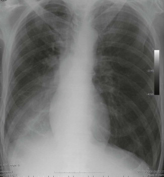
In the context of a patient attending the Emergency Department with abdominal pain there are just two questions to ask of the CXR:
Does the erect CXR show any evidence of free air under the diaphragm?
Is there any evidence of pneumonia?
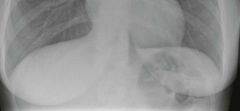
This erect CXR shows: normal lung bases and no free air under the diaphragm. The normal appearance of each lung is described on pp. 308–313 .
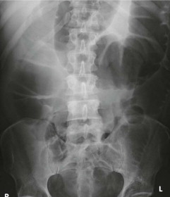
When analysing an AXR some normal features can be listed:
Gas within the stomach may be seen as gas outlining the gastric folds in the left upper quadrant; there is rarely any gas seen in the small bowel.
Normal gas patterns in the colon and rectum will vary. Sometimes segments of gas will be seen but no long continuous segments that are distended.
No calcification overlying the liver or gall bladder, nor over the pancreas, kidney, ureters or urinary bladder.
Calcification in the aorta may be age related and is common in the elderly; it will not show any localised bulge.
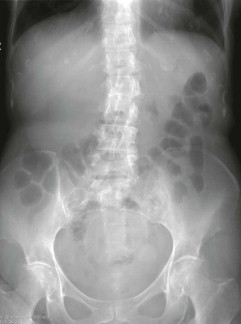
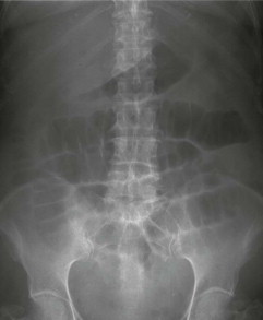
Elderly patient. Predominantly normal features on this AXR. Gas in an undistended large bowel (ascending, transverse, and descending colon); some age related calcification in the aorta. (The liver shadow might be prominent, but possible hepatomegaly is better assessed on palpation during the clinical examination.)
Both the likelihood of a particular diagnosis and the level of clinical concern will determine the specific radiological investigation that is selected. In some instances no imaging will be requested. The local availability of equipment or particular skills may also influence the choice of the first line test.
The following descriptions refer solely to plain film radiography; ie when a supine AXR or a CXR is obtained in the Emergency Department.
Become a Clinical Tree membership for Full access and enjoy Unlimited articles
If you are a member. Log in here