Physical Address
304 North Cardinal St.
Dorchester Center, MA 02124
This chapter is designed to introduce the instrumentation commonly used to perform routine dental extractions and other basic oral surgical operations. The instruments illustrated and described are used for a wide variety of purposes, including soft and hard tissue procedures. This chapter primarily provides a description of instruments; their use is discussed in subsequent chapters.
Many surgical procedures begin with an incision. The primary instrument for making incisions is the scalpel, which is composed of a handle and a sterile, very sharp blade ( Fig. 7.1 ). Scalpels are available as single-use instruments with plastic handles and fixed blades; scalpel handles to which disposable blades can be attached are also available. The most commonly used handle for oral surgery is the No. 3 handle. The tip of a scalpel handle is configured to receive a variety of differently shaped scalpel blades that are inserted onto the slotted portion of the handle.

The most frequently used scalpel blade for intraoral surgery is the No. 15 blade ( Fig. 7.2 ). The blade is small and is used to make incisions around teeth and through soft tissue. The blade is similar in shape to the larger No. 10 blade, which is used for large skin incisions in other parts of the body. Other commonly used blades for intraoral surgery include the No. 11 and No. 12 blades. The No. 11 blade is a sharp-pointed blade that is used primarily for making small stab incisions as for incising an abscess to establish drainage. The hooked No. 12 blade is useful for mucogingival procedures in which incisions are made on the posterior aspects of teeth or in the maxillary tuberosity area.

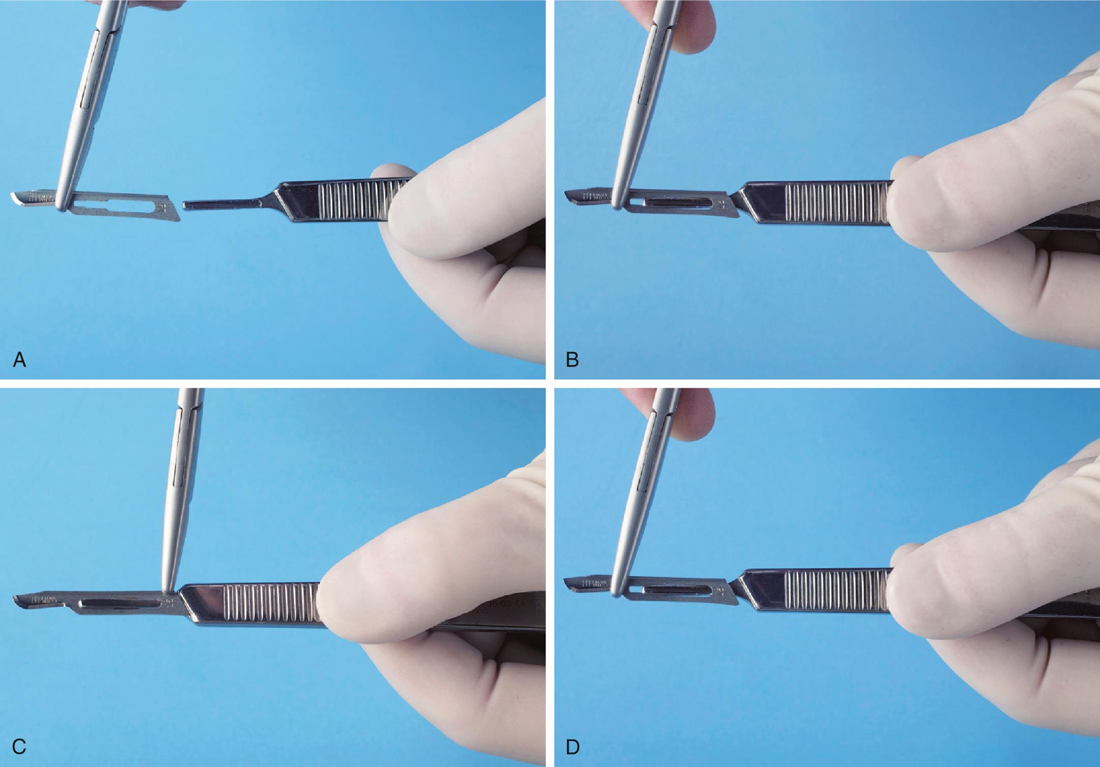
The scalpel blade must be carefully loaded onto the handle, ideally holding the blade with a needle holder. This lessens the chance of injuring one's fingers. The blade is held along the unsharpened edge, where it is reinforced with a small rib, and the handle is held so that the male portion of the fitting is pointing upward ( Fig. 7.3A ). The scalpel blade is then slowly slid onto the handle along the grooves in the male portion until it clicks into position (see Fig. 7.3B ).

The scalpel is unloaded in a similar manner. The needle holder grasps the end away from the blade (see Fig. 7.3C ) and lifts it to disengage it from the male fitting. The scalpel is then slid off the handle, always away from the body and anyone nearby (see Fig. 7.3D ). The used blade is immediately discarded into a specifically designed rigid-sided sharps container (see Fig. 5.6C ).
When using the scalpel to make an incision, the surgeon typically holds the handle in the pen grasp ( Fig. 7.4 ) to allow maximal control of the blade as the incision is made. For maximum cutting efficiency, mobile tissue should be held firmly in place under some tension so that as the incision is made, the blade will incise and not just push away the mucosa. When incising depressible soft tissue, an instrument such as a retractor should be used to hold the tissue taut while incising. When a full-thickness mucoperiosteal incision is desired, the blade should be pressed down firmly so that the incision penetrates the mucosa and periosteum with the same stroke.

Scalpel blades are designed for single-patient use. Blades dull easily when they come into contact with hard tissue such as bone or teeth and even after repeated strokes through keratinized tissue. If several incisions through the mucoperiosteum to bone are required, it may be necessary to use additional blades during a single operation. Dull blades do not make clean, sharp incisions in soft tissue and therefore should be replaced before they become overly dull.
The tissue plane between periosteum and bone is relatively bloodless and well defined. When an incision is made through the periosteum, ideally the periosteum should be reflected from the underlying cortical bone in a single subperiosteal layer with a periosteal elevator. The instrument that is most commonly used in oral surgery is the No. 9 Molt periosteal elevator ( Fig. 7.5 ). This instrument has a sharp, pointed end and a broader, rounded end. The pointed end is used to begin the periosteal reflection and to reflect dental papillae from between teeth, whereas the broad, rounded end is used to continue the elevation of the periosteum from bone.

The No. 9 Molt periosteal elevator is typically used to reflect tissue by two methods. In the first method, the pointed end is used in a twisting, prying motion to elevate soft tissue, most commonly when elevating a dental papilla from between teeth or the attached gingiva around a tooth to be extracted or when beginning to elevate a full thickness mucoperiosteal flap. The second method involves the push stroke in which the side of the pointed end or the broad end of the instrument is slid underneath the periosteum, separating it from the underlying bone. This is the most efficient stroke that results in the cleanest reflection of periosteum.
There are other types of periosteal elevators for use by periodontists, orthopedic surgeons, and other surgeons involved in work on bones.
Good access and vision are critical to performing excellent surgery. A variety of retractors have been specifically designed to retract the cheek, tongue, and mucoperiosteal flaps to provide access and visibility during surgery. Retractors are also used to help protect soft tissue from sharp cutting instruments.
The two most popular cheek retractors are (1) the right-angle Austin retractor ( Fig. 7.6 ) and (2) the broad offset Minnesota retractor ( Fig. 7.7 ). These retractors can also be used to retract the cheek and a mucoperiosteal flap simultaneously. Before the flap is created, the retractor is held loosely in the cheek. Once the flap is reflected, the retractor edge is placed on bone and is then used to retract the flap.


The Henahan and Seldin retractors are other types of instruments used to retract oral soft tissue ( Fig. 7.8 ). Although these retractors may look similar to a periosteal elevator, the leading edge is not sharp but, instead, smooth; these instruments are not typically used to elevate the mucoperiosteum. The No. 9 Molt periosteal elevator can also be used as a retractor for small flaps. Once the periosteum has been elevated, the broad blade of the periosteal elevator is held firmly against bone, with the mucoperiosteal flap elevated into a reflected position.

The instrument most commonly used to retract the tongue during routine exodontia is the mouth mirror. This is usually part of every basic setup because it is useful for examining the mouth and for indirect visualization during dental procedures. The Weider tongue retractor is a broad, heart-shaped retractor that is serrated on one side so that it can more firmly engage the tongue and retract it medially and anteriorly ( Fig. 7.9A ). When this retractor is used, care must be taken not to position it so far posteriorly as to cause gagging or to push the tongue into the oropharynx (see Fig. 7.9B ).

A towel clip (see Fig. 7.28 ) can also be used to hold the tongue in certain circumstances. When a biopsy procedure is to be performed on the posterior aspect of the tongue, the most positive way to control the tongue is by holding the anterior tongue with a towel clip. Local anesthesia must be profound where the clip is placed, and, if anticipated, it is wise to mention to the patient that this method of retraction may be used.
Various oral surgical procedures require the surgeon to grasp soft tissue to incise it, to stop bleeding, or to pass a suture needle. The instrument most commonly used for this purpose is the Adson forceps (or pickup; Fig. 7.10A ). These are delicate forceps, with or without small teeth at the tips, that can be used to hold tissue gently while stabilizing it. When this instrument is used, care should be taken not to grasp the tissue too tightly to avoid crushing it. Toothed forceps allow tissue to be securely held with a more delicate grip than untoothed forceps.

When working in the posterior part of the mouth, the Adson forceps may be too short. Longer forceps that have a similar shape are the Stillies forceps. These forceps are usually 7 to 9 inches long and can easily grasp tissue in the posterior part of the mouth, still leaving enough of the instrument protruding beyond the lips for the surgeon to hold and control it (see Fig. 7.10B ).
Occasionally it is more convenient to have an angled forceps. These include the college, or cotton, forceps (they are also called cotton pliers ) (see Fig. 7.10B ). Although these forceps are not especially useful for handling tissue, they are an excellent instrument for picking up loose fragments of tooth, amalgam, or other foreign material and for placing or removing gauze packs.
In some types of surgery, especially when removing larger amounts of tissue or doing biopsies, such as in an epulis fissurata, forceps with locking handles and teeth that will firmly grip the tissue are necessary. In this situation, the Allis tissue forceps are used ( Fig. 7.11A–B ). The locking handle allows the forceps to be placed in the proper position and then to be held by an assistant to provide the necessary tension for proper dissection of the tissue. The Allis forceps should never be used on tissue that is to be left in the mouth because they cause a relatively large amount of tissue crushing (see Fig. 7.11C ). However, the forceps can be used to grasp the tongue in a manner similar to a towel clamp.

When incisions are made through tissue, small arteries and veins are incised, causing bleeding. For most dentoalveolar surgery, pressure on the wound is usually sufficient to control bleeding. Occasionally pressure does not stop the bleeding from a larger artery or vein. When this occurs, an instrument called a hemostat is useful ( Fig. 7.12A ). Hemostats come in a variety of shapes; they may be small and delicate or larger and are either straight or curved. The hemostat most commonly used in surgery is the curved hemostat (see Fig. 7.12B ).

A hemostat has long, delicate beaks that are used to grasp tissue and a locking handle. The locking mechanism allows the surgeon to clamp the hemostat onto a vessel and then let go of the instrument or let an assistant hold it. The tip of the hemostat will remain clamped onto the tissue. This is useful when the surgeon plans to place a suture around the vessel or to cauterize it (i.e., use heat to sear the vessel closed).
In addition to its use as an instrument for controlling bleeding, the hemostat is especially useful in oral surgery to remove granulation tissue from tooth sockets and to pick up small root tips, pieces of calculus, amalgam, fragments, and any other small particles that have dropped into the wound or adjacent areas. However, it should never be used to suture.
The instrument most commonly used for removing bone in dentoalveolar surgery is the rongeur forceps. This instrument has sharp blades that are squeezed together by the handles, cutting or pinching through bone. Rongeur forceps have a rebound mechanism incorporated so that when hand pressure is released, the instrument reopens. This allows the surgeon to make repeated bone-trimming actions without manually reopening the instrument ( Fig. 7.13A ). The two major designs for rongeur forceps are (1) a side-cutting forceps and (2) the side- and end-cutting forceps (see Fig. 7.13B ).

The side-cutting and end-cutting rongeurs are more practical for most dentoalveolar surgical procedures that require bone removal. The end-cutting forceps can be inserted into sockets for the removal of interradicular bone and can also be used to remove sharp edges of bone. Rongeurs can be used to remove large amounts of bone efficiently and quickly. Because a rongeur is a delicate instrument, the surgeon should not use it to remove large amounts of bone in single bites. Rather, smaller amounts of bone should be removed in multiple bites. Likewise, the rongeur should never be used to remove teeth because this practice will quickly dull and destroy the instrument and risks losing a tooth in the patient's throat because a rongeur is not designed to hold an extracted tooth firmly. Rongeurs are expensive, so care should be taken to keep them sharp and in working order.
Another method for removing bone is with a burr in a handpiece. This is the technique that most surgeons use when removing bone for the surgical removal of teeth. Moderate-speed, high-torque handpieces with sharp carbide burrs remove cortical bone efficiently ( Fig. 7.14 ). Burrs such as No. 557 or No. 703 fissure burr and No. 8 round burr are used. When large amounts of bone must be removed, as in torus reduction, a large-bone burr that resembles an acrylic burr is typically used.

Any handpiece that is used for oral surgery must be completely sterilizable. When a handpiece is purchased, the manufacturer's specifications must be checked carefully to ensure that they can be met. The handpiece should have high speed and torque. This allows rapid bone removal and efficient sectioning of teeth. The handpiece must not exhaust air into the operative field, which would make it improper to use the typical high-speed air-turbine drills employed in routine restorative dentistry. The reason is that the air exhausted into the wound may be forced into deeper tissue planes and produce tissue emphysema, a dangerous occurrence.
Occasionally bone removal is performed using a mallet and chisel ( Fig. 7.15 ), although the availability of high-speed handpieces for removing bone and sectioning teeth has greatly limited the need for mallets and chisels. The mallet and chisel are sometimes used in removing lingual tori. The edge of the chisel must be kept sharp if it is to function effectively (see Chapter 13 ).

Become a Clinical Tree membership for Full access and enjoy Unlimited articles
If you are a member. Log in here