Physical Address
304 North Cardinal St.
Dorchester Center, MA 02124

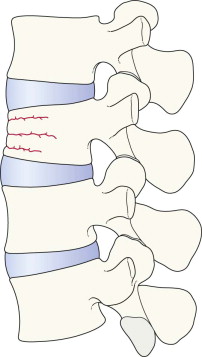
Transverse process fractures.
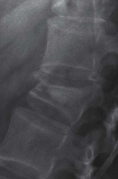
Lateral and AP views.
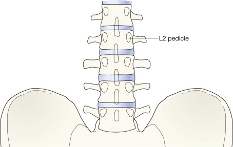
AP, anterior-posterior; L1, the 1st lumbar vertebra; T6, the 6th thoracic vertebra.
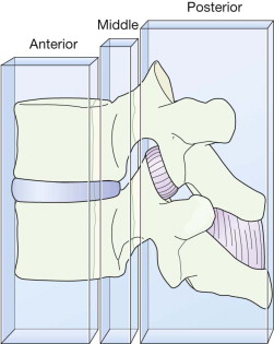
The concept of a three column spine is a familiar one when evaluating a CT or MRI examination. This anatomical concept can also be applied to the lateral radiograph.
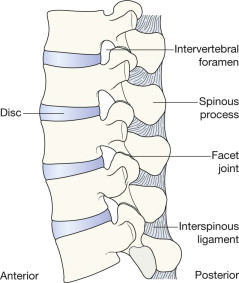
Anterior column The anterior longitudinal ligament, the anterior part of the annulus fibrosus, and the anterior two thirds of the vertebral body.
Middle column The posterior longitudinal ligament, the posterior part of the annulus fibrosus, and the posterior margin of the vertebral body.
Posterior column The facet joints, the pedicles, and the posterior ligaments.
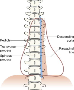
In the thoracic spine the soft tissue shadow of the left paraspinal line (aka paravertebral stripe) should be closely applied to the vertebral bodies. This line is produced by the interface between the paravertebral soft tissues and the adjacent lung.
On the right side there is no visible paraspinal line .
A step-by-step analytical approach when evaluating the plain films may provide an immediate indication that a potentially catastrophic injury has occurred.
The AP projection can provide useful information, but the lateral radiograph is invariably the more useful. 70–90% of detectable plain film abnormalities will be shown on the lateral projection . Also, it is the lateral radiograph to which the three column stability principle is applied:
“Instability is present if any two of the three columns are disrupted”
Look for:
Loss of height or wedging of a vertebral body: this is evidence of a compression fracture. Wedging may be associated with loss of the normal concavity of the posterior aspect of the vertebral body . This loss indicates significant posterior displacement of the middle column.
Fragment(s) of bone detached from the anterior aspect of a vertebral body.
More than one abnormality. The importance of recognising all of the radiological abnormalities that may be present is explained under stability (pp. 209–210).
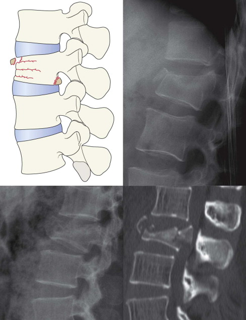
Top left: two fragments have been detached. Two columns are disrupted… the anterior and middle columns. Unstable.
Top right: Several fragments. The posterior margin of L1 is disrupted and bulges into the spinal canal. Two column disruption. Unstable.
Bottom left and right: Large fragment in canal. Two columns disrupted. Unstable.
Become a Clinical Tree membership for Full access and enjoy Unlimited articles
If you are a member. Log in here