Physical Address
304 North Cardinal St.
Dorchester Center, MA 02124

While imaging of the abdomen is now largely performed utilizing CT, ultrasound, or MRI, many patients still have conventional radiographs (“plain films”) of the abdomen as a first step before other imaging studies are performed or as a method of following-up on findings demonstrated by other modalities. Many of the principles that guide the interpretation of conventional radiographs also apply to the modalities of CT, MRI, and ultrasound.
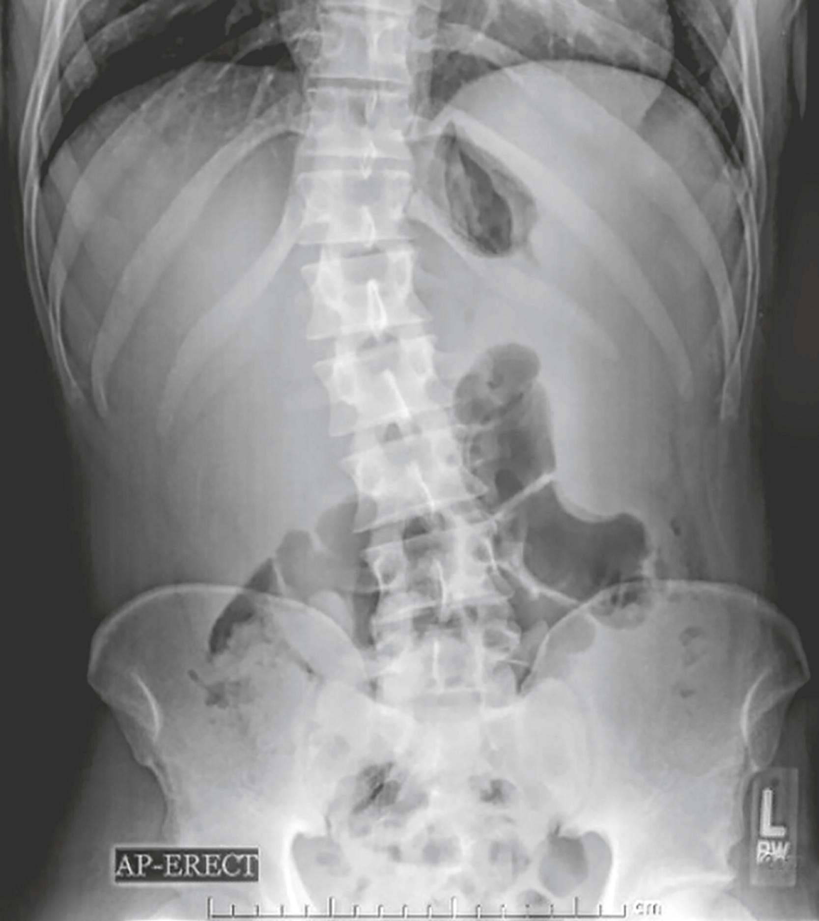
This is a 45-year-old male with a known history of leukemia. Conventional radiographs of the abdomen were obtained for abdominal discomfort and the upright abdominal view is shown. Palpation of the abdomen was abnormal. What abnormalities does this radiograph suggest? See the answer at the end of this chapter.
In order to recognize abnormal findings on conventional radiographs of the abdomen, you must familiarize yourself with the appearance of normal first.
First , look at the overall gas pattern.
You are looking for the overall pattern, so don’t spend too much time trying to identify every bubble of bowel gas you see.
Second, check to see if there is extraluminal air.
Third, look for abnormal abdominal calcifications.
Fourth, look for any soft-tissue masses.
Virtually all gas in the bowel comes from swallowed air. Only a fraction comes from the bacterial fermentation of food. For the purposes of this chapter, the terms gas and air are used interchangeably to refer to the contents of the bowel.
Loops of bowel that contain a sufficient amount of air to fill the lumen completely are said to be distended. Distension of bowel is normal.
Loops of bowel that are filled beyond their normal size are said to be dilated. Dilatation of the bowel is abnormal.
Stomach
There is almost always air in the stomach , unless:
The patient has recently vomited, or
There is a nasogastric tube in the stomach and the tube is attached to suction.
Small bowel
There is usually a small amount of air in about two or three loops of nondilated small bowel ( Fig. 12.1 ).

The normal diameter of small bowel is less than 2.5 cm, which is about 1 inch or the diameter of one United States quarter.
Large bowel
There is almost always air in the rectum or sigmoid. There may be varying amounts of gas in the remainder of the colon ( Fig. 12.2 ).

Use this rule to decide whether the large bowel is dilated or not:
The large bowel can normally distend to about the same size as it does on a barium enema examination. To give you an idea of how large that is, look at Fig. 12.3 .

Stool is recognizable by the multiple, small bubbles of gas present within a semisolid appearing soft-tissue mass. Recognizing the appearance of stool will help in localizing the large bowel ( Fig. 12.4 ).

 Important Points
Individuals who swallow excessive quantities of air may develop aerophagia, accompanied by belching, bloating, and abdominal distension. A gas pattern characterized by numerous polygonal-shaped , air-containing loops of bowel, none of which is dilated, may be seen ( Fig. 12.5 ).

Stomach
There is almost always air and fluid in the stomach so there is almost always an air-fluid level visible in the stomach on either an upright abdomen, a study done with the patient in the decubitus position, or an upright chest radiograph.
To see an air-fluid level , the path of the x-ray beam must be directed horizontally —parallel to the floor.
Small bowel
Two or three air-fluid levels in small bowel may be seen normally on an upright or decubitus view of the abdomen.
Large bowel
The large bowel functions, in part, to remove fluid so there are usually no or very few air-fluid levels in the colon ( Fig. 12.6 ).

 Important Points
There may be many air-fluid levels present in the colon if the patient has had a recent enema or if the patient is taking medication with a strong anticholinergic, antiperistaltic effect.
The normal distribution of bowel gas and fluid is summarized in Table 12.1 .
| Organ | Normally Contains Gas | Normally Has Air/Fluid Levels |
|---|---|---|
| Stomach | Yes | Yes |
| Small bowel | Yes, 2–3 loops | Yes |
| Large bowel | Yes, especially rectosigmoid | No |
Recognizing large bowel
Large bowel is peripherally placed around the perimeter of the abdominal cavity except for the right upper quadrant, which is occupied by the liver ( Fig. 12.7 ).

Haustral markings usually do not extend completely across the large bowel from one wall to the other. If they should connect one wall with another, haustral markings are spaced more widely apart than the valvulae conniventes of the small bowel ( Fig. 12.8 ).

Recognizing small bowel
Small bowel is centrally placed in the abdomen. Valvulae markings typically extend across the lumen of small bowel from one wall to the other. The valvulae are spaced much closer together than the haustra of the large bowel ( Fig. 12.9 ).

Small bowel can achieve a maximally dilated diameter of about 5 cm. Large bowel can dilate to several times that size.
Almost every Department of Radiology has a series of radiographic images (i.e., a protocol) that is routinely obtained in patients who have acute abdominal pain. These series are sometimes called obstruction series or complete abdominal series or acute abdominal series or something similar. We will call such a combination of views an acute abdominal series.
The views that make up such a series will vary not only between facilities but also based on each patient’s ability to accommodate to the positioning needed to obtain the views. In general, views may include:
A supine view of the abdomen is almost always obtained (sometimes called a flat plate or KUB [for kidneys, ureters, and bladder]).
A prone or lateral rectum view is the most variable as to whether it is acquired.
An upright view (or left lateral decubitus view) may depend on the patient’s condition.
A frontal chest x-ray (upright or supine) may also be included.
Table 12.2 summarizes what to look for on each of the views of an acute abdominal series.
| View | Look for |
|---|---|
| Supine abdomen | Overall bowel gas pattern, calcifications, masses |
| Prone abdomen or lateral rectum | Gas in the rectosigmoid |
| Upright abdomen | Free air, air-fluid levels in the bowel |
| Upright chest | Free air, pneumonia, pleural effusions |
Why it is important
For the overall appearance of the bowel gas pattern , its general location, volume, and configuration is more important than identifying every small bubble of air on the radiograph.
In identifying the presence or absence of calcifications.
For identifying the presence of soft-tissue masses.
How it is acquired
The patient lies on their back on the x-ray table or stretcher, and the x-ray beam is directed vertically downward ( Fig. 12.10 ).

Substitute view
There is really no other view that substitutes for a supine view of the abdomen. Virtually all patients, regardless of their condition, can tolerate this part of the examination.
Become a Clinical Tree membership for Full access and enjoy Unlimited articles
If you are a member. Log in here