Physical Address
304 North Cardinal St.
Dorchester Center, MA 02124

6- to 10-mm increased lateral tibiofemoral joint opening 20 degrees of flexion.
≥15 degrees increased external tibial rotation 30 degrees, 90 degrees of flexion.
± Varus recurvatum, standing and supine.
± Hyperextension gait abnormality.
Double or triple varus knee, after osteotomy.
Acute injuries, bony avulsions amendable to internal fixation.
The primary soft tissue–stabilizing structures of the posterolateral (PL) aspect of the knee joint are the fibular collateral ligament (FCL) and popliteus muscle-tendon-ligament unit (PMTL), including the popliteofibular ligament (PFL) and posterolateral capsule (PLC) shown in Figure 17-1 . These structures function together to resist lateral joint opening (LJO), posterior subluxation of the lateral tibial plateau with tibial rotation, knee hyperextension, and varus recurvatum.

The mechanism of injury may be contact or noncontact and usually involves a combined varus and hyperextension joint displacement. The proper management of injuries involving the PL structures requires knowledge of the complex anatomy and potential variations that may exist, the function of the major soft tissue stabilizers, appropriate diagnostic techniques, and surgical options for reconstruction. Isolated PL injuries are rare; however, on occasion an avulsion fracture at the femoral attachment occurs requiring internal fixation. PL injuries are frequently accompanied by anterior cruciate ligament (ACL) or posterior cruciate ligament (PCL) ligament ruptures.
Although the incidence of PL injury is unknown (owing to misdiagnosis or failure to detect the injury), the consequences of untreated PL ruptures are readily apparent. Chronic deficiency of the PL structures may be a factor in the failure of cruciate reconstructions and may also play a role in the development of gait abnormalities and giving-way. The detection and proper treatment of these problems is critical, because failure to properly treat all of the abnormalities may result in a poor outcome. The patient will complain of a varus type of instability with LJO during stance phase and show either a neutral or valgus alignment. The abnormal LJO during stance phase is always greater than that detected on the varus stress test. The patient may demonstrate the abnormal LJO by producing a varus loading at the knee joint while standing.
Knees that fulfill the double or triple varus diagnosis criteria (varus osseous malalignment with increased LJO, external tibial rotation, varus recurvatum, and knee hyperextension [see Chapter 26 ]) require high tibial osteotomy (HTO) first, followed approximately 6 months later, with an appropriate PL reconstruction. In many instances, an ACL or PCL deficiency also exists, which is corrected at the time of the PL reconstruction.
There are different surgical options available for acute knee injuries, dislocated knees with multiple ligament ruptures, chronic knees, and revision knees. The decision-making process for determining the appropriate PL procedure is discussed in detail under “Operative Treatment of Acute Posterolateral Ruptures” and “Operative Treatment of Chronic Posterolateral Ruptures” later in this chapter.
<5-mm increased lateral tibiofemoral joint opening 20 degrees of flexion.
<10 degrees increased external tibial rotation 30 degrees, 90 degrees flexion.
Double varus knee, in which valgus osteotomy and subsequent adaption (decreased laxity) of posterolateral structures eliminate abnormal lateral joint opening and external tibial rotation.
Triple varus knee without correction of varus malalignment.
Prior joint infection.
Patient noncompliant with rehabilitation, bracing, weight-bearing restrictions.
Hyperextension gait abnormality with no preoperative gait retraining.
Advanced joint arthritis, <2 mm remaining joint space.
Contraindications to PL reconstruction are findings of less than 12 mm of absolute increased lateral tibiofemoral joint opening and less than 15 degrees of increased external tibial rotation. These findings are frequently noted in knees with associated varus osseous malalignment (double varus knees) that are candidates for HTO (see Chapter 26 ).
Patients with varus malalignment who do not undergo HTO and have associated chronic insufficiency of the PL structures are not candidates for a PL procedure. Untreated varus osseous malalignment is a frequent cause of failure of PL reconstructions. In many cases, a knee hyperextension gait abnormality also exists, which must be corrected before surgery with a specific gait retraining program described in Chapter 29 . Failure to correct a hyperextension gait abnormality places PL reconstructions at risk for failure owing to the excessively high tensile forces placed on the PL soft tissues with weight bearing after surgery. Gait retraining usually decreases abnormally high knee extension and adduction moments to normal values.
Patients with a history of prior joint infection or who are obese (body mass index > 30) are not candidates for PL reconstruction. Patients with muscle atrophy of the lower extremity undergo preoperative rehabilitation before PL reconstruction.
Knees that demonstrate a loss of lateral tibiofemoral compartment joint space, with less than 2 mm remaining on 45-degree posteroanterior (PA) weight-bearing radiographs, are usually not candidates for PL reconstruction.
Common injury mechanism blow to anteromedial tibia causing excessive knee hyperextension, external tibial rotation, lateral tibiofemoral joint opening.
Most posterolateral injuries occur with anterior cruciate ligament or posterior cruciate ligament ruptures.
Knee flexion, extension
Joint effusion
Patellofemoral (medial and lateral subluxation, Q-angle, crepitus, compression pain)
Tibiofemoral crepitus, joint line pain, compression pain
Recurvatum (standing, supine)
Gait (severe hyperextension stance phase)
Muscle strength
Diagnosis of posterolateral injury is based on final position of the lateral tibial plateau.
Subluxation of the medial and lateral tibial plateaus separately at 30 degrees, 90 degrees of knee flexion.
Produce maximal external tibial rotation, determine change in position of medial and lateral tibial plateaus separately.
Qualitatively determine if anterior or posterior subluxation occurred in each tibial plateau.
External rotation recurvatum
Lateral and medial tibiofemoral joint opening 5 degrees, 20 degrees of flexion
Pivot shift, Lachman
Reverse pivot shift
Posterior drawer, 90 degrees of flexion
KT-2000 20 degrees of flexion, 134 N
Lateral, 30 degrees of flexion
Posteroanterior, weight-bearing, 45 degrees of flexion
Patellofemoral axial
Lateral stress, neutral tibial rotation select knees
Posterior cruciate ligament ruptures: posterior stress lateral, 90 degrees of flexion, neutral tibial rotation
Varus malalignment: full standing radiographs, mechanical axis and weight-bearing line
Sports Activity and Function Form
Occupational Rating Form
Symptom Rating Form
The PL structures are injured when excessive varus, external tibial rotation, and hyperextension forces are applied to the lower extremity. A blow to the anteromedial tibia during sports participation appears to be one of the most common injury mechanisms. These injuries frequently involve rupture of other knee ligament structures, complicating the diagnosis. An isolated complete PL rupture is rare because, usually, the injury is accompanied by an ACL or PCL rupture. In some cases, the PL structures are only partially disrupted and do not require surgical restoration. It is important to correctly determine the increases in LJO, external tibial rotation, and knee hyperextension of the injured knee (compared with the contralateral knee) preoperatively and intraoperatively. The decision of whether surgical restoration of the PL structures is indicated is based on the abnormal knee motion limits, joint subluxations, and the tissues disrupted.
One frequent patient presentation is a failed ACL or PCL reconstruction owing to untreated PL insufficiency. Another patient presentation is a chronic varus osseous malalignment and underlying ACL insufficiency in which, over time, interstitial stretching and slackening of the PL structures occurred. In these cases, HTO unloads the PL soft tissues to the extent where physiologic remodeling and shortening may subsequently occur in some knees and a PL reconstruction is not required.
A comprehensive physical examination is required, including assessment of knee flexion and extension, patellofemoral indices, tibiofemoral crepitus, tibiofemoral joint line pain, and gait abnormalities. Pain in the medial tibiofemoral compartment occurs owing to increased compressive forces related to varus osseous malalignment. Pain in the PL soft tissues may occur from increased soft tissue tensile forces caused by a varus thrusting gait pattern. The abnormal knee hyperextension involves increased extension in the sagittal plane and is often accompanied by a varus alignment in the coronal plane, which has been described as a varus recurvatum alignment. Together with a varus osseous malalignment, this is referred to as a triple varus knee (see Chapter 26 ). Patients with chronic PL insufficiency have varying amounts of altered gait mechanics and knee hyperextension. Some individuals may present with a markedly abnormal gait that is severely disabling and limits ambulation. Other patients may have a less noticeable alteration because the abnormal knee hyperextension occurs only after prolonged walking and muscle fatigue. The abnormal gait pattern is characterized by excessive knee hyperextension during the stance phase, which does respond to gait retraining that initiates normal stance phase flexion (see Chapter 29 ). Subjective complaints of giving-way during routine daily activities, along with severe quadriceps atrophy, often accompany this gait abnormality.
The surgeon must determine all of the abnormal translations and rotations in the knee joint. The ligament injuries that result in knee hyperextension and varus recurvatum frequently involve not only the PL structures, but also other ligament and capsular structures. The biomechanic and kinematic studies that form the basis for the interpretation and diagnosis of the manual stress tests are described in Chapter 15 .
The increases in LJO and external tibial rotation shown in Table 17-1 are only approximations of what would be expected with clinical injury to the PL structures. Importantly, an increase of only a few millimeters (2-5 mm) in LJO occurs with complete rupture of the FCL, whereas an increase of 5 to 9 mm occurs with complete rupture of all the PL structures (FCL, PMTL, and PFL). These values are based on biomechanic studies discussed in Chapter 15 . LaPrade and colleagues conducted a cadaveric study in which lateral stress radiography was applied at 12 N-m (on an experimental apparatus) and the increase in LJO over the intact state was compared with that measured during a clinician-applied load after an isolated FCL rupture and a combined FCL, PMTL, and PFL rupture. Compared with the intact state, LJO induced by the clinician-applied load increased by 2.7 mm (isolated FCL rupture) and 4.0 mm (combined PL rupture). However, the mean values showed a wide standard deviation and variation among specimens, making extrapolation to the clinical setting difficult. In addition, the lateral joint space measurement showed wide confidence intervals (CI). For an isolated FCL rupture, the mean lateral gap distance was 10.99 mm (CI, 7.8-14.3 mm) and for the combined PL rupture, the mean distance was 12.2 mm (CI, 9.3-15.2 mm). This amount of overlap indicates that it would not be possible to accurately separate a FCL rupture alone from a combined PL injury. The measurements are important and useful in providing the clinician with a baseline in interpreting lateral stress radiographs. The gap test is based on the joint separation between articular cartilage seen at arthroscopy, and not the tibiofemoral separation on a stress radiograph. Even so, the measurements are somewhat equivalent as to the increase in the amount of millimeters with PL injuries. For example, Figure 17-2 shows an approximate normal lateral gap of 4 mm at the closest point of the lateral compartment at arthroscopy. An increase of only 6 mm results in 10 mm of absolute opening at the closest point or 12 mm at the periphery, which is viewed as a positive gap test and indicative of injury to the PL structures. Fortunately in most knees, these are the lesser values and it is more common that the lateral gap exceeds these measurements, indicating that concurrent PL reconstruction is necessary.
| Examination | Technique | Illustration | Grading | Significance |
|---|---|---|---|---|
| Dial test 30 degrees | Supine position, palpate anterior tibial prominence, medial and lateral joint, maximum external rotation, posterior position, lateral tibia (PL) subluxation, anterior position medial tibia (anteromedial subluxation). |
|
Compare external rotation between knees. | External tibial rotation with PL subluxation. Increase 3-5 degrees FCL tear. Increase 6-10 degrees FCL tear, partial PMTL. Increase ≥15 degrees FCL, PMTL, PLC. |
| Dial test 90 degrees | Maximum external tibial rotation. Determine PL tibial subluxation. |
|
Compare external rotation between knees. | Increase at 30 and 90 degrees means PCL and PLC injury. With PCL tear, degrees of external rotation difficult to estimate owing to posterior tibial position. |
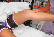
| PL external rotation test | Knee at 90 degrees, posterior and external rotation loading on tibia, palpate posterior subluxation lateral tibiofemoral joint. |
|
Qualitative PL tibial subluxation 90 degrees (less accurate at 30 degrees of flexion). | Similar to dial test but foot held stationary. Dial test allows tibia to externally rotate fully, providing better estimate of increased tibial rotation. PL subluxation 90 degrees, combined PCL, PLC injury. |
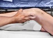
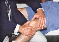
| Posterior drawer test | Knee at 90 degrees position, posterior load proximal tibia, no tibial rotation. Palpate medial tibiofemoral step-off. |
|
Partial PCL: increase 0-9 mm translation; complete PCL tear: increase >10 mm translation. | Stress radiography more accurate. Increases >10 mm indicate secondary restraints torn or physiologic slack (combined injury). |
| Quadriceps active test | Knee position at 70-90 degrees. Foot stabilized by examiner, patient activates quadriceps by pushing foot against table or attempting to extend knee. |
|
Qualitative. Observe, palpate tibiofemoral position. | Confirms posterior tibial subluxation, PCL injury at resting position. Quadriceps contraction produces anterior translation knee position ≥70 degrees. |
| Lachman (anterior drawer, 30 degrees of flexion) | Anterior load proximal tibia. |
|
Observe anterior translation tibia; compare with opposite knee. | Estimate increase translation in millimeters. Soft endpoint, ACL not resisting anterior translation, indicates ACL tear. Increase 3- to 5-mm ACL tear. >5 mm ACL plus secondary restraints. |
| Pivot shift | Knee position 10-30 degrees of flexion. Anterior load tibia, gentle internal tibial rotation (subluxation) followed by posterior load, gentle external rotation (reduction). |
|
Qualitative. Grade: I slipping. II thud, clunk with reduction. III gross anterior subluxation lateral tibiofemoral joint, anterior impingement tibia limits reduction event. | Grade I physiologic laxity, no or partial ACL tear. II ACL tear. Grade III ACL tear plus secondary restraints lax. |
| Reverse pivot shift test | Similar loading as pivot shift. |
|
PL subluxation with external rotation confused for reduction in pivot shift. No abnormal anterior tibial subluxation. | Observe obvious PL tibial subluxation with posterior and external rotation loading. Dial test more accurate. |
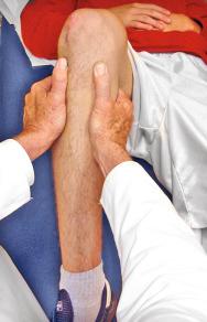
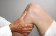
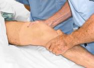
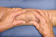
| Varus stress testing | Thigh supported on examination table. Knee position 0 degree, 30 degrees. Varus load with no external-internal tibial rotation. Palpate lateral joint line opening. |
|
Subtle 30 degrees increase LJO. 2-4 mm complete FCL tear, further increase LJO with PLC injury. | Stress radiography more accurate. 30 degrees of flexion. Increase 2- to 4-mm FCL tear. Increase 5- to 9-mm complete PLC tear (also perform valgus stress test). |
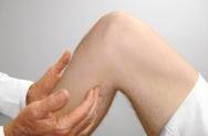
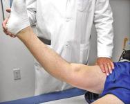
| External rotation recurvatum test | Grasp and hold both feet above table, allow gravity knee hyperextension. |
|
Qualitative. Tibia externally rotates, varus position caused by PL joint opening, knee. | Hyperextension, indicates PLC injury, >10 degrees frequently associated ligament injury (ACL, PCL). |
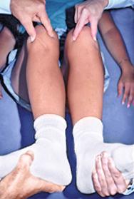
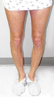
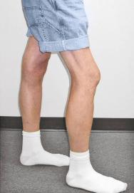
| Standing recurvatum test | Patient stands, feet together pushes knees backward into hyperextension, compare knees. |
|
Qualitative, observe varus hyperextension position (can be measured with goniometer). Increased loading over supine recurvatum test brings out deformity. | 10 degrees hyperextension with varus alignment PLC disruption, lateral, PL joint abnormal opening, often combined PLC, ACL tear, confirm with other tests. |
| Standing frontal alignment | Standing 0-5 degrees of flexion. Avoid hyperextension. |
|
Confirm varus alignment. Hip-knee-ankle radiographs 0-5 degrees of flexion. | Classify primary, double, triple varus based on all tests (see Chapter 26 ). |
| Knee hyperextension gait or varus thrust | Observe gait walking to and from examiner. |
|
Qualitative. Knee goes into hyperextension on stance phase. Knee has varus thrust without hyperextension. | Two hyperextension patterns (see Chapter 29 ). Forward trunk position, loss of quadriceps control, ankle dorsiflexion push-off, requires gait retraining. Varus thrust increases medial compartment loads and tensile forces lateral ligaments, osteotomy may be required. |
| Range of motion | Perform passive flexion-extension. |

|
Normal 3-0-135 degrees. Hyperextension to neutral to flexion | 10 degrees hyperextension posterior capsule possible ACL or PCL injury. ≥15 degrees multiple ligament injury. |
| Effusion, soft tissue swelling, pain | Palpate joint for effusion, tenderness, meniscus, ligament attachments. |
|
Qualitative. Complex examination necessary + meniscus tests. | Partial to complete tears PLC. FCL local tenderness, pain varus, dial tests. |
| Patellofemoral examination | Comprehensive examination. All tests. Alignment, PF crepitus, medial/lateral translation, patella height. |
|
See Chapter 35 . | Increased external tibial rotation 30 degrees, PLC tear, produces abnormal lateral shifting tibial tubercle, increases Q-angle. |
| Neurovascular examination | Complete examination. Both lower extremities, PT, DP pulses, lower extremity muscle function. |
|
Peroneal nerve injuries associated with severe PLC disruption (10%-30%). Arterial studies indicated multiple ligament injuries, dislocations. |

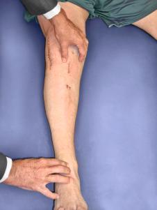
An increase in external tibial rotation may occur with anterior subluxation of the medial tibial plateau, posterior subluxation of the lateral tibial plateau, or a combination of both subluxations. The dial test, which the senior author (F.R.N.) published, allows a diagnosis of tibial rotational subluxations of the medial and lateral tibiofemoral compartments at 30 and 90 degrees of knee flexion (see Table 17-1 ). Other variations of this test have been described.
The position of the medial and lateral tibial plateau is assessed at the starting position (neutral tibial rotation) with the knee flexed to 30 and 90 degrees and at the final position with the tibia in maximal external rotation. The examiner palpates the position of the medial and lateral tibial plateau, which is compared with the normal knee to assess whether a subluxation (anterior or posterior) of the medial or lateral tibial plateau is present. An increase in internal tibial rotation occurs with both lateral ligament, medial ligament, and PCL disruption (see Chapter 15 ). The axis of tibial rotation is observed in the involved knee and compared with the normal knee to detect a shift in the medial or lateral tibiofemoral compartment during tibial rotation. It is not recommended that the dial test be performed in the prone position because the tibiofemoral joint cannot be accurately palpated to distinguish an anteromedial from a PL tibial subluxation.
It is not possible to clinically determine the actual millimeters of translation of the medial and lateral tibial plateaus in reference to the femoral condyle. A qualitative determination is made of the anterior or posterior subluxation of the medial or lateral tibiofemoral joint. With PL injuries, there is an abnormal lateral deviation of the tibial tubercle in the dial test compared with the opposite knee.
The use of the dial test in knees with PCL ruptures requires maintenance of a normal anatomic tibiofemoral position. This is accomplished by applying an anterior translation, loading the ACL in both limbs, during the external tibial rotation. It is still necessary to use the supine position so that the examiner can palpate the tibiofemoral position. The dial test is less accurate with a PCL rupture because it is difficult to compare limbs, and other tests to be described (LJO, gap test at arthroscopy, varus recurvatum) for the integrity of the PL structures need to be carefully assessed.
When a posterior subluxation of the lateral tibial plateau is positively identified by the tibiofemoral rotation test, additional tests must be conducted to determine the integrity of other ligament structures. The amount of LJO at 5 and 20 degrees of knee flexion should be determined to further assess the integrity of the FCL and other secondary ligament restraints. The posterior tibial subluxation of the central tibial and medial tibiofemoral joint determines the amount of increased translation because of a PCL injury, which adds to the maximum posterior subluxation to the lateral compartment with external tibial rotation.
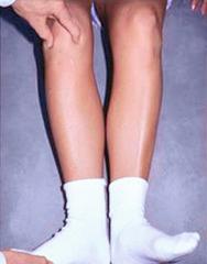
The presence of a varus recurvatum in both the supine and standing positions must be carefully assessed. Often, the varus recurvatum reaches its maximum position when the patient is standing and asked to maximally hyperextend both knees.
The appropriate tests to determine the integrity of the ACL and PCL are performed, including KT-2000 (MEDmetric) arthrometer testing at 20 degrees of flexion (134 N) to quantify total anteroposterior (AP) displacement. The pivot shift test is recorded on a scale of 0 to III (grade 0, no pivot shift; grade I, slip or glide; grade II, jerk or clunk; grade III, gross subluxation with impingement of the PL aspect of the tibial plateau against the femoral condyle). A misdiagnosis of a positive pivot shift test may occur with PL injuries as the lateral tibial plateau is brought to a reduced position (starting from a posterior subluxated position) with knee extension and then posteriorly subluxates with knee flexion (reverse pivot shift test). The medial posterior tibiofemoral step-off on the posterior drawer test is done at 90 degrees of flexion.
Radiographs taken during the initial examination include AP, lateral at 30 degrees of knee flexion, weight-bearing PA at 45 degrees of knee flexion, and patellofemoral axial views. Lateral stress radiographs may be required of both knees (20 degrees of flexion, neutral tibial rotation, and 67-N varus force). A comparison is made of the millimeters of lateral tibiofemoral compartment opening between knees.
A lateral radiograph is used to determine the approximate length required for FCL anatomic grafts. The distance from the anatomic femoral insertion site to the anatomic fibular insertion site is measured and adjusted for magnification. A measurement of the patellar tendon length is also made when a bone-patellar tendon-bone (B-PT-B) FCL autograft is planned; however, in most knees, a B-PT-B allograft is used, as will be discussed.
Posterior stress radiographs are obtained in patients with PCL ruptures, especially those in which the distinction of a partial versus complete PCL deficiency is difficult to determine on clinical examination. A lateral PCL stress radiograph is taken of each knee at 90 degrees of flexion. The limb is placed in neutral rotation with the tibia unconstrained and the quadriceps relaxed, and 89-N force applied to the proximal tibia. Measurement is made of the millimeters of posterior tibial translation in both knees. Knees with 10 mm or more of increased posterior tibial translation are considered candidates for PCL reconstruction.
Full standing radiographs of both lower extremities, from the femoral heads to the ankle joints, are done in knees with varus lower extremity alignment. The mechanical axis and weight-bearing line are measured to determine whether HTO is indicated.
Patients complete questionnaires and are interviewed for the assessment of symptoms, functional limitations, sports and occupational activity levels, and patient perception of the overall knee condition according to the Cincinnati Knee Rating System (CKRS; see Chapter 41 ).
The classification and treatment of first-, second-, and third-degree acute PL injuries are detailed in Table 17-2 . It is important to diagnose partial tears of the PL structures, with a mild to moderate increase in LJO and external tibial rotation, to allow protection and maintain lateral tibiofemoral joint closure in the initial 3 weeks to allow “stick-down” and healing of lateral soft tissues. This is a program similar to that recommended for medial ligament ruptures (see Chapter 19 ).
| First Degree | Second Degree | Third Degree * | |||
|---|---|---|---|---|---|
| Anatomic lesion | Minor tearing fibers | Partial tears, one third to two third fibers | FCL tear † | FCL tear, partial tear PMTL, PL capsule | FCL tear, PMTL tear, PL capsule tear |
| Signs | Minor tenderness and swelling | Tenderness and swelling lateral tissues | Tenderness and swelling lateral tissues | ||
| Increase in lateral joint opening ‡ | |||||
| 30 degrees | None | None | 2-3 mm | 2-5 mm | 5-9 mm |
| 0 degree | None | None | None | None | 3-5 mm |
| Increase in external tibial rotation (dial test, 30 degrees) ‡ | None | None | 3-5 degrees | 6-10 degrees | 15 degrees |
| Treatment | Progress per symptoms, no crutches | Progress per symptoms, soft support brace | Bivalved cylinder cast 3 wk ROM 0-90 degrees 2 wk Support brace 3-6 week Wean crutches 3-6 wk |
Operative repair, reconstruction; usually associated ACL, PCL | |
* Avulsion FCL, popliteus tendon: surgical indication to reattach.
† Even though FCL shows complete tear, adjacent lateral tissues maintain ligament continuity for healing. Bivalved cylinder cast with protected motion, maintain lateral tibiofemoral joint closure.
‡ See Chapter 15 . Increases related to degrees of knee flexion, minor opening may be less under clinical conditions with lower joint loading.
Golden period acute surgical repair: 7-14 days after injury.
Lower extremity venous ultrasound, vascular consult for multiple ligament ruptures.
Delay surgery 5-7 days, observe neurovascular status, soft tissue swelling, skin integrity.
Soft hinged full-leg brace, well-padded compression dressing.
PCL rupture: bivalved cast with posterior plaster shell, posterior calf pad.
Verify tibiofemoral reduction with lateral radiograph in multiligament injuries.
MRI for location of major ligament disruptions.
Protected knee motion, patellar mobilization, isometrics.
Contraindications to acute surgery in dislocated knees: excessive soft tissue swelling, hemorrhage, edema. Delay reconstruction until swelling resolved, muscle function and knee motion restored.
Muscle atrophy requires preoperative rehabilitation.
Hyperextension gait abnormality requires gait-retraining program before PL reconstruction.
Varus osseous malalignment requires osteotomy before PL reconstruction.
Absent lateral meniscus, early tibiofemoral arthritis, consider lateral meniscus transplant.
Ensure B-PT-B, Achilles tendon allografts available.
Determine appropriate grafts for ACL or PCL reconstruction.
ACL , Anterior cruciate ligament; B-PT-B , bone-patellar tendon-bone; MRI , magnetic resonance imaging; PCL , posterior cruciate ligament; PL , posterolateral.
There is a distinct advantage for repairing completely disrupted PL structures and meniscal attachments in acute injuries ( Fig. 17-3 ). At the time of surgery, extensive disruption of these structures is observed. Careful dissection is required to identify anatomic tissue planes and maintain an intact vascular and neural supply. The so-called golden period to perform an acute surgical repair is within 7 to 14 days of the injury. After this time, scar tissue will obliterate tissue planes and make the dissection and repair difficult.

A lower extremity venous ultrasound is obtained before surgery in acute multiligament knee injuries that have swelling and soft tissue damage to detect occult venous thrombosis that requires urgent treatment and contraindicates surgery. An initial delay in surgery for 5 to 7 days allows for observation of the neurovascular status, soft tissue swelling, skin integrity, and some clearing of hemorrhage in soft tissues in the injured extremity.
During this time, the lower extremity is supported in a soft-hinged full-leg brace in extension with a well-padded compression dressing. In knees with extensive damage to the PL structures and PCL, a bivalved cylinder cast with a posterior plaster shell and posterior foam calf pad may be required to provide added stability and prevent posterior tibial subluxation. Reduction of the tibiofemoral joint is verified by a lateral radiograph. Lower limb elevation, ice, and compression are important. The physical therapist initiates early protected knee motion, patellar mobilization, active quadriceps function, and electrical muscle stimulation. Dislocated knees scheduled for surgery require vascular consultation, ankle/brachial studies (ankle/brachial index ≥90%), and possible arteriography to exclude arterial injuries, even when intact peripheral pulses are present.
Contraindications to acute surgical repair are excessive soft tissue swelling, hemorrhage, and edema that are frequently present in dislocated knees with multiple ligament ruptures. The operative procedure adds to the injury by increasing edema and soft tissue swelling, risk of infection, vascular problems (including compartment syndromes), and skin flap necrosis. In these cases, it is preferable to treat the acute injury and perform ligament reconstructive procedures later after tissue swelling is resolved and muscle function and knee motion have been restored.
In addition, there is a significant incidence of knee arthrofibrosis after acute surgical treatment of knee dislocations, which is lessened with a staged approach. In our experience, the majority of multiligament disruptions in dislocated knees are not candidates for acute surgical procedures. A delay in surgical reconstruction results in a decreased incidence of knee arthrofibrosis and markedly improves surgical outcomes. Other obvious contraindications include open wounds and skin abrasions.
Magnetic resonance imaging (MRI) provides important information regarding ligament ruptures, articular cartilage damage, and meniscus tears. Frequently, the sites of rupture to the FCL, popliteus muscle and tendon, PFL, and meniscal attachments may be identified before surgery. One note of caution is the edema and swelling in the PL tissues leads to a conclusion of greater tissue damage and disruption than what is actually encountered at surgery.
Become a Clinical Tree membership for Full access and enjoy Unlimited articles
If you are a member. Log in here