Physical Address
304 North Cardinal St.
Dorchester Center, MA 02124

A child's developing skeleton is vulnerable to specific elbow injuries unlike those that affect an adult. Paediatric elbow injuries are dealt with separately in Chapter 7 .
AP view in full extension.
Lateral with 90° flexion.
Routine in some departments : the radial head–capitellum view ( p. 121 ).
Fracture of the radial head or neck.
Monteggia injury .
AP, anterior-posterior; RC, radiocapitellar.
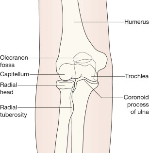
The olecranon is obscured by the humerus. The capitellum is lateral and articulates with the radial head. The trochlea is medial and articulates with the ulna.
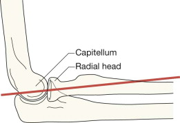
On the lateral view: the line to draw is along the long axis of the proximal 2–3 cm of the shaft of the radius. This line should pass through the capitellum.
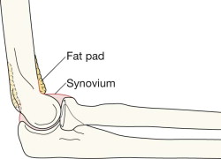
Two pads of fat are situated close to the cortex of the distal humerus. Referred to as the anterior and posterior fat pads, these are external to the synovial lining of the joint. The fat pads are never visualised on the AP projection. Look for them on the lateral view. The fat is seen as a dark streak in the surrounding grey soft tissues.
The anterior fat pad will be seen in most (but not all) normal elbows and is closely applied to the humerus.
The posterior fat pad is not visible on the radiograph of a normal elbow because it is situated deep within the olecranon fossa. Because the elbow is radiographed in the flexed position this fat shadow is hidden by the overlying bone.
Become a Clinical Tree membership for Full access and enjoy Unlimited articles
If you are a member. Log in here