Physical Address
304 North Cardinal St.
Dorchester Center, MA 02124
On completion of this chapter, you should be able to:
• Describe the advantages of ultrasound-guided procedures
• Describe the benefits of sonographer involvement in procedures
• Discuss the advantages and disadvantages of free-hand and needle-guided techniques
• List potential complications of ultrasound-guided interventional techniques
• Discuss techniques for finding the needle tip
• Discuss indications and contraindications for the procedures discussed in this chapter
Ultrasound has been used to assist in interventional procedures since the 1970s by means of specially designed A-mode and B-mode transducers ( Fig. 18.1 ). Ultrasound-guided procedures have come a long way since those early days of A-mode and static B-scan units with the development of real time imaging and color Doppler. Ultrasound is being used to guide a variety of invasive procedures on various organs and masses located in the neck, chest, abdomen, retroperitoneum, musculoskeletal (MSK) system, and pelvis and on the fetus and gravid uterus. Ultrasound is also being used to drain a variety of fluid collections and abscesses.
The use of ultrasound to guide procedures continues to grow, with procedures being performed by a wide variety of medical personnel. Besides radiologists and maternal fetal medicine physicians, other health care providers have started to use ultrasound to help them in their practice. Anesthesiologists place ultrasound-guided intravenous (IV) lines and perform nerve blocks, the orthopedic department performs ultrasound-guided joint and nerve injections as well as tap joint effusions, some physical therapists do ultrasound-guided dry needling, urologists perform prostate biopsies, pain management physicians perform ultrasound- guided nerve blocks, the IV team does ultrasound-guided IV placement, hospitalists use ultrasound-guided aspiration techniques to tap ascites and pleural effusion, and endocrinologists as well as cytopathologists perform thyroid and neck node biopsies. There are other medical practices that use ultrasound for procedures, and there are other uses for ultrasound-guided procedures that these specialties may perform that are not listed. There are multiple factors contributing to this growth: the development of low-cost compact and laptop units, hands-on training with simulated biopsy phantoms provided by the ultrasound vendors, and training offered by various medical societies.
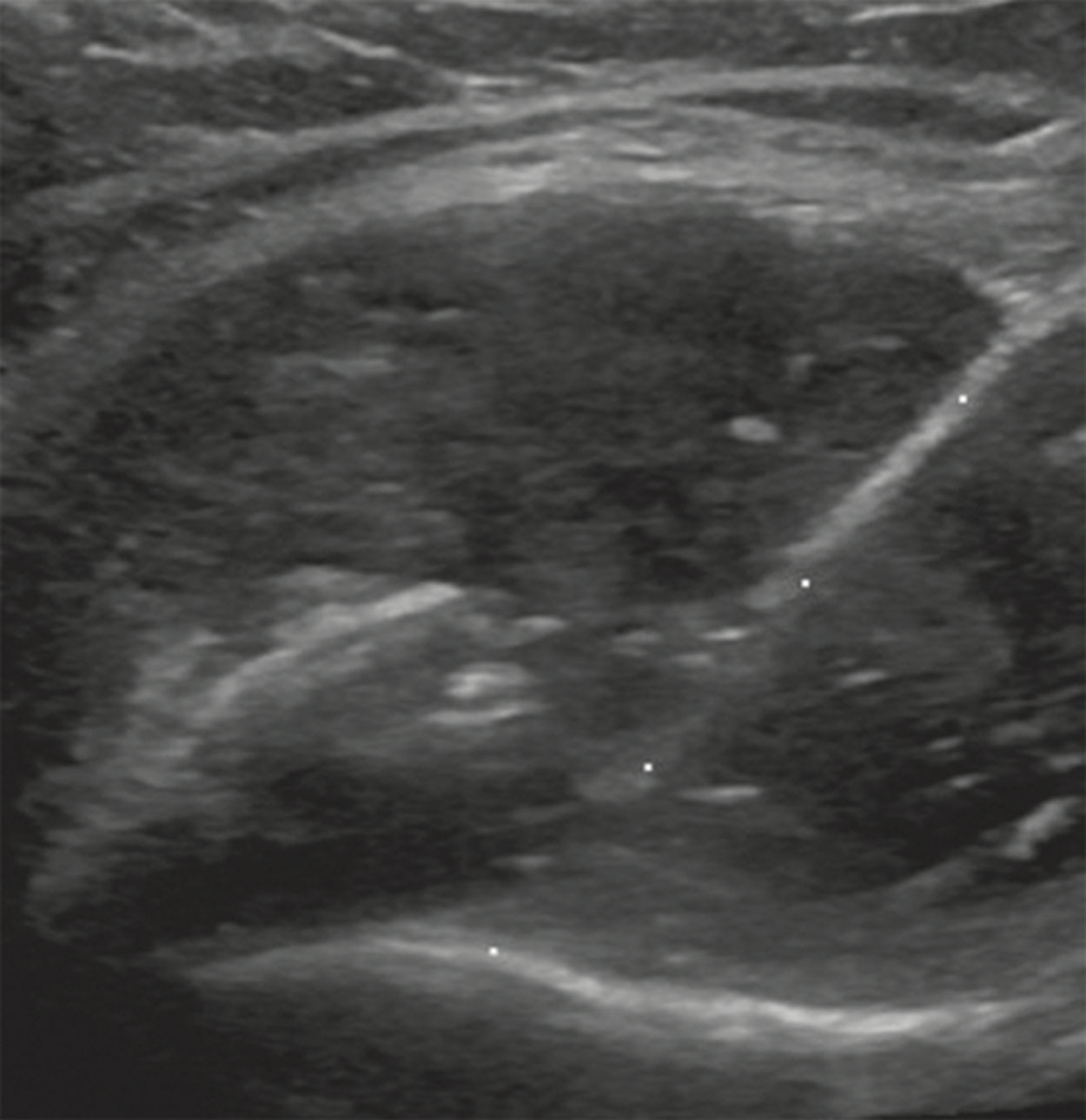
In recent years, there has been a movement to perform more and more procedures under ultrasound guidance. Retroperitoneal masses, pleural-based masses, deep masses in the liver, and MSK masses that were once typically biopsied with computed tomography (CT) guidance or in open surgical biopsies are now being successfully performed using ultrasound guidance ( Fig. 18.2 ).
Ultrasound can be used to:
Biopsy malignant or benign masses.
Biopsy organs for parenchymal disease or transplant rejection.
Drain various fluid collections, including cysts, joint effusions, ascites, or pleural fluid.
Drain or obtain samples of abscesses to determine the type of organism, especially on patients who are not responding well to antibiotic therapy.
Mark spots for fluid taps to be performed without direct sonographic guidance.
Guide needles for nerve injections.
Help place IV lines, especially on patients that are difficult sticks.
The main advantage of using ultrasound for guidance is that it has continuous real-time visualization of the needle, which allows adjustment of the needle as needed during the procedure. When used to guide biopsies, the needle tip can be watched in real time to ensure that it does not slip outside the mass. This is especially important for small masses or if the patient has trouble holding their breath. Ultrasound also has the advantage over CT of allowing different patient positions and approaches to be considered. The patient may be turned into a decubitus or oblique position to allow safe access to the mass ( Fig. 18.3 ). Liver masses in the dome of the liver can be accessed by a subcostal approach using a steep angle with the needle directed cephalad, reducing the risk of a pneumothorax , which could happen with an intercostal approach. Using ultrasound, the patient can be placed in a comfortable position. For example, the patient’s head may be slightly elevated, or the patient can move slightly between passes to relieve back or joint pain. Another benefit is the ability to comfort and reassure the patient, because the sonologist, sonographer, and nurse are with the patient during the entire procedure. Even the most anxious patients can be coached to cooperate when the team is by their side and not constantly in and out of the room. Other advantages include the ability to perform the biopsy in a single breath hold, portability, lack of radiation, and shorter procedure times.
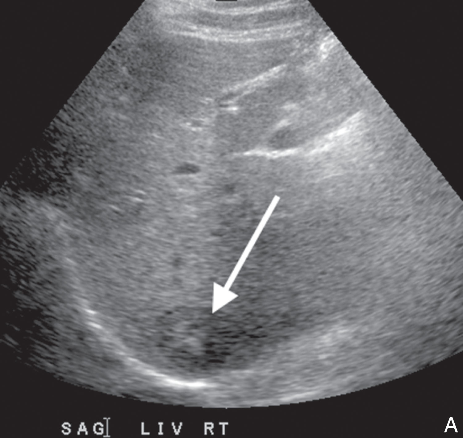
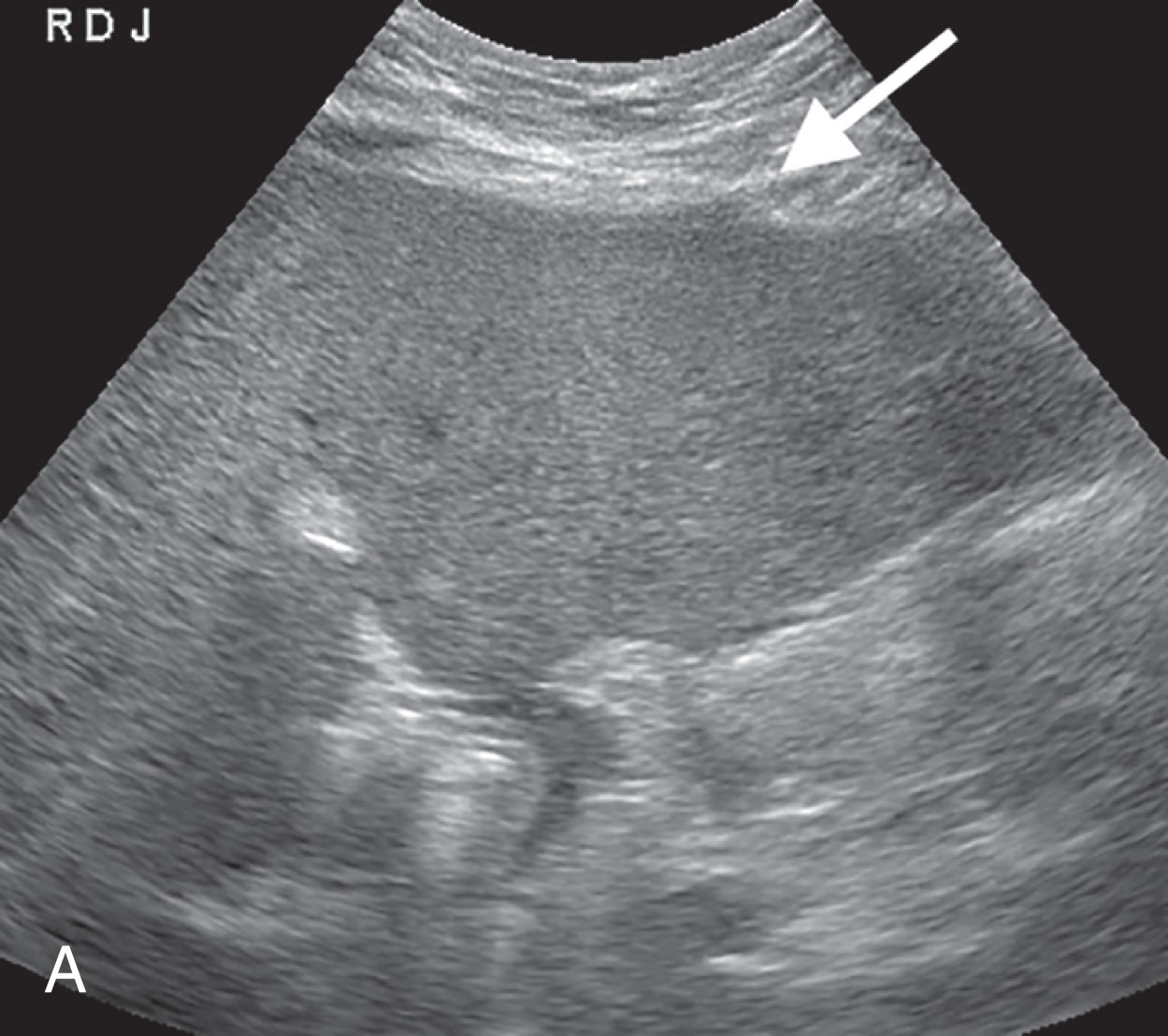
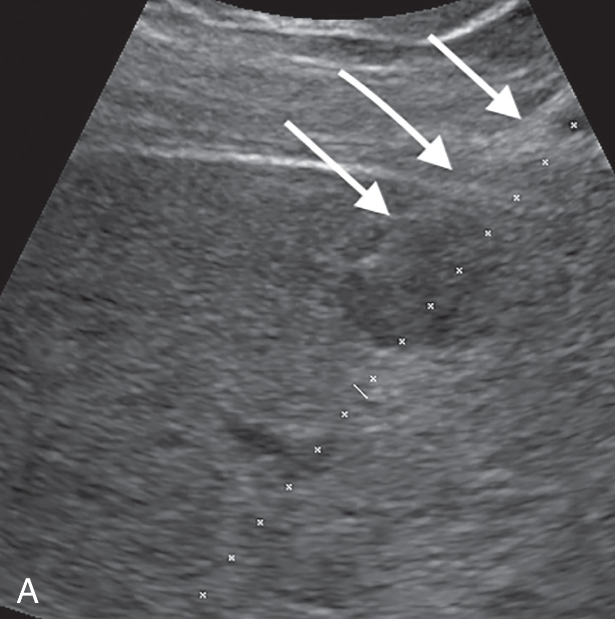
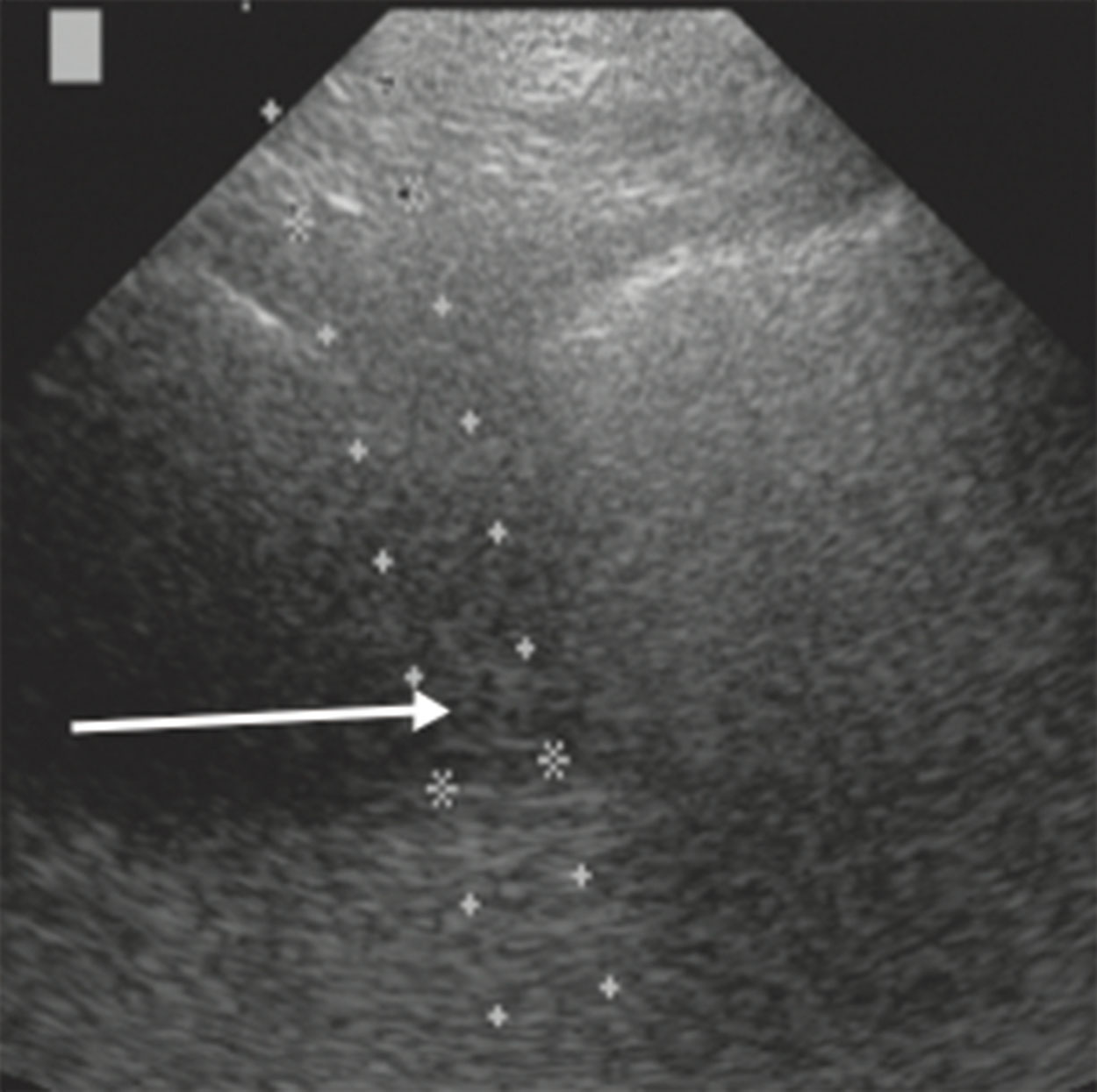
Despite all its benefits, ultrasound guidance does have some limitations. Not all masses can be visualized with ultrasound, because the mass may be isoechoic to the normal tissue. When this is the case, the sonographer should look for indirect signs of the presence of a mass, such as displaced vessel capsule bulges or the presence of tumor vessels ( Fig. 18.4 ). These isoechoic masses may potentially be biopsied using fusion technology or contrast imaging. Abdominal masses may be obscured by bowel gas, or gas may move during the procedure, causing difficulty in seeing the mass. The sonographer can try to press the gas out of the way with the transducer. If this does not work, the biopsy may be canceled under ultrasound and either performed under CT or rescheduled for another day. There may be issues seeing the needle tip, especially if it deviates from the projected path. This problem relies on the sonographer’s scanning skills to maneuver the transducer to find the needle tip or to correct for needle deviation. By understanding the cause of the deviation, the sonographer can determine how to correct for it, allowing for a successful biopsy ( Fig. 18.5 ). It sometimes felt like some of our best samples occurred when we could not see the needle tip. A major limitation can be the comfort level of both the sonographer and the sonologist. Without proper training, they both can feel intimidated and not confident, especially if the radiologist has a comfort level using CT guidance.
The most common indication for a biopsy is to confirm if a mass is malignant. The mass may be a primary tumor in a patient or a metastatic mass in a patient with an unknown primary. The liver is a common place for metastatic disease, and these masses can be easily biopsied using ultrasound. With a biopsy of the metastatic tumor, the primary tumor can be determined. Other indications include the need to differentiate between a metastatic mass and a second primary, to differentiate recurrent tumor from postoperative or therapy scarring, to differentiate malignancy from inflammatory or infectious disease, to determine metastatic lymph adenopathy from lymphoma, or to characterize a benign mass. Procedures that are not mass related include obtaining a sample of the parenchyma of an organ to determine the severity or progression of a disease such as fatty liver, hepatitis, or renal failure or to obtain a sample of tissue to determine the cause of rejection in a transplanted organ ( Fig. 18.6 ).
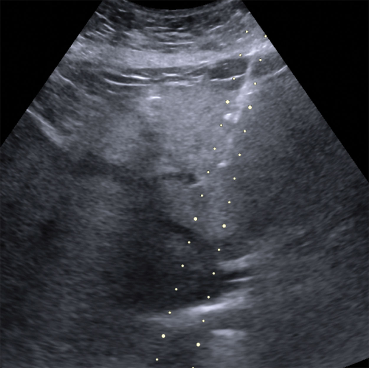
Contraindications of ultrasound-guided procedures are few because of the procedure’s minimally invasive nature. Most contraindications include an uncorrectable bleeding disorder, the lack of a safe needle pathway ( Fig. 18.7 ), or an uncooperative patient. Patient cooperation is needed so that the mass may be biopsied safely. If the patient will not hold still, is jumpy, or cannot control their breathing, the risk of a complication for the patient increases significantly, as does the risk to the sonographer or sonologist of being stuck by a contaminated needle.
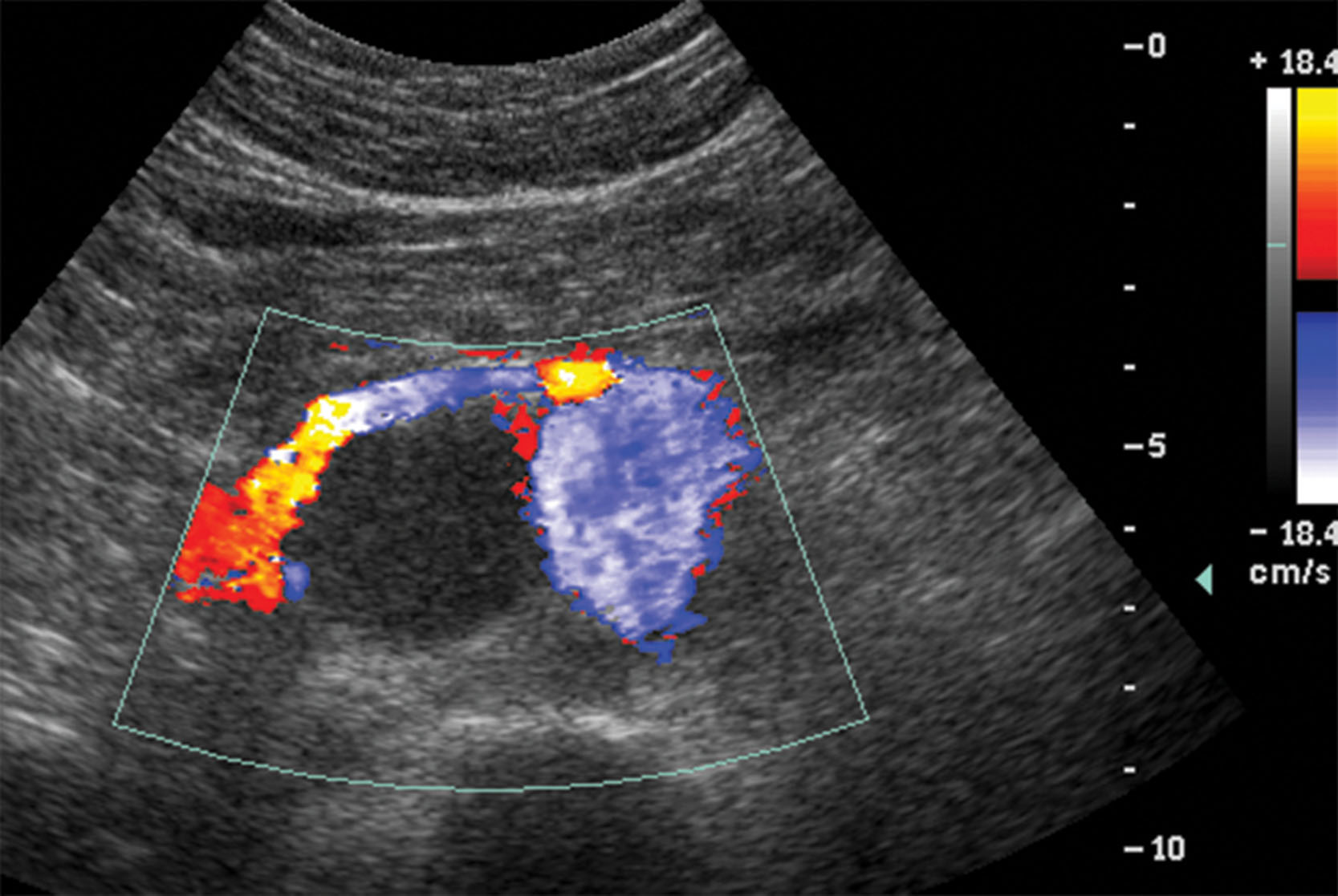
With the exception of bleeding times, blood or urine tests are not needed before an ultrasound-guided procedure. An abnormal laboratory value will be part of the patient’s work-up that led to the biopsy. Some abnormal values that may trigger a request for a biopsy or aspiration include elevated alpha-fetoprotein (AFP) in the presence of a liver lesion, elevated prostate-specific antigen (PSA) to evaluate the prostate for cancer, changes in thyroglobulin levels in a patient with a history of thyroid cancer, and increased white blood cell count, leukocytosis, when an abscess is suspected.
It is important to know that the patient will stop bleeding after the needle is removed and does not have a blood clotting problem. Therefore blood tests that determine the patient’s bleeding times should be reviewed before most procedures. These tests measure the time it takes the blood to form a clot. This is especially needed for patients who are on blood thinners such as warfarin or Coumadin, heparin, or aspirin therapy. Because vitamin K is essential in the blood clotting process, patients with liver disease can be at a risk for prolonged bleeding times. To eliminate patient rescheduling or cancellation, test results should be obtained as close to the date of the procedure as possible, although results may be acceptable up to 3 to 4 weeks before the scheduled procedure, depending on the type of procedure and the guidelines of the lab where the procedure is being performed.
At least a dozen factors are needed to form a blood clot to stop bleeding; they all interact through a complex series of reactions called the coagulation cascade. There are three pathways in the blood clotting process: intrinsic, extrinsic, and common. To evaluate all three pathways, both prothrombin time (PT) and partial thromboplastin time (PTT) may be evaluated. PTT measures the time it takes a clot to form and is also used to evaluate the effects of heparin therapy. PTT evaluates factors found in the intrinsic and common pathways. PTT values may vary depending on the method and activators used, with normal values typically between 30 and 45 seconds. PT also tells the physician how likely the patient is to have a bleeding or clotting problem during or after surgery and is used to evaluate factors found in the extrinsic pathway, which may be affected by patients on Coumadin. Normal values are typically between 10 and 13 seconds. Because of the variability of PT results between laboratories, a method of standardization was developed called the international normalized ratio (INR) . The INR test was created in 1983 by the World Health Organization (WHO) to account for the various thromboplastin reagents used to determine PT. The INR is a calculation that adjusts for the variations in PT processing and values so that test results from different laboratories can be compared. The INR is expressed as a number. Values of less than 1.4 are needed to ensure a safe procedure. The INR/PT is not used on patients with liver disease or on heparin. It is evaluated on patients taking anticoagulants, especially Coumadin. In addition to PT, PTT, and INR, the patient’s platelet count may be required. Patients with a defect in their blood clotting mechanism, coagulopathy , will need to have a platelet transfusion just before and during the procedure to ensure that their body can form a blood clot to stop any bleeding. Some departments may not require a hemostatic evaluation for fluid aspirations and superficial biopsies with a low risk of bleeding such as of the thyroid, neck nodes, or prostate.
Anticoagulants should be discontinued before the biopsy to reduce the risk of postprocedural bleeding. Patients should be off their blood thinners before the procedure as follows: 4 to 6 hours for heparin, 5 days for Coumadin, and 5 days for aspirin, although these values can be different for different hospitals or imaging centers. Patients need to discuss stopping these drugs with their physician. Recent studies have shown that patients taking omega-3 and fish oils do not have an increased risk for postprocedural bleeding and that they may be actually beneficial, so stopping them is no longer required. The reader is encouraged to stay current on medicines and supplements that might put the patient at an increased risk of prolonged bleeding after a procedure.
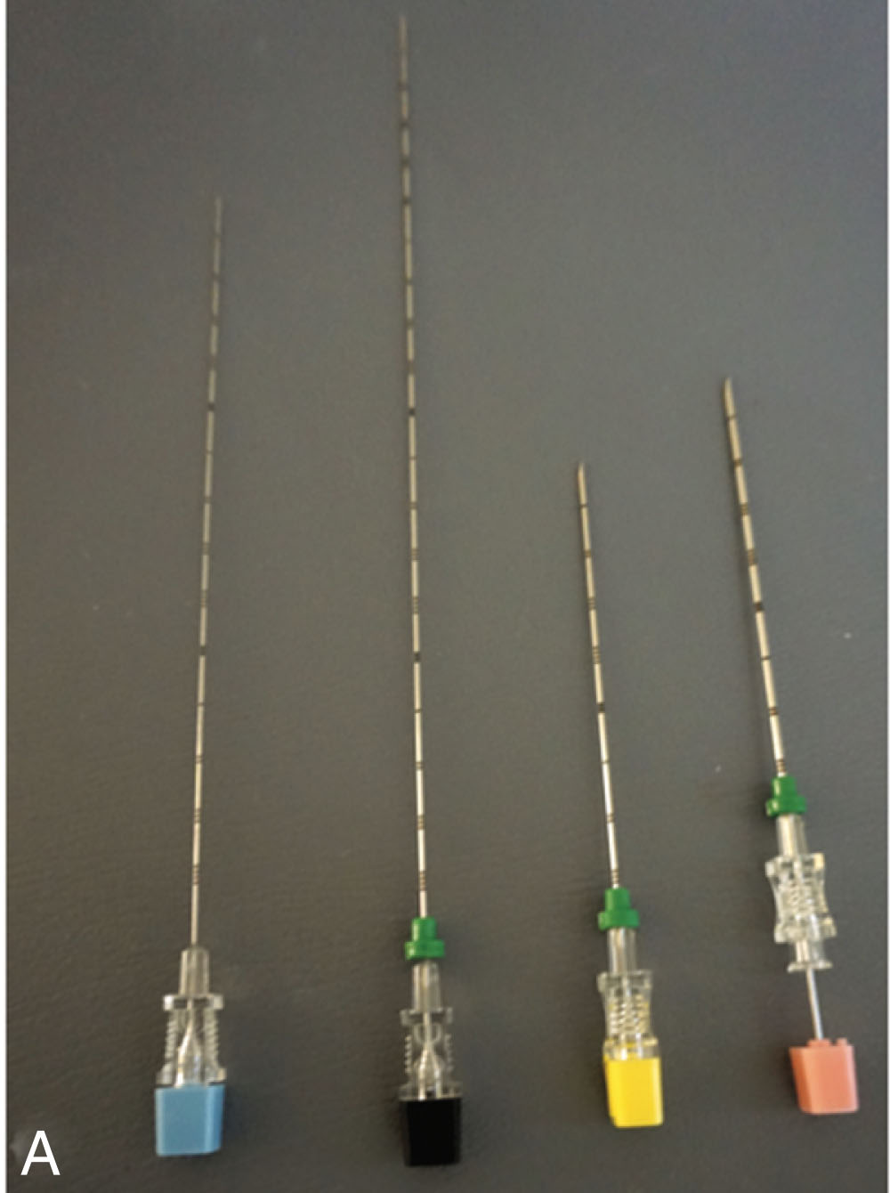
Biopsies are used to confirm if a mass is benign, malignant, or infectious. Most biopsies are easily and safely performed as an outpatient procedure. Good success rates have been reported using ultrasound for biopsy guidance. Cell type is often needed to determine treatment type and options, as specific tumors respond better to certain types of chemotherapy or to radiation therapy. Fine-needle aspiration (FNA) uses thin-gauge needles to obtain cells from within the mass. FNAs are performed using a 20- to 25-gauge needle with a cutting tip, such as a Franseen, Chiba, or spinal needle ( Fig. 18.8 ). These types of needles have the least risk associated with their use, allowing multiple passes as needed. The number of the gauge corresponds to the diameter of the needle, the higher the number, the smaller the diameter, and refers to the inner measurement or opening of the needle. For example, a 20-gauge needle has a 0.6-mm internal diameter, and an 18 gauge has a 0.8 mm internal diameter. Needles are described by their length and gauge. For example, if the physician asks for a 20 × 15 needle, they want a 20-gauge needle that is 15 cm in length. If different types of needles are available, the physician might ask for a specific type of needle for that procedure. For example, they may ask for a Franseen 20, 15 meaning they want a Franseen style needle that is 20 gauge and 15 cm long. The needles are color coded so that by looking at the color of the hub you know the needle size. 25 Gauge needles are blue, 22 gauge are black, 20 gauge are yellow, and 18 gauge are pink (see Fig. 18.8A ). This is a universal coloring system. Seeing the length of the needle may cause a patient to become quite apprehensive. The sonographer should explain to the patient that most of the needle will be inside the needle guide and not in their body. I would also tell the patient that it is the diameter of the needle that causes pain and not the length, and we usually use needles that are smaller than used to take their blood. The specimen is obtained by using a capillary action technique. This involves a steady, quick up-and-down motion of the needle, after the stylet is removed, which obtains the needed cells through a scraping or cutting action. As the needle is removed from the body, the physician will place their thumb over the open hub so that the cells are not sucked back into the body. An FNA technique reduces the trauma to the cells and decreases the amount of background blood. If the sample is scant, suction techniques can be used. Suction technique involves using a syringe and tubing attached to the needle. As the needle is being moved up and down, suction is applied to draw up the cells into the needle. Once the needle is removed, the physician will place the tip of the needle over the slide to deposit the cells so that they can be smeared for staining and evaluation. The physician may also express the cells into a container holding special fluid to preserve the cells. Because of its thin size, this type of needle can safely go through the gastrointestinal tract and near vascular structures. FNA in conjunction with onsite cytopathology can help ensure that the procedure is diagnostic and minimize the number of passes.
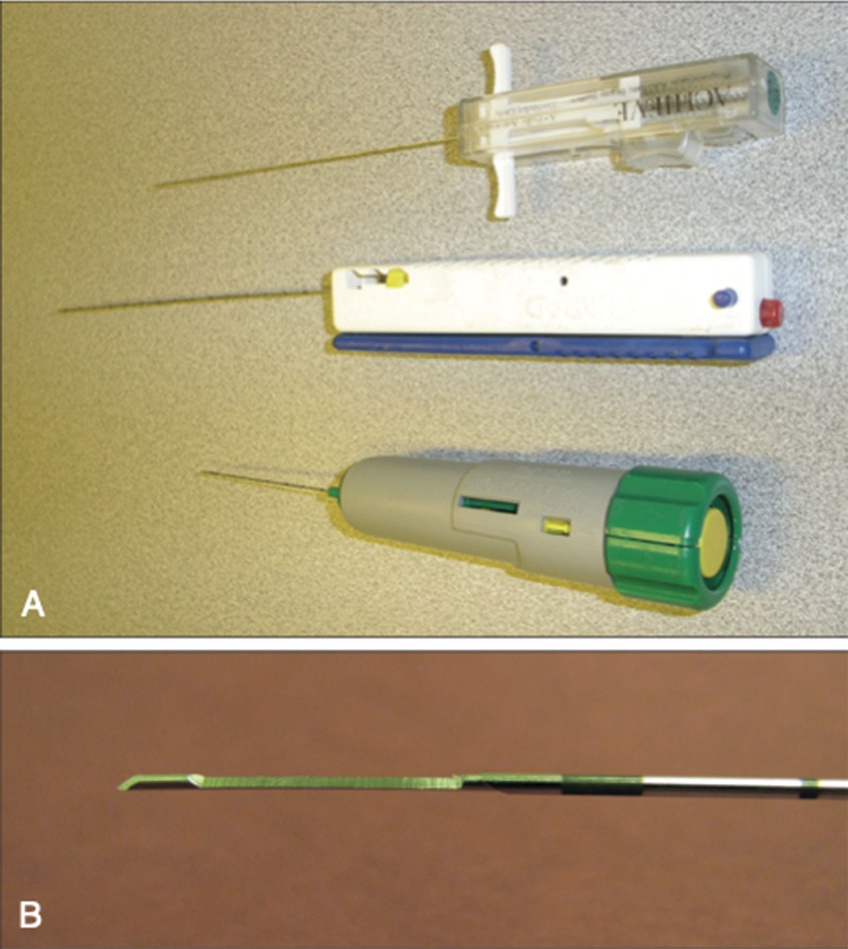
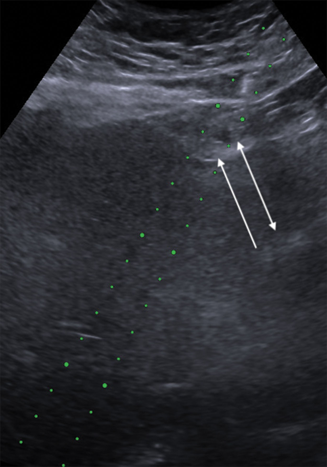
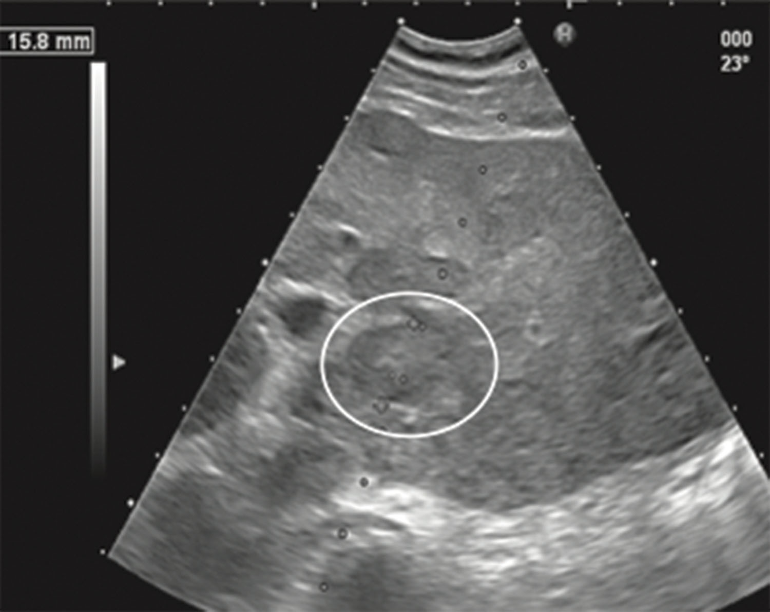
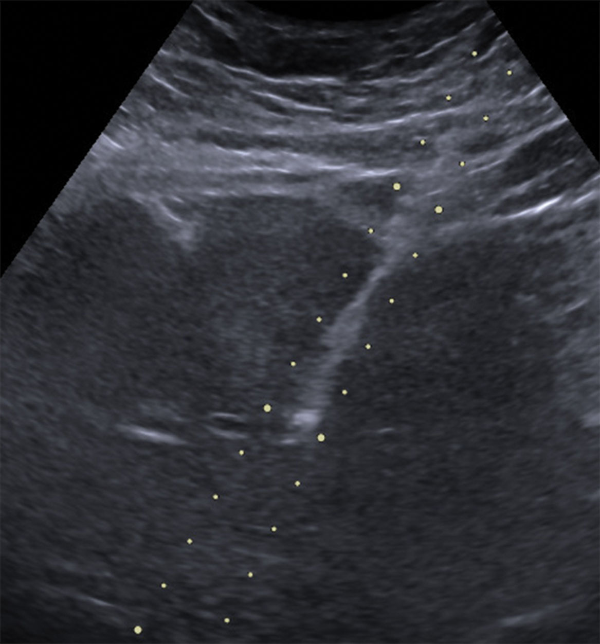
A core biopsy uses an automated, spring-loaded device, termed a biopsy gun , to obtain a core of tissue for histologic analysis ( Fig. 18.9 ). The biopsy device is cocked, and the needle tip is placed just inside the mass or on the outside edge of the mass. For a biopsy of an organ for parenchymal disease, the needle tip can be placed just inside the organ itself. Sometimes if the needle tip is just on the outside of the organ and then fired, going through the capsule could sometimes deflect the needle as the capsule offers some resistance. The Glisson capsule is a good example because you can see the needle pushing the capsule out of the way but never piercing it when positioning the needle. This is called tenting ( Fig. 18.10 ). The button is then pushed, and the cutting needle is thrown, obtaining a core of tissue, which is deposited into the slot on the inner needle (see Fig. 18.9B ). Various throw lengths are available, ranging from 10 to 23 mm, which will correspond to the length of the specimen that is obtained. The throw length is the distance that the cutting needle will advance when fired. The inner needle that receives the specimen is stationary. The proper throw distance needs to be determined so that the needle does not go through the back wall of the mass and damage underlying structures or vessels ( Fig. 18.11 ). The snapping sound that the device makes can be startling to the patient, and it is advised to let the patient hear the sound before obtaining the specimen. This also ensures that the device is not defective. Core biopsy needles are larger in diameter and range in size from 14 to 20 gauge. A core biopsy can be used in conjunction with FNA techniques, especially if a definitive diagnosis could not be determined from the FNA or if the cytopathologist requires more tissue for special stains to confirm or make the diagnosis. Core biopsies are used to diagnose diffuse parenchymal disease of the liver, kidney, and transplanted organs ( Fig. 18.12 ), as well as masses in the breast and prostate.
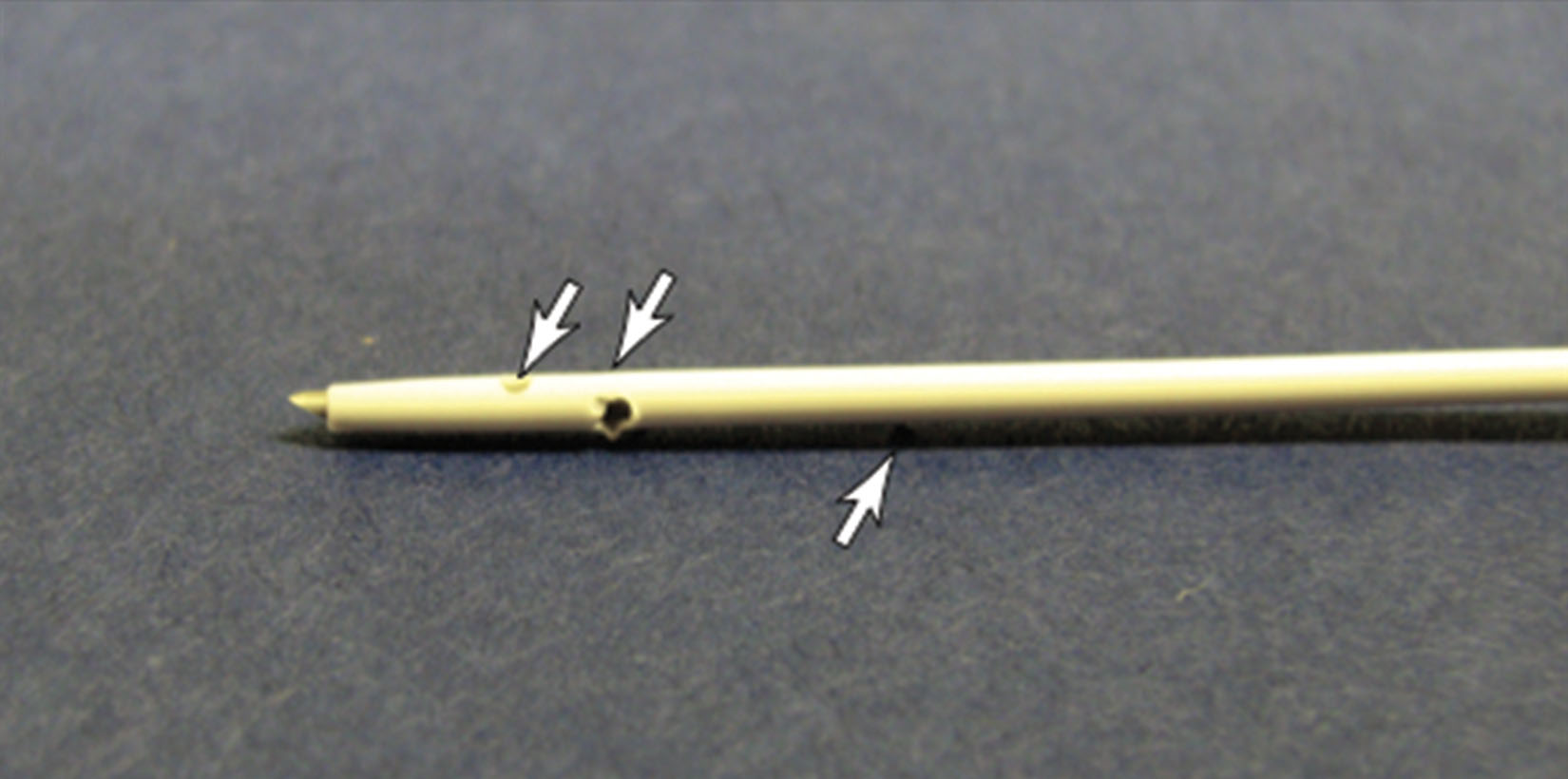
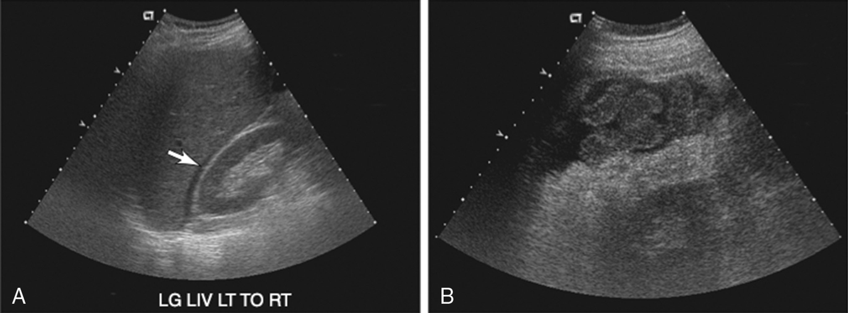
Ultrasound is routinely used to guide needle placement to drain or obtain samples from ascites or pleural effusions. Usually, these procedures are performed without the use of a needle guide unless the fluid is multiloculated, is only a small amount, or is unsafe to drain by free-hand techniques. If a small amount of fluid is needed, a 22- or 20-gauge needle may be used. If the fluid is viscous, an 18- or a 16-gauge needle may be required. If the goal is to drain as much fluid as possible, a special catheter, called a centesis catheter, is used ( Fig. 18.13 ). After the needle is properly placed, the stylet is removed, leaving the catheter which will have multiple side holes to drain the fluid. For large volume drainage, a 1-L vacuum bottle is used to drain the fluid as the vacuum pressure will draw the fluid out. As the bottle gets full, it is replaced with another empty bottle. This continues until no more fluid is draining into the bottle. If the vacuum becomes broken, the fluid will not drain. This will occur if the draining tubing is not closed off when inserting the spike into the collection jar. Ultrasound can be used periodically to check the amount of fluid remaining or when the fluid stops draining to help reposition the catheter or to free it from bowel that may have been sucked against the wall of the catheter ( Fig. 18.14 ). The sonographer can usually scan outside the sterile field on these exams ( Fig. 18.15 ). In a patient with massive ascites, a large volume of fluid may be drained for patient comfort. It is usually recommended that no more than 4 to 6 L be drained because draining more than that can place the patient at risk for electrolyte imbalance, hypovolemia, hypotension, and hepatorenal syndrome. If more than 5 L are to be removed, the patient is usually given IV albumin to decrease the chance of these complications.
Fluid or abscess collections are usually performed using a needle guide. Abscess or fluid collections may be located in or around the liver, in the peripancreatic, perinephric, intra-abdominal, pelvic, or intramuscular regions, or in the prostate gland. The needle gauge used to drain the fluid depends on the thickness of the fluid. For pelvic collections, depending on their location, an endovaginal approach can be considered and endorectal approaches in both men and women. For prostatic abscess drainage, an endorectal technique is used. Fluid is obtained to determine the type of organism present, so that the correct antibiotics can be administered to the patient. For a large abscess, catheters may be left in place to drain the collection. Patients may have follow-ups to monitor that the cavity is getting smaller and to check that the catheter is still in the correct position. These follow-up examinations are performed under fluoroscopy in a procedure called a sinogram, which uses iodinated contrast media to outline the cavity. It can be common for the person calling to schedule the patient’s exam incorrectly as a sonogram as opposed to a sinogram because the names are only one letter different and sound similar. Asking the reason for the exam will uncover that a sinogram is needed and not a sonogram.
Ultrasound is used in the radiology interventional lab to guide placements of catheters and lines in various vessels, including the subclavian, jugular, brachial, and femoral vessels, to assist in transjugular intrahepatic portosystemic shunt procedures, and to place nephrostomy tubes in obstructed kidneys.
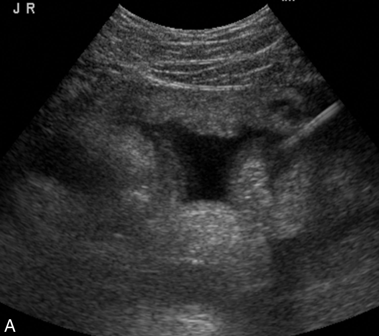
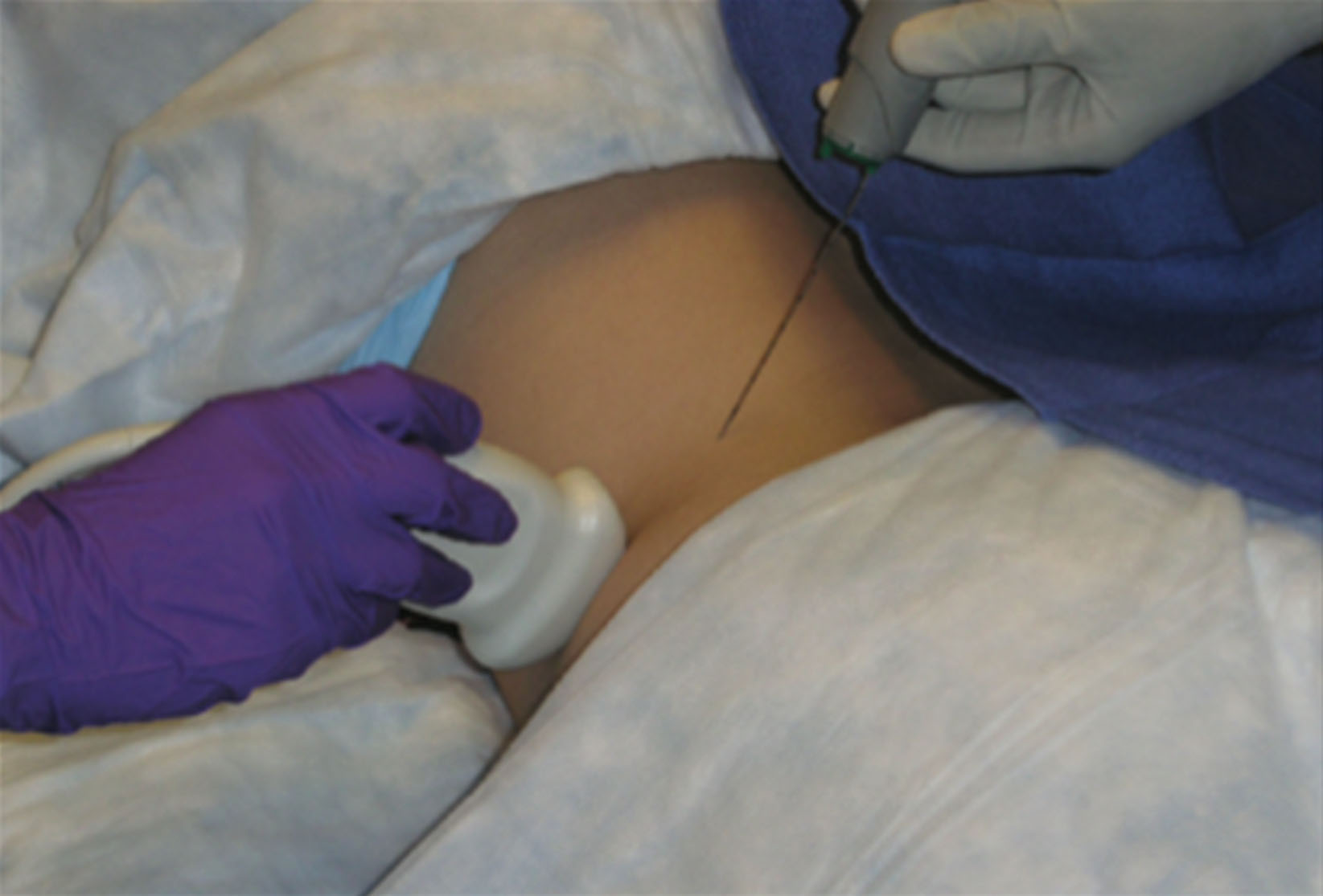
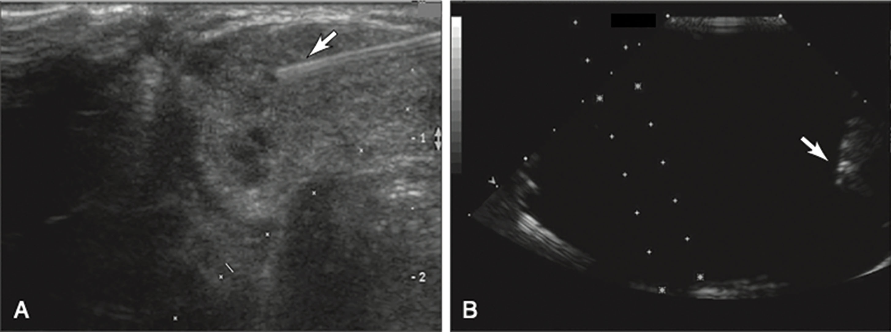
There are three different methods to perform an ultrasound-guided procedure: in-plane and out-of-plane free-hand techniques and using needle guides. The free-hand technique is performed without the use of a needle guide on the transducer. The transducer is placed in a sterile cover, and the radiologist, or the physician who performs the biopsy, will hold the transducer in one hand and the needle in the other hand. The in-plane technique is similar to using a guide in that the needle will approach from the short end of the transducer so that the shaft of the needle can be seen as it advances through the tissue ( Fig. 18.16 ). Care must be taken to align the needle with the soundbeam, using the transducer as a guide, or else the needle tip will not be seen as it traverses the soundbeam. If the needle tip disappears while advancing the needle on the image, the transducer should be repositioned in alignment with the needle path to bring the needle tip back into view ( Fig. 18.17 ). If this causes the area of interest to no longer be seen, the needle should be withdrawn and inserted again. It is important to remember that to see the needle tip, the transducer must be aligned to the needle path. If deviation continues to be an issue, the last resort would be to attach a needle guide.
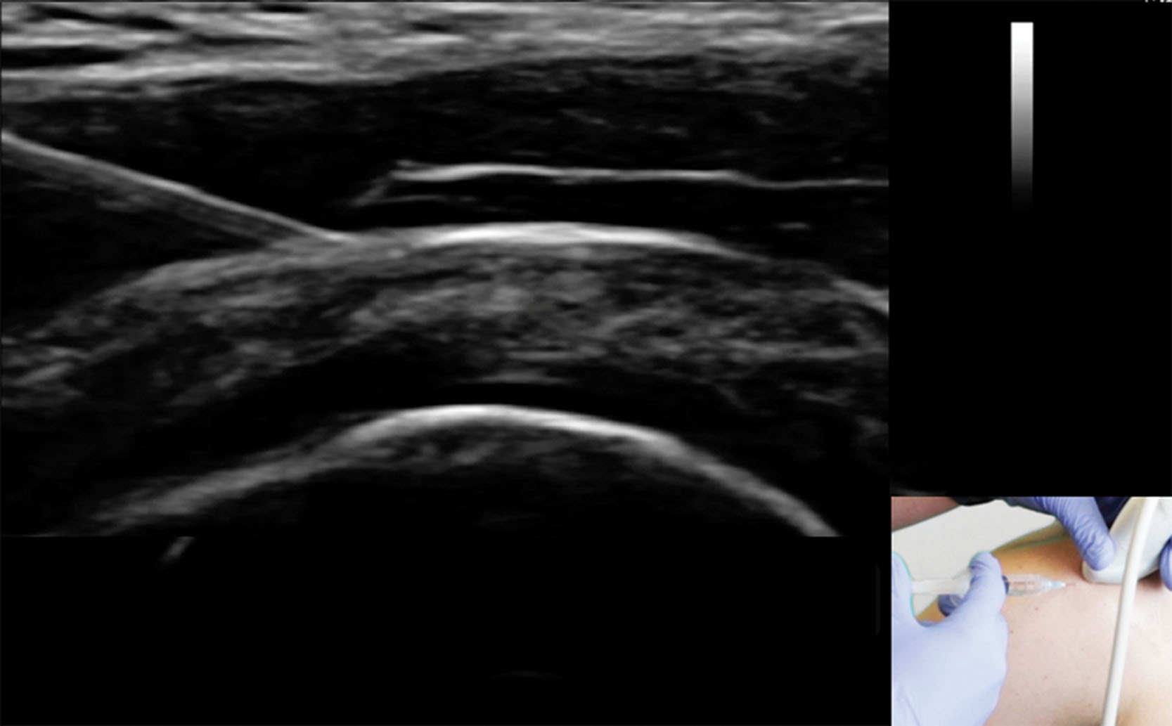
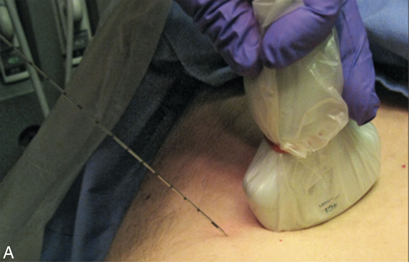
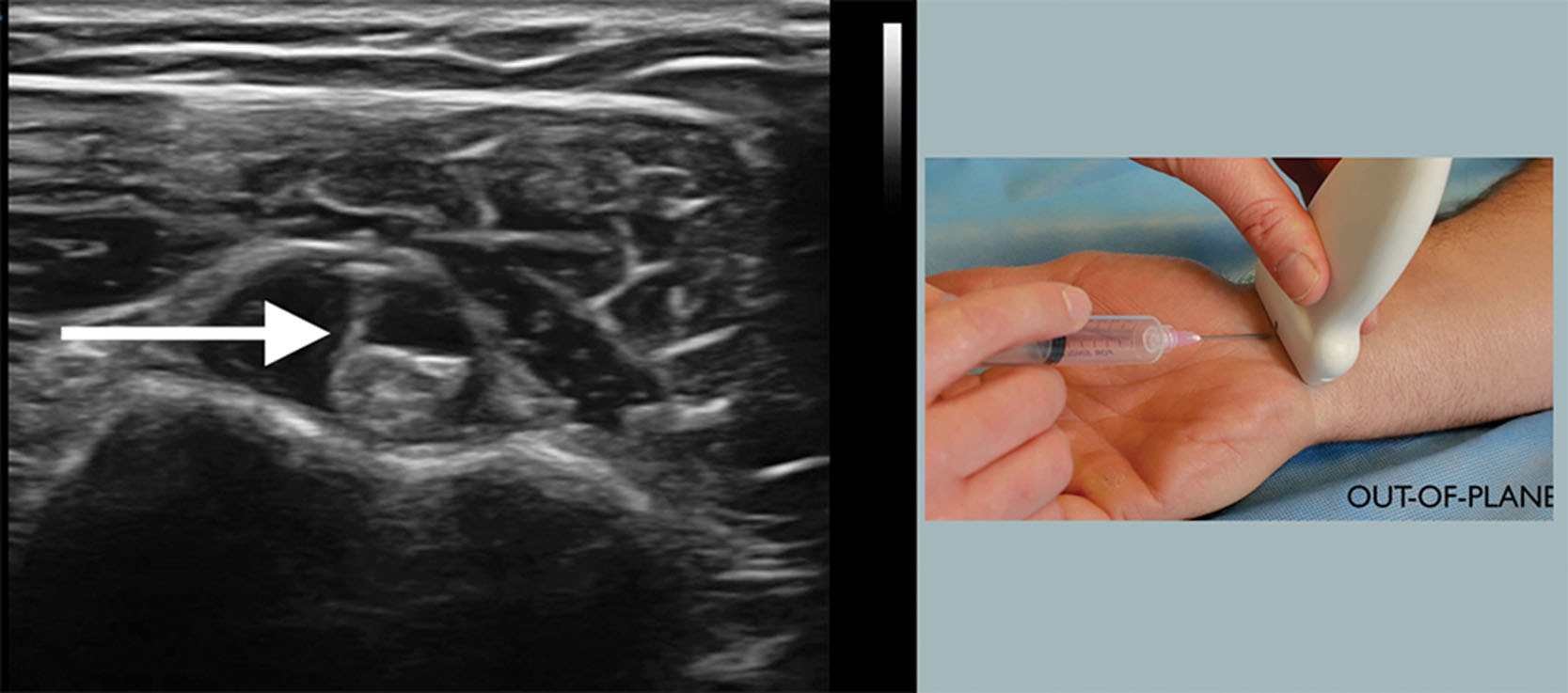
The out-of-plane technique is favored for superficial procedures in which the needle would not be seen until it is in the area of interest, such as starting an IV line. With this technique the needle approaches the long side of the transducer and the needle is not visualized until the tip intercepts the soundbeam, causing a bright white echo to appear ( Fig. 18.18 ). It is important that the needle approaches the transducer at a perpendicular angle and stays on course for a successful procedure. If the needle approaches the transducer at a slight angle, the needle will miss the target area and be either lateral or medial to the target. Although the needle may start out perpendicular to the transducer before it pierces the skin, it is easy to slightly angle the needle while shifting your focus to observe the monitor. This is a drawback of this approach because you cannot see that the needle is not heading to the target until it has missed it.
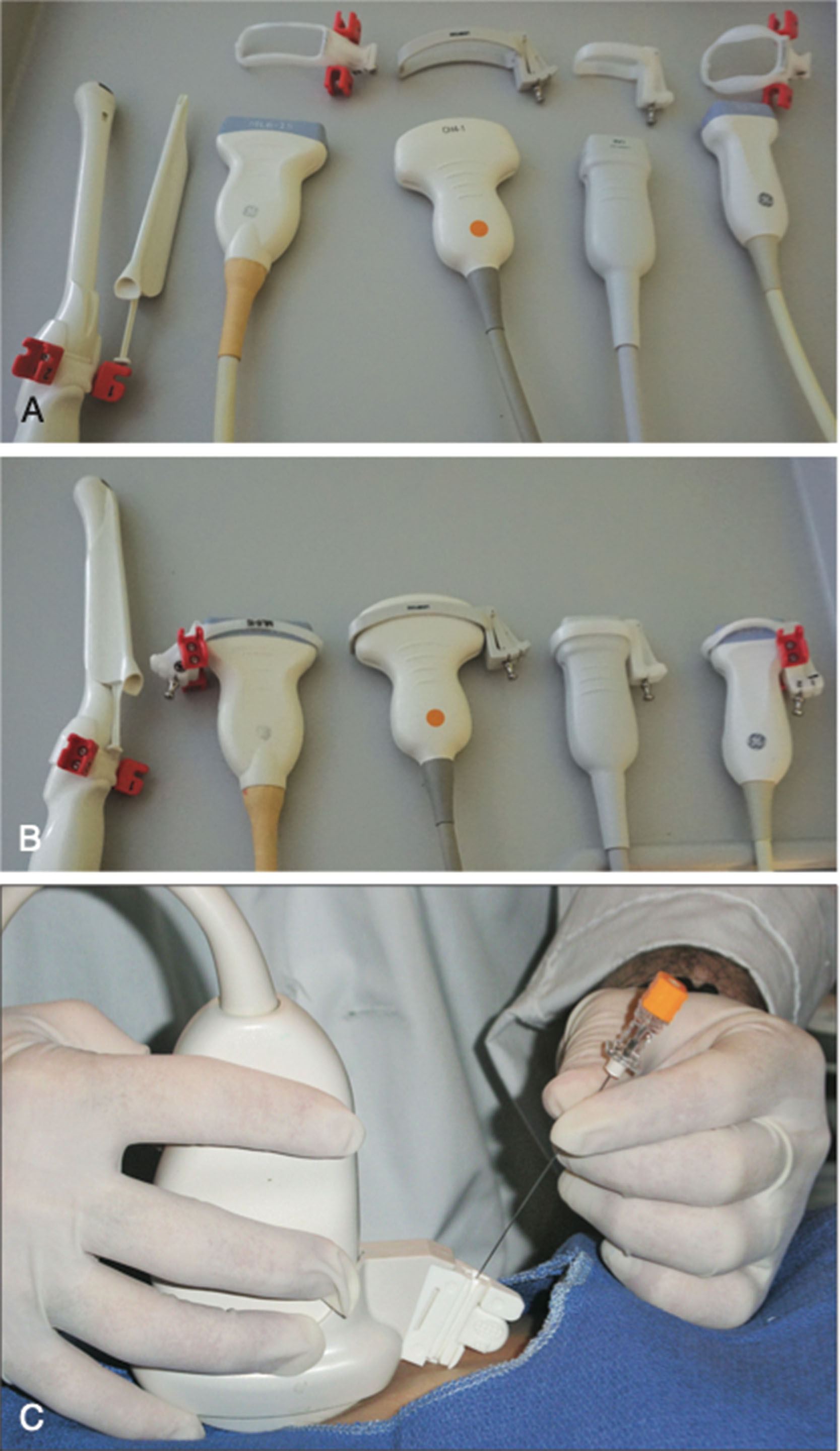
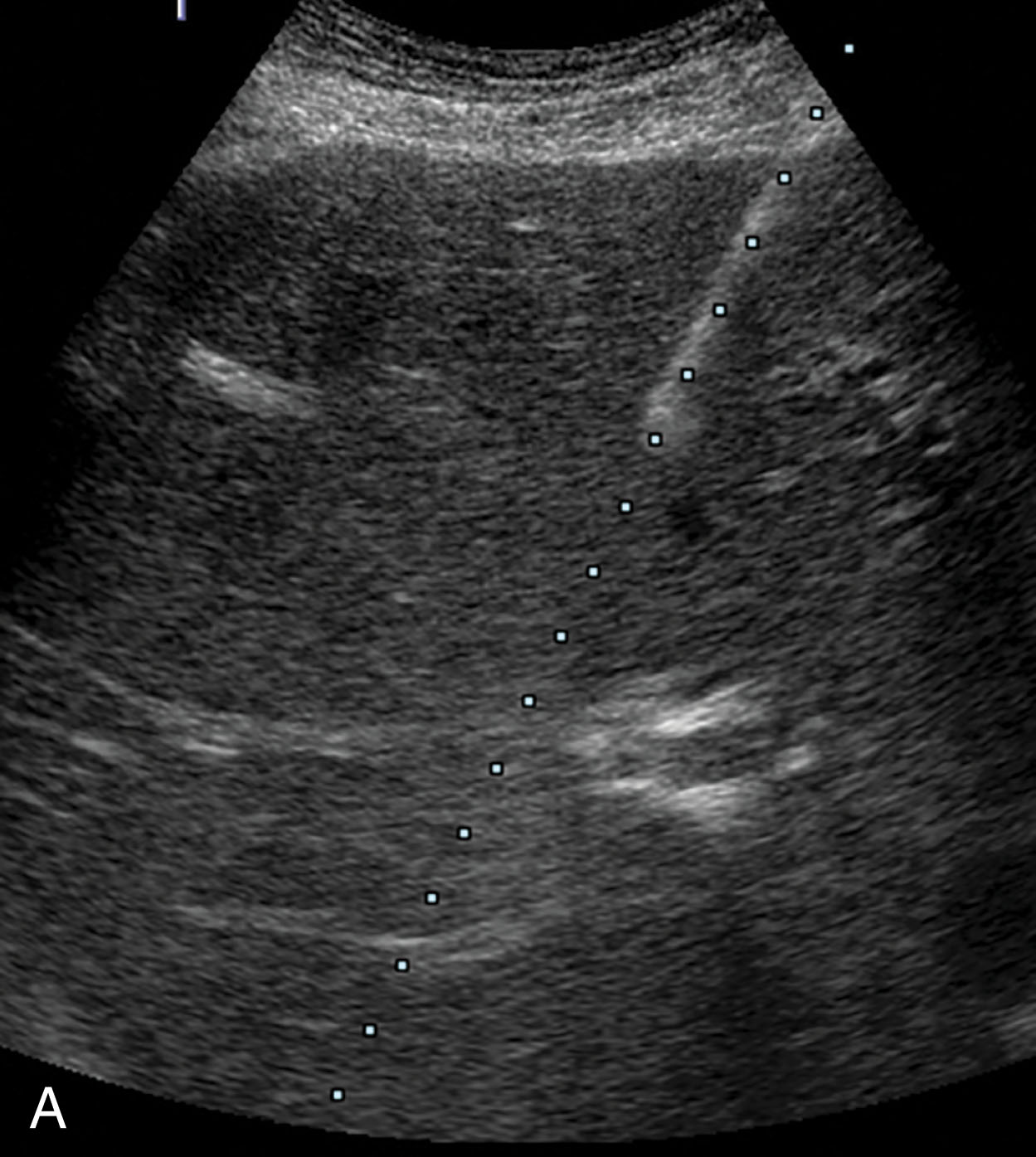
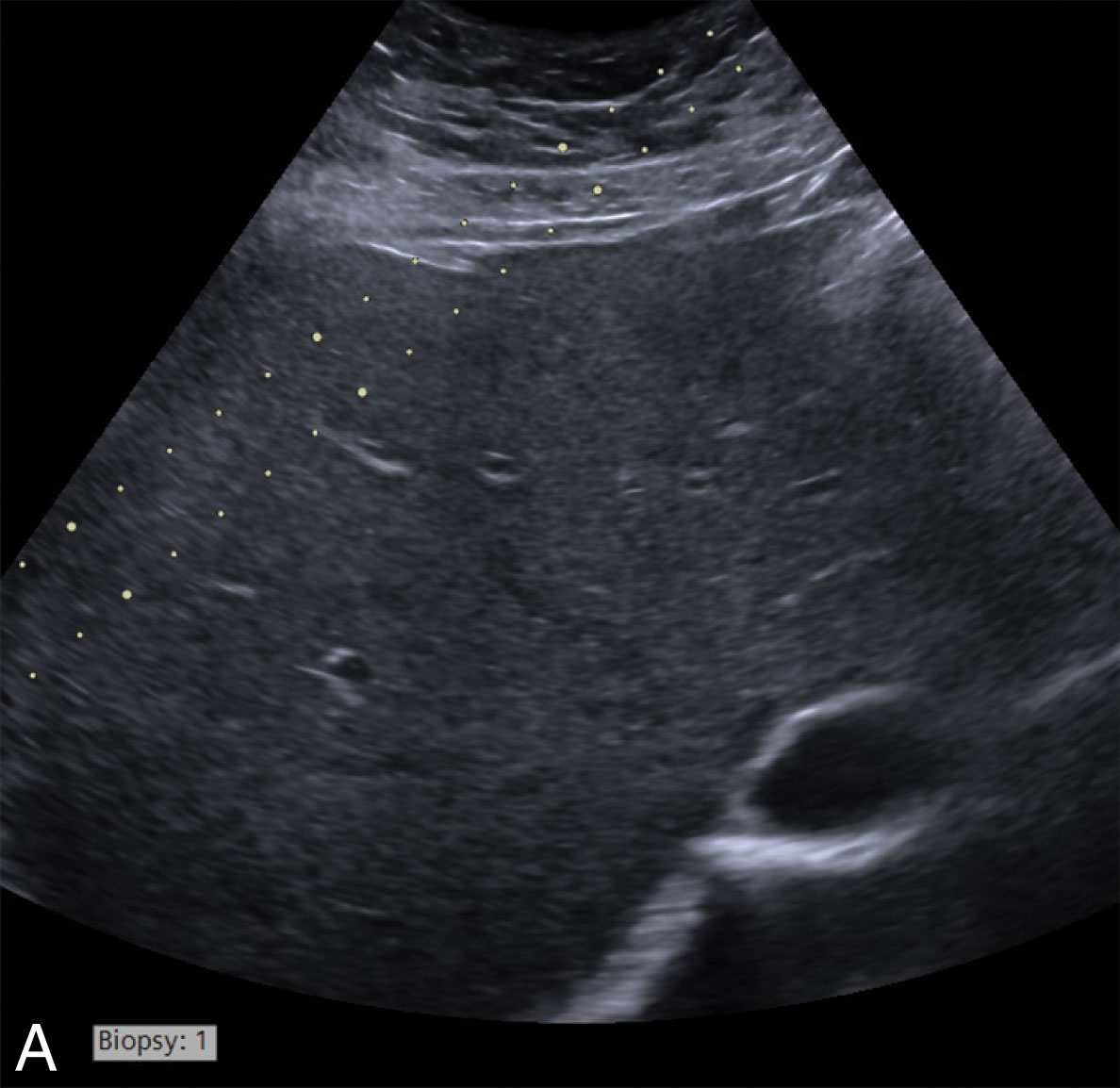
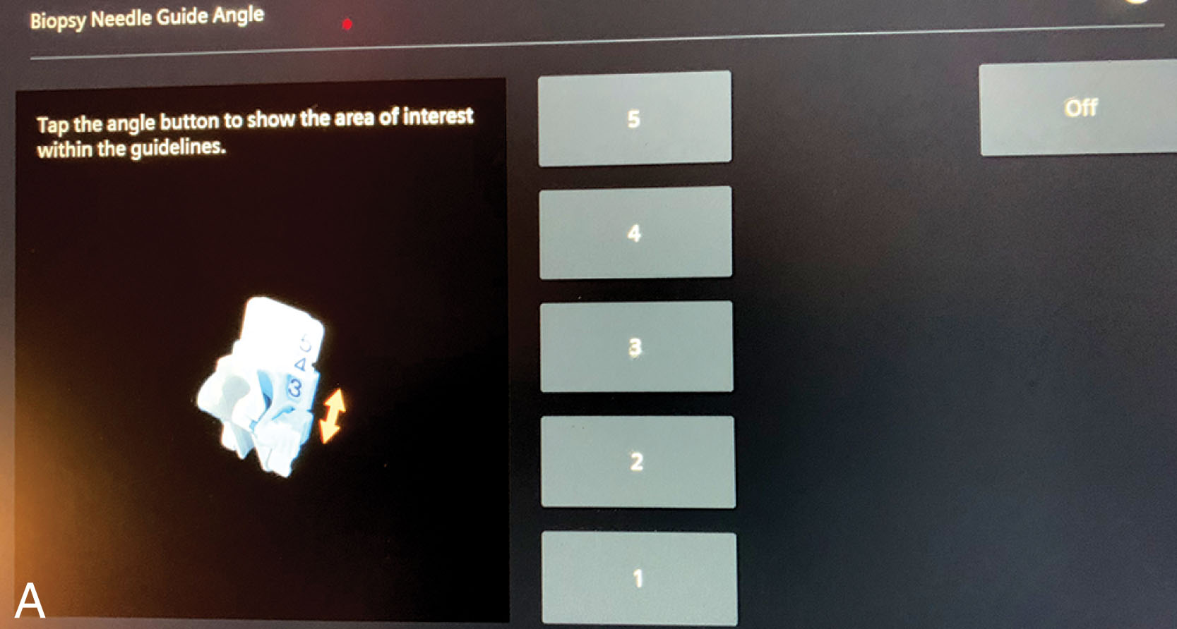
The third method involves using a needle guide that is attached to the transducer. Each transducer type will have its own needle guide ( Fig. 18.19 ). The predicted needle path is displayed on the screen either as a single line or as two parallel lines ( Fig. 18.20 ). The mass is then lined up along the path, either on the single line or between the two lines. Some transducers offer a choice of angles, usually a steep angle, a shallow angle, and one or three angles in between ( Fig. 18.21 ). This gives some flexibility for finding the best approach and to avoid vessels and other structures as needed. There are many benefits to using a needle guide, which may include a faster learning curve especially for the sonologist, more accurate placement of the needle, and the ability to keep the needle going through the anesthetized area when multiple passes are required. The use of needle guides has expanded the role of ultrasound-guided procedures by allowing biopsies of pleural-based lung lesions ( Fig. 18.22 ), deep retroperitoneal lesions, and small masses. When the sonographer attaches the needle guide to the transducer, it is important to attach it correctly and set the angle on the guide to the same angle as on the screen ( Fig. 18.23 ). The needle guides are precision-made devices and need to be handled with care and attached properly. If the angle that is set on the transducer is not the same angle as on the screen, the needle will appear to deviate, as it is not following the projected path on the screen; if the guide is put on backward, the needle will come in from the opposite side of the guide ( Fig. 18.24 ).
All methods have their pros and cons, and the physician performing the biopsy or procedure will decide which technique is the best and offers the safest approach.
Some ultrasound departments collaborate with the cytopathology department by having the cytopathology team present during the procedure. Usually, one to three passes are made, placed on the sterile slides, and stained in the ultrasound room. The cytopathologist looks at the slides to determine if the material is diagnostic. This can ensure that enough diagnostic material is obtained and helps to minimize the number of passes. If it is a necrotic mass, the cytopathologist can determine the best area to obtain good material. They might say “pass number 3 was good,” so the sonographer should try to remember the areas biopsied so that they can return to that location. The cytopathologist may request additional material for special stains and flow cytometry. (Flow cytometry may be needed when lymphoma is suspected.) The cytopathologist may also request that a core sample be obtained to enhance the chances of obtaining a diagnosis for the patient. It typically takes 3 to 5 minutes to stain and evaluate the slides per pass or group of passes. It is important to tell the patient why the radiologist is waiting before the next pass. The benefit of having cytopathology onsite is that it increases the percentage of successful biopsies, helps to minimize the number of passes, and possibly reduces overall procedure time. For some biopsies, such as a thyroid biopsy, only a cytopathology technician may be present to evaluate the specimen. They will not give a diagnosis but have the skills to determine if the pass is diagnostic. With the changes in health care, having a cytopathologist or even a cytopathology technician in the room is now a rare occurrence.
Unfortunately, not all biopsies yield sufficient material to provide a diagnosis. There are multiple reasons that can lead to an inconclusive result, including insufficient material, necrotic areas in the lesion making it difficult to find viable cells, and not sampling the area where there are malignant cells. The possibility of an inconclusive result should have been explained to the patient during the consent process. The patient should be given the option of a repeat biopsy. The radiologist may prefer to repeat the biopsy with CT, although that is not a guarantee of a successful outcome.
Complications from an ultrasound-guided biopsy are usually minor and may include postprocedural pain or discomfort, vasovagal reaction , and hematoma. Serious complications, although rare, include hemorrhage, pneumothorax, pancreatitis, infection, and possibly death. Another complication is tumor seeding of the needle track, allowing tumor dissemination outside of the biopsied mass. If this occurs, it can change the staging of the tumor or cause metastatic disease. It is estimated to occur in approximately 1 in 20,000 patients. These potential complications must be explained to the patient during the consent process. It is important for the sonographer to look for any sign of a complication by observing the patient and scanning the area where the biopsy took place. The sonographer should scan after every pass to look for any sonographic sign of bleeding both with gray scale and color Doppler ( Fig. 18.25 ). The sonographer should observe the patient for any indication of a vasovagal reaction or active bleeding from the biopsy site. Any complication should be reported to the physician immediately. Applying pressure with the transducer over the area of bleeding can treat both internal and external bleeding. For bleeding from the skin at the biopsy site, cover the puncture site with a sterile 4 × 4 gauze and press with the transducer. The sterile 4 × 4 gauze will absorb the blood so that it does not drip down the patient’s side and will lend stability to the transducer so that it does not slide. Vasovagal symptoms include paleness, profuse sweating, complaining of feeling faint or lightheaded, passing out, and nausea. The vasovagal syncope occurs as a reaction to something and causes the heart rate and blood pressure to drop suddenly. It is thought that this can occur during a biopsy due to the stress of the situation or the body’s reaction to pain even though the patient does not feel any. A vasovagal reaction causes reduced blood flow to the brain, causing the patient to lose consciousness. The patient should be placed in a Trendelenburg position or have their feet elevated with a stack of sheets to help restore blood back to the brain. A cold cloth or compress can also be placed on the forehead to help cool the patient down. After these feelings pass, the biopsy may continue.
Become a Clinical Tree membership for Full access and enjoy Unlimited articles
If you are a member. Log in here